Block 1 Histology of the Muskuloskeletal System
1/37
There's no tags or description
Looks like no tags are added yet.
Name | Mastery | Learn | Test | Matching | Spaced |
|---|
No study sessions yet.
38 Terms
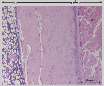
Going from left to right, label the regions shown in the image:
Bone marrow, compact bone, periosteum and muscle.
Name the pale pink structure in this image that runs around the outside of the bone:
The cortex.

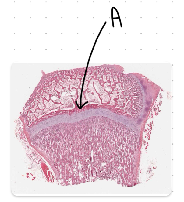
The the structure labeled A in this image:
Trabecula (spongy) bone.

Name the region labeled B in this image:
The proximal epiphysis.

Name the region labeled C in this image:
The diaphysis.

Name the structure labeled D in this image:
The articular cartilage.

Name the structure labeled E in this diagram:
The epiphyseal line.

Name the structure labeled F in this diagram:
The periosteum.

Name the structure labeled G in this image:
Compact bone.

Name the structure labeled H in this image:
The medullary cavity.
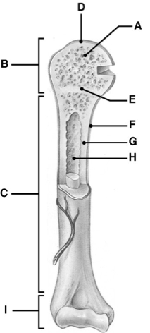
Name the region labeled I in this image:
The distal epiphysis.

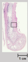
What region of a long bone does this slide show?
The metaphysis.

What is the name of the structures labeled T in this image?
Trabecular bone
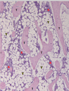
What is the name of the structures labeled R in this image?
Red (hematopoetic) marrow

What is the name of the structures labeled Y in this image?
Yellow (fatty) marrow.

What are the structures labeled A in this image?
Trabeculae.
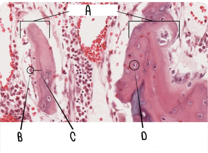
What are the structures labeled B in this image?
Osteoblasts.

What is the structure labeled C in this image?
Osteoid.

What is the structure labeled D in this image?
Osteocyte.
What is the structure labeled A in this image?
Epithelial line

What is the structure labeled A in this image?
Hyaline cartilage
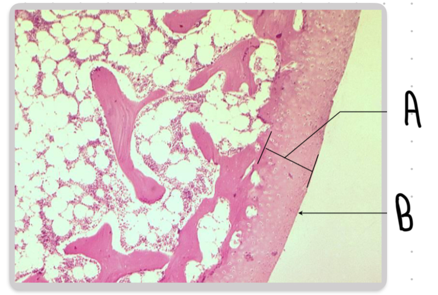
Name the structure labeled B in this image:
The articular surface.

What is the structure labeled A in this image?
Epimysium

Name the structure labeled B in this image?
The endomysium.
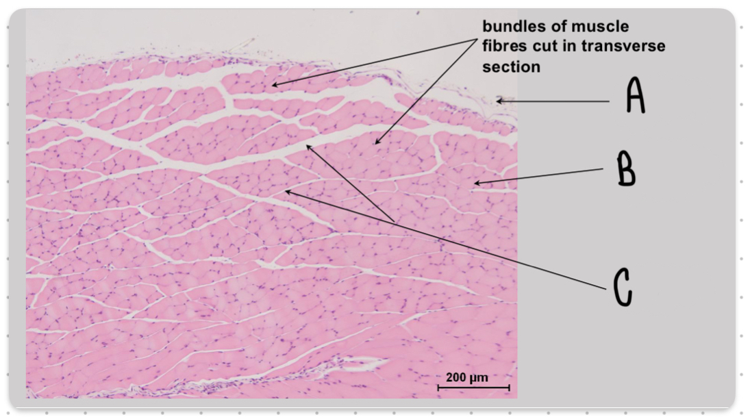
Name the structure labeled C in this image:
The perimysium.

What is the structure labeled A in this image?
The A band

Name the structure labeled B in this image:
The M line.
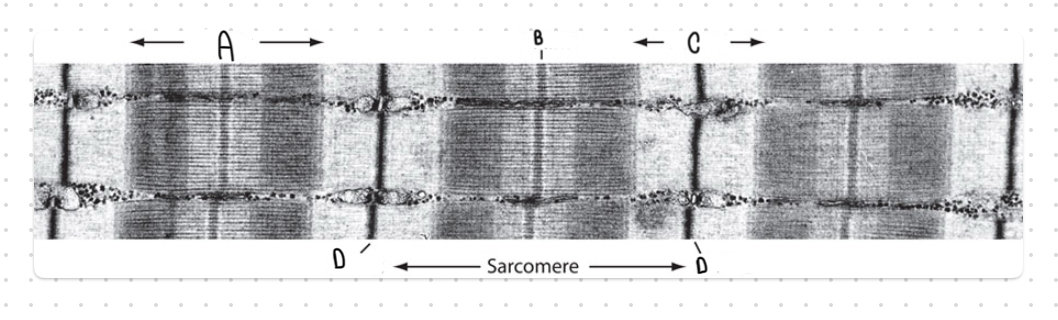
Name the structure labeled C in this image:
The I band.

Name the structures labeled D in this image:
Z lines.

What are the structures labeled A?
Intercalated discs
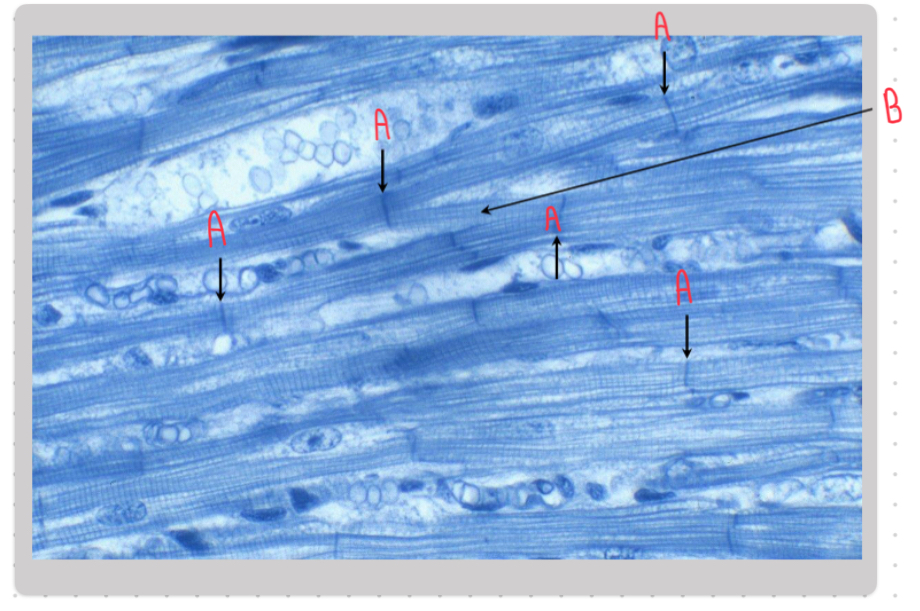
Name the structures labeled B in this image:
Branching fibres.
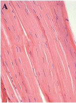
What type of muscle is shown in this image?
Smooth muscle.
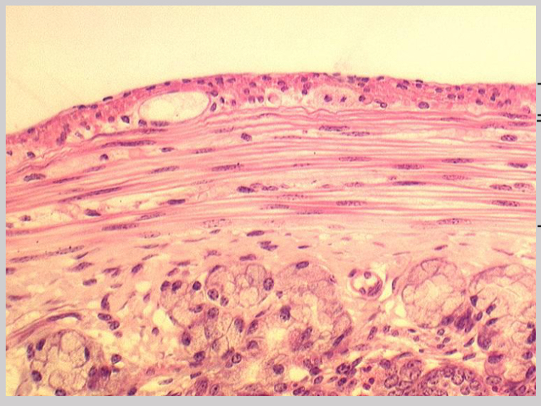
What structure is shown in this image?
A tendon.
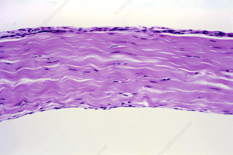
What structure is shown in this?
A ligament.

What is the structure labeled A?
Matrix
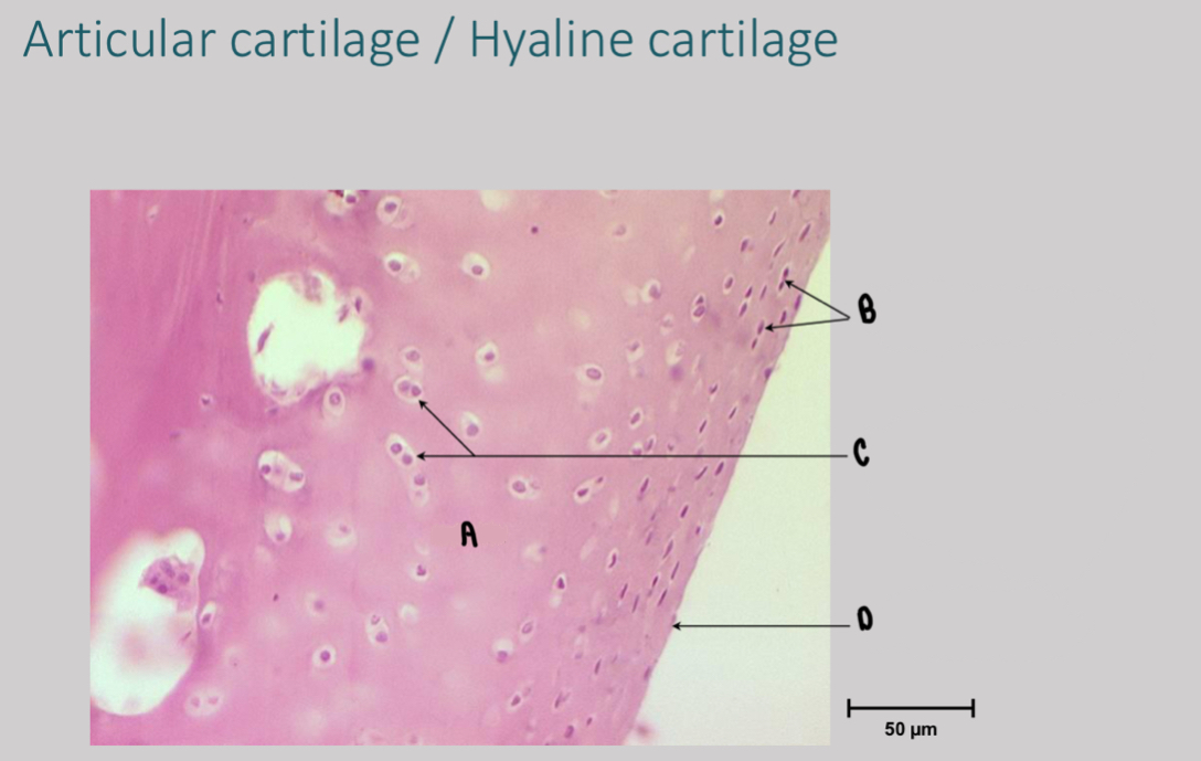
What are the structures labeled B?
Flattened chondrocytes

What are the structures labeled C?
Multiple chondrocytes in single lacunae

What is the structure labeled D?
Articular surface