Lecture 5 B: Electrons
1/12
There's no tags or description
Looks like no tags are added yet.
Name | Mastery | Learn | Test | Matching | Spaced |
|---|
No study sessions yet.
13 Terms
Electrons are generally used to treat:
Skin lesions
boost to surgical scars
Where is the patient simulated?
Simulation is most often performed on the treatment unit
On occasion, a CT Sim is performed
Why is Simulation is most often performed on the treatment unit?
if there is no conventional simulator available
or if the particular gantry or table angle required is not possible on a simulator.
Why is a CT SIM performed?
to get the depth of the lesion → to select the proper electron energy
Basic simulation of a skin lesion includes:
Position patient with immobilization devices in a reproducible way
Dr. draws field size & shape on the patient’s skin
Position gantry en face to tx area
100 SSD (as close as possible)
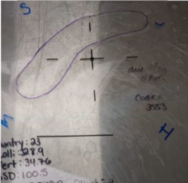
trace shape onto an insert in the electron cone
On the insert, note anatomical direction in relation to the insert
Photograph
Create a skin template using film or plastic sheeting
take skin measurement
Notate

Mnemonic: People Drive Fast. Speeding In Peaceful, Safe & Modest Neighborhoods.
shape Dr drew traced on electron cone insert & anatomical directions in direction to the insert
Position patient with proper immobilization devices in a reproducible way. How do we position them in a reproducible way?
With as few of the following as possible:
immobilization devices
table angles
gantry angles
term. en face
dfef. the plane of the beam is parallel to the plane of the surface to be treated
How many photos do we take? What photos
patient-cone interface
gantry position
the field
general position
What materials do we use to create a skin template?
film
other plastic sheeting
What do we note on the skin template? Why?
direction
3 landmarks (e.g. mole, tattoo, scar, eye, ear, mask, edge etc)
to reorient in subsequent days
What skin measurement must we obtain?
Measure the size of the field on the skin in 2 axis: long and wide
notate
name
what we are treating
orientation
ssd
gantry angle
collimator angle
table angle
hint: Never wait or stay