Biology 30 AP - Nervous System & Senses
1/90
There's no tags or description
Looks like no tags are added yet.
Name | Mastery | Learn | Test | Matching | Spaced | Call with Kai |
|---|
No analytics yet
Send a link to your students to track their progress
91 Terms
Central nervous system (CNS)
the body’s coordinating center for mechanical and chemical actions
brain & spinal cord
Outer region of CNS
white matter
protected by oligodendrocytes which have a myelin sheath
Inner region of CNS
does NOT have a myelin sheath
Functions of CNS
relays nerve impulses to and from the brain
uses sensory and motor neutrons
controls spinal reflexes
Peripheral nervous system (PNS)
all parts of the nervous system excluding brain and spinal cord
relay information between the CNS and other parts of the body (muscles & glands) for a voluntary (somatic) or involuntary (autonomic) response
Glial (neuroglial) cell
non-conducting cells
important for structural support and metabolism of nerve cells
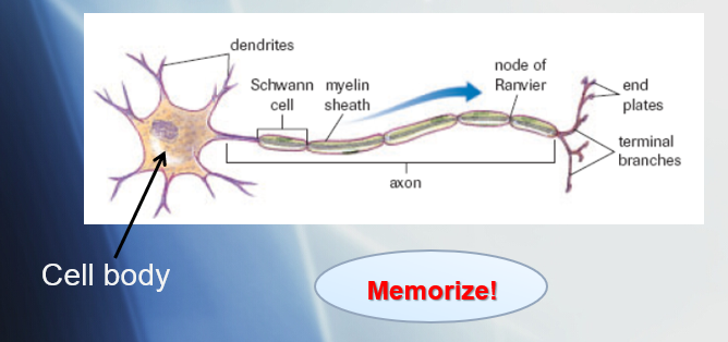
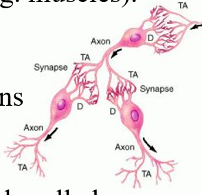
Neurons
functional units of nervous system
conducts nerve impulses

Dendrites
receive information from either environment or other neurons
projection of cytoplasm that carries impulses towards cell body

Axon
extension of cytoplasm that carries the nerve impulses away from the cell body

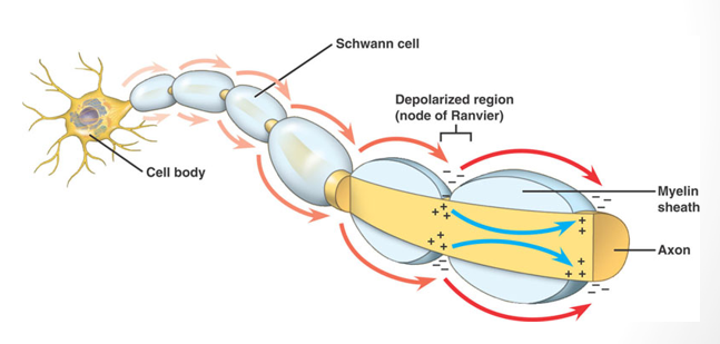
Myelin sheath
white coat of fatty protein
acts as insulation for the neurons

Schwann cell
special type of glial cell that produces myelin sheath

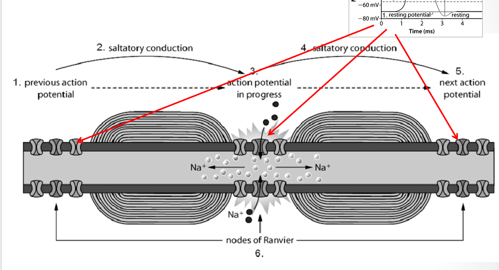
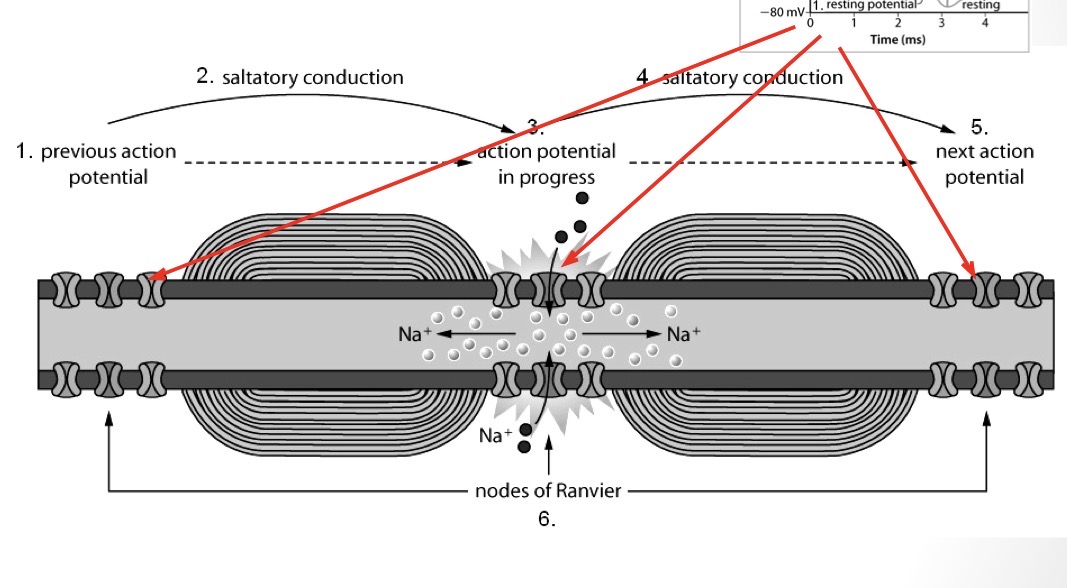
Nodes of Ranvier
regularly occurring gaps between section of myelin sheath along axon
Neurilemma
delicate membrane
surrounds axon of SOME nerve cells
promotes the regeneration of damaged axons
not all nerve cells that have a myelin sheath have a neurilemma (CNS lacks neurilemma)
Types of neurons
sensory (afferent)
interneuron
motor (efferent)
Sensory (afferent)
relay information or stimuli received by sensory receptors about the internal/external environment to the CNS for processing
Interneurons
links neurons to other neurons
found only in brain and spinal cord
integrate and interpret the sensory information and connect to outgoing motor neurons
Motor (efferent)
relay information to the effectors (cell/organ that responds)
effectors: muscles, organs, glands
Reflex arc
simplest nerve pathway
involuntary/unconscious
do not require coordination by the brain
stimulus → receptor → sensory neuron → interneuron → motor neuron → effector → response
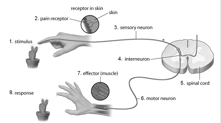
Steps of the reflex arc
The stimulus is detected by receptors in the skin
Receptors initiate nerve impulses in the sensory neurons leading from them to the spinal cord
Impulses enter the spinal cord and initiate impulses in one or more association/interneurons
Association/interneurons initiate impulses in the appropriate motor neurons
When these impulses reach the junction between the motor neurons and the muscles, the muscles are stimulated to contract and the hand is withdrawn
Resting potential
voltage difference across a nerve cell membrane when it is not transmitting a nerve impulse
more negative inside
average: -0.70 millivolts (mV)
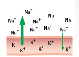
Polarization
generating resting potential
unequal distribution of positively charged ions during a resting membrane
Factors to achieve polarization/resting potential
large, negatively charged protein molecules found in intracellular fluid (too big to exit)
membrane is impermeable to some smaller negatively charged ions such as Cl-
sodium-potassium exchange pump (most important)
3 Na+ out → 2 K+ = positive charge accumulates outside the cell
Sodium-potassium exchange pump
uses ATP to actively transport sodium ions out of the cell and potassium ions into the cell
Action potential
the voltage difference across a nerve cell membrane when the nerve is excited
reversal of electric charges inside the cell membrane
Nerve impulse
series of action potentials
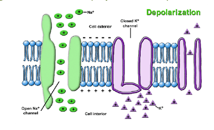
Depolarization
neuron receives a stimulus, membrane becomes more permeable to Na+ than K+
Na+ channels opened, while K+ stays closed
Na+ rush into the cell by diffusion and charge attraction
charge reversal occurs (depolarization)
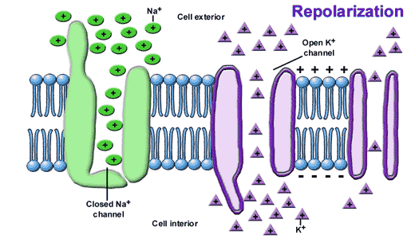
Repolarization
positive inside causes Na+ channels to close, stopping Na+ inflow
K+ channels open → K+ diffuse out, charge outside cells becomes positive again
restoring original polarity (repolarization)
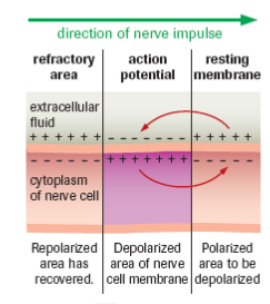
Refractory period
K+ gates close too slowly, outside cell becomes more positively charged than resting potential (hyperpolarization)
sodium-potassium pump restores resting membrane potential
membrane cannot generate another action potential during this time (refractory period)
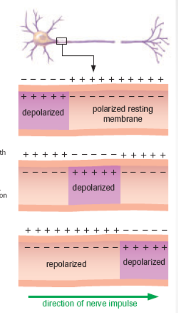
Does action potential move?
No
Wave of depolarization
many action potentials in a row
stimulus triggers first action potential → generates depolarization → triggers next depolarization → and so on
Why is the refractory period crucial/important?
stops the action potential from happening in reverse (via hyperpolarization)
What does a stimulus have to reach?
threshold to initiate an action potential
below this value do not initiate a response (all-or-none response)
increasing intensity of stimuli above threshold will NOT produce an increased response → intensity and speed of transmission are the same
neurons either fire maximally or not at all (no in-between)
Threshold
different for each neuron
What does the brain recognize for each nerve impulse?
the more intense the stimulus, the higher the frequency of impulses
the greater the frequency of impulses is a difference the brain can recognize
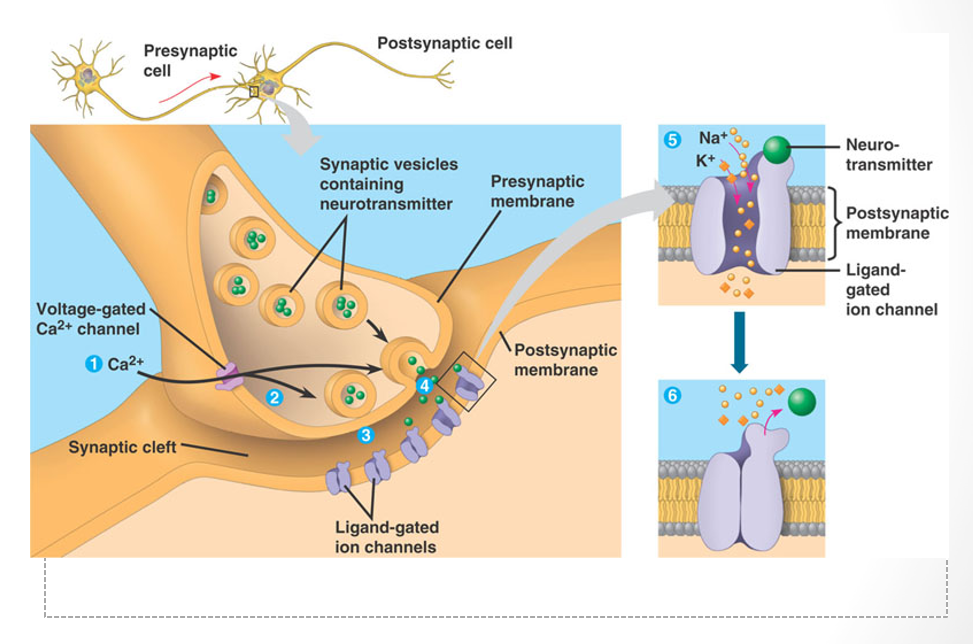
Synapses
small spaces between neurons or between neurons and effectors (e.g. muscles)
terminal branches of one neuron may join with many different neurons (rarely just two)
Neurotransmitters
small vesicles containing chemicals
released from end plates of axon terminals
Pathway of neurotransmitters from presynaptic neuron
released from presynaptic neuron
diffuses across synaptic cleft
bind to receptors
depolarize dendrites on post synaptic
Is diffusion across synapses fast or slow?
relatively slow
the more synapses over a specified distance = slower speed of transmission
Acetylcholine
found in most nerve cells
acts as an excitatory neurotransmitter by opening Na+ channels
Na+ channels open → Na+ rush into postsynaptic neuron → depolarization → action potential → nerve impulse
Cholinesterase
enzyme released by presynaptic membrane to destroy acetylcholine
Na+ channels close → neuron recovery stage
Inhibitory neurotransmitters
make postsynaptic membrane more permeable to K+
more K+ gates open → more K+ diffusion out of cell → hyperpolarization → action potentials inhibited
Steps for impulse to be transmitted through synaptic cleft
Action potential reaches presynaptic terminal
Voltage-gated Ca2+ channels open
Influx of Ca2+
Synaptic vesicles fuse with membrane (exocytosis)
Neurotransmitters released into synaptic cleft and diffuse to postsynaptic terminal
Neurotransmitter binds to receptor on postsynaptic membrane
Excitatory or inhibitory
If threshold reached → action potential initiated
Neurotransmitters broken down by specific enzymes in synaptic cleft
Summation
effect in the postsynaptic neuron produced by accumulation of neurotransmitters from two or more neurons
Neurotoxins
bacterial proteins that are exocytosed into nerve endings
inhibit the release of essential neurotransmitters
e.g. botulinum, tetanus
Why do nerve impulses move faster along myelinated nerve fibres?
myelin sheath acts as an electrical insulator, forcing the impulse to jump b/w the nodes of Ranvier instead of moving along/down the axon
Meninges
protective layers surrounding the brain and spinal cord
3 layers
3 layers of meninges
outer: dura mater
middle: arachnoid mater
inner: pia mater
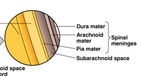
Cerebrospinal fluid
circulates between innermost and middle meninges
circulates through central canal of spinal cord
acts as a shock absorber
transports medium (carrying nutrients to/removing wastes from)
Meningitis
inflammation of meninges
bacterial/viral infection
can spread to underlying brain tissue
Blood-brain barrier (right)
tight seal
only allows certain substances to pass through
Foramen magnum
opening from the skull
where the spinal cord extends downward through a canal within the backbone
Central grey matter nerve tissue
non-myelinated interneurons
White matter nerve tissue
myelinated nerve fibers from sensory and motor neurons
Dorsal root
brings sensory info into the spinal cord
Ventral root
carries motor info from the spinal cord to the peripheral nervous system
Forebrain
consists of: cerebrum, thalamus, hypothalamus, olfactory bulbs

Cerebrum
largest part of brain
left and right hemispheres - act as coordinating centers from which sensory info and motor actions originate

Right side of brain
visual patterns
spatial awareness
Left side of brain
linked to verbal skills
Corpus callosum
allows for communication b/w left and right hemispheres
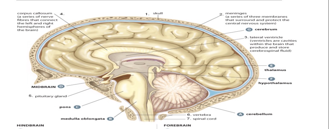
Cerebral cortex
surface of cerebrum
composed of grey matter
many fissures (folds or “sulci”) - increase surface area
bumps or hills = “gyri”
Thalamus
relay station for sensory info going to cerebrum

Hypothalamus
small size
big role in maintaining body’s internal equilibrium
direct connection w/ pituitary glands unites nervous and endocrine systems

Lateral ventricle
produce and store cerebrospinal fluid
Olfactory bulbs
receive and interpret information about smell
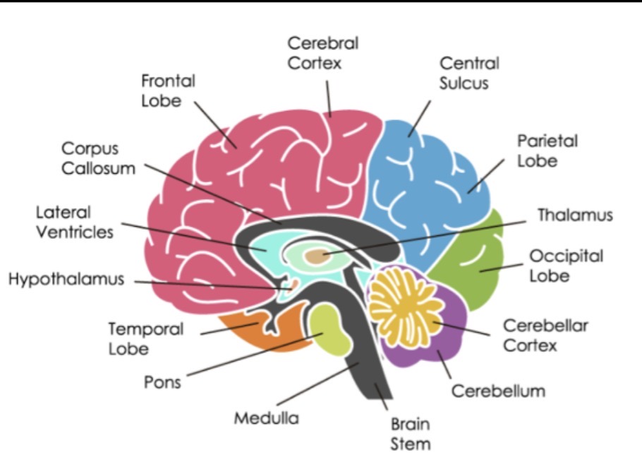
Midbrain
lies just below thalamus
relay center for some eye/ear reflexes
Hindbrain
posterior to midbrain
joins with spinal cord
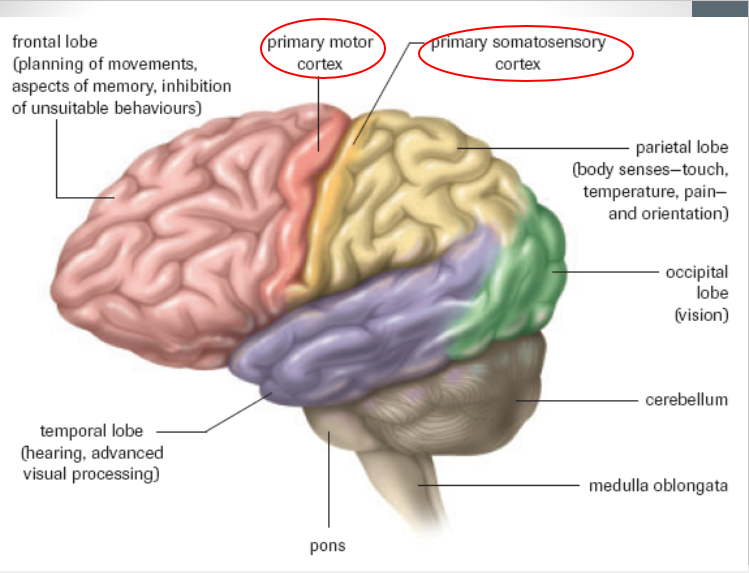
consists of: cerebellum, pons, and medulla oblongata

Cerebellum
controls limb movements, balance, and muscle tone

Pons
relay station for regions of cerebellum and b/w cerebellum and medulla

Medulla oblongata
connection b/w CNS and PNS
controls involuntary muscle action (e.g. breathing, heart rate)
coordinating center for the autonomic nervous system

What are MRIs, CTs, & PETs used for?
non-invasive tools
map human brain function
screen for diseases
Frontal lobe
motor area control movement to voluntary muscles
linked to intellect, memory, emotions, and personality
inhibits unsuitable behaviors

Temporal lobe
process hearing (and some visual) information
linked to visual and verbal memory, emotions, learning, and interpretations of language

Parietal lobe
associated with touch and temperature awareness from the skin
linked to emotions and interpretations of language
body senses & orientation

Occipital lobe
associated with vision
interpret visual information
Somatic vs. autonomic
both consist of sensory and motor neurons
somatic: responds to external stimuli
autonomic: responds to internal stimuli
Sensory somatic system (SSNS)
info about external enviro to CNS
sends messages to skeletal muscles (voluntary)
somatic reflex arcs do NOT fit
12 pairs of cranial nerves
31 pairs of spinal nerves
What do cranial nerves control?
vision
hearing
balance
taste & smell
facial & tongue movements
muscles of the head & neck
Spinal nerves
operate skeletal actions that we consciously make to cope with our surroundings
e.g. putting on sunglasses
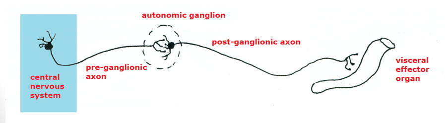
Autonomic nervous system (ANS)
info about body’s internal enviro to CNS
body responds to maintain homeostasis (involuntary)
controls smooth/cardiac muscle and internal organs/glands
2 systems: sympathetic and parasympathetic
2 groups of motor neurons of PNS
stimulate target effectors
Preganglionic neurons
Postganglionic neurons
Preganglionic neurons
run from the CNS to a ganglion (mass of nerves) where they connect with a second group

Postganglionic neurons
run to target organ, muscle, or gland
Sympathetic system
fight or flight
prepares the body for stress
Parasympathetic system
rest and digest
restore the body to normal
Vagus nerve
important cranial nerve of the parasympathetic system
wandering “vagabond”
innervates the heart, bronchi of the lungs, liver, pancreas, and the digestive tract
Why might meninges not be visible?
extremely thin and tightly attached to the nervous system
pulled/cut off during shipping
Function of the meninges
protection: cushion and protect the brain and spinal cord by enclosing CSF (absorbs chock and reduces injury)
delivery and regulation of the CSF
dura mater: outer layer that supports large veins
arachnoid mater: middle layer that provides a cushioning effect for CNS
pia mater: inner layer where blood vessels travel between brain and meninges
What makes the corpus callosum lighter in colour than the cerebrum?
made mostly of white matter - myelinated axons - that appears pale
cerebrum outer layer made mostly of grey matter
Why are arteries and veins not visible inside the brain? What replaces the blood?
no arteries or veins inside the brain
replaced by cerebrospinal fluid
What type of nerve conducts impulses to the olfactory bulbs?
sensory neuron in the olfactory nerve