Regulation of breathing
1/42
There's no tags or description
Looks like no tags are added yet.
Name | Mastery | Learn | Test | Matching | Spaced |
|---|
No study sessions yet.
43 Terms
Cerebrum function (2)
largest part of brain
Voluntary breathing
Medulla Oblongata function (3)
Involuntary breathing
Rhythmicity centres
Respiratory control centres
Motor Innervation of Muscles Involved in Breathing (2)
Breathing involves the contraction and relaxation of specific muscles controlled by somatic motor neurons in the spinal cord
These neurons receive inputs from both voluntary and involuntary pathways
Motor Innervation of Muscles (3)
Diaphragm
Muscles of the Rib Cage
Abdominal Muscles
Diaphragm - Motor Innervation (2)
Nerve: Phrenic nerve
Cell Body Location: Cervical level of the spinal cord (gray matter)
Muscles of the Rib Cage - Motor Innervation (2)
Includes external intercostals, internal intercostals, and parasternal intercostals
Cell Body Location: Gray matter at various levels of the thoracic spinal cord
Abdominal Muscles - Motor Innervation
Cell Body Location: Thoraco-lumbar parts of the spinal cord
Somatic Motor Neurons (3)
responsible for controlling respiratory muscles
located in the gray matter (central H-shaped region) of the spinal cord
controlled by descending tracts from Medulla Oblongata + Cerebral Cortex
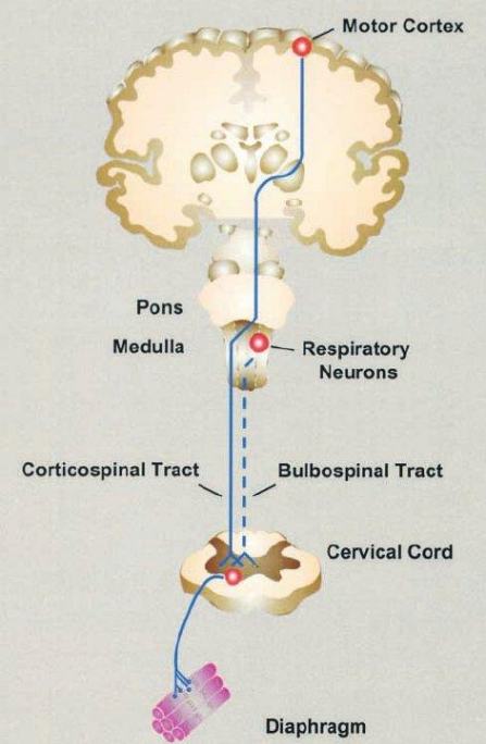
Descending (Tracts) pathways (2)
Voluntary Pathway
Involuntary Pathway
Voluntary Pathway / Breathing (3)
Origin: Cerebral Cortex
Path: Axons descend in the corticospinal tracts
Allows conscious control of respiration (e.g., holding breath)
Involuntary Pathway / Breathing (3)
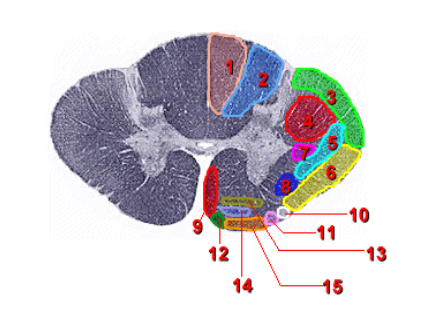
Origin: Respiratory control centers in the Medulla Oblongata (brainstem)
Path: Axons descend in the lateral (4) and ventral (9) white matter of the spinal cord
Responsible for rhythmic + automatic control of breathing (e.g., during sleep)
Involuntary, Automatic Breathing (3)
Controlled by neurons in the ventrolateral region of the medulla oblongata
Rhythmicity Centre generates the rhythm
Pacemaker neurons in the Rhythmicity Centre responsible for the rhythmic firing of inspiration and expiration neurons
Pacemaker neurons (3)
Located within the Rhythmicity Centre of the medulla oblongata
Responsible for the rhythmical firing of inspiration (I) and expiration (E) neurons
Show spontaneous, cyclical changes in membrane potential, similar to the pacemaker cells of the heart
Ventrolateral (Rhythmicity Centre) components (2)
I neurons (inspiration): 4 types
E neurons (expiration): 2 types
Ventrolateral Centre of the medulla oblongata parts (2)
Dorsal Respiratory Group
Ventral Respiratory Group
Dorsal Respiratory Group
Stimulates spinal motor neurons of the phrenic nerve to innervate the diaphragm
Ventral Respiratory Group (2)
I neurons stimulate spinal interneurons activating spinal motor neurons
E neurons inhibit motor neurons of the phrenic nerve during expiration
Respiratory Control Centres in Pons (2)
Apneustic Centre
Pneumotaxic Centre
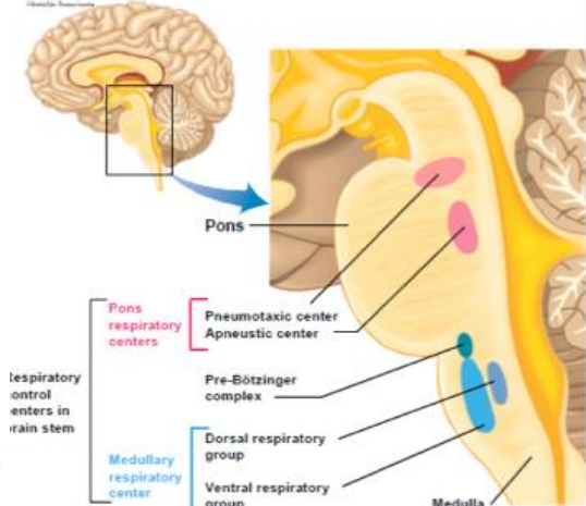
Apneustic Centre
Promotes inspiration by stimulating I neurons in the medulla oblongata
Pneumotaxic Centre
Inhibits inspiration by antagonizing the apneustic centre
Chemoreceptors (2)
sensory receptors that respond to chemical stimuli in the environment
changes in the concentration of certain molecules such as carbon dioxide (CO₂), oxygen (O₂), and hydrogen ions (H⁺)
Sensory feedback from Chemoreceptors sensitive to (5)
Arterial blood:
PCO2
PO2
pH
PCO2 + pH changes in Brain interstitial fluid + Cerebrospinal fluid
Central chemoreceptors + Peripheral chemoreceptors similarity (3)
sensitive to PCO2 + PO2 + pH in Arterial blood
detect changes in blood gas levels (PCO2, PO2) and pH
thus regulating ventilation to maintain homeostasis
Central Chemoreceptors location
Medulla Oblongata, near the exit of the IX and X cranial nerves, on the ventrolateral surface
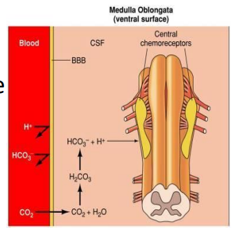
Central Chemoreceptors Stimulated by (2)
Arterial PCO2 (most)
Cerebrospinal fluid PCO2 and pH
Central Chemoreceptors Function (4)
Responsible for 70-80% of the increase in ventilation in response to a sustained rise in arterial PCO2
Communicate with the rhythmicity centre neurons in the medulla
Their response to a rise in PCO2 takes several minutes
The immediate response to PCO2 elevation is mediated by peripheral chemoreceptors
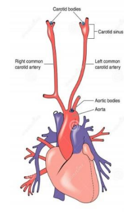
Peripheral Chemoreceptors Location (2)
Carotid Bodies
Aortic Bodies
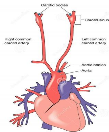
Carotid Bodies (2)
Small nodules at the bifurcation of the common carotid artery into the internal and external carotid arteries
Send information via the glossopharyngeal nerve
Aortic Bodies (2)
Small nodules around the aortic arch
Send sensory information to the medulla oblongata via the vagus nerve
Peripheral Chemoreceptors Stimuli (3)
Decrease in pH in arterial blood due to an increase in H+ ions
Immediate response
PCO2 rise in hypoventilation produces a Fall (acidic) in pH which stimulates the peripheral chemoreceptors

PCO2 and Respiratory Regulation
PCO2 is a stronger stimulus for the reflex control of ventilation
Respiratory rate and depth are adjusted to maintain arterial PCO2 at 40 mmHg
PO2 content in the blood fluctuates less with changes in ventilation because oxygen is attached to hemoglobin
Blood PCO2 and pH are affected more quickly by changes in ventilation
Oxygenation of the blood occurs as a side product
Hypercapnia (2)
Refers to a rise in PCO2 due to hypoventilation
Chemoreceptors respond by stimulating an increase in ventilation
Hypocapnia (2)
Refers to a fall in PCO2 due to hyperventilation
Chemoreceptors respond by decreasing ventilation
Chemoreceptor Input
Chemoreceptor input to the brainstem modifies the rate and depth of breathing to maintain relatively constant levels of PCO2 + pH + PO2
The Effect of Arterial PO2 on Ventilation (5)
The effect of PO2 on breathing is indirect
PO2 influences chemoreceptor sensitivity to changes in PCO2
Low PO2 increases chemoreceptor sensitivity to PCO2
High PO2 decreases chemoreceptor sensitivity to PCO2
Breathing 100% oxygen blunts the response to PCO2 decreasing the ventilation rate
Hypoxic Drive (5)
PO2 in the blood falls below 70 mmHg (Hypoxemia)
Ventilation increases significantly due to a direct effect on the carotid bodies
sensitive to plasma oxygen (not the oxygen bound to hemoglobin in red blood cells)
Not normally occur at sea level
Relevant in Chronic Obstructive Pulmonary Disease (COPD)
Chronic Obstructive Pulmonary Disease (COPD) - Hypoxic Drive (3)
Chronic retention of PCO2 due to inadequate ventilation results in blunting of the chemoreceptor response to increased PCO2
Over time, chronic hypoxia reduces the sensitivity of carotid bodies to low PO2, and even hypoxic drive is no longer effective
Breathing problems are exacerbated
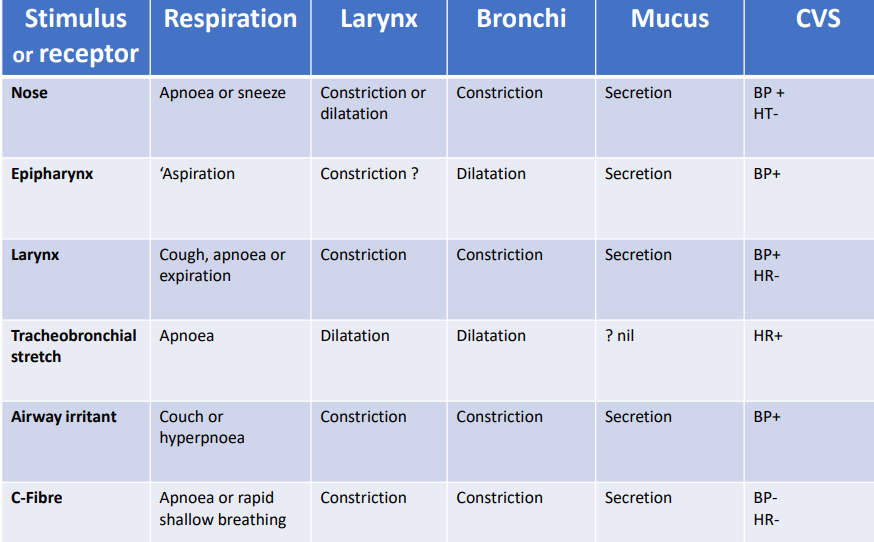
Pulmonary Receptors
Influence brainstem respiratory control centres via sensory fibers in the vagus nerve
Pulmonary Receptors Types (3)
Rapidly Adapting Receptors (Irritant receptors)
Pulmonary Stretch Receptors
Unmyelinated C fibres
Rapidly Adapting Receptors (Irritant receptors) (5)
Found in the walls of the larynx and lungs
Stimulated by an increase in pulmonary interstitial tissue fluid
Cause coughing in response to irritants like smoke, inhaled fine particles, asthma, and smog
Pulmonary Stretch Receptors (3)
Inhibit respiratory control centres during inspiration
Prevent overdistention of lungs
Involved in the Hering-Breuer reflex
Unmyelinated C fibres (2)
Stimulated by capsaicin (chemical in hot peppers)
Initial apnoea, followed by rapid shallow breathing
Motor responses to airway pulmonary receptors (photo!)