Bacteria and antibacterial agents introduction 6.2:2:
1/45
Earn XP
Description and Tags
introduction 1. 6.2
Name | Mastery | Learn | Test | Matching | Spaced | Call with Kai |
|---|
No analytics yet
Send a link to your students to track their progress
46 Terms
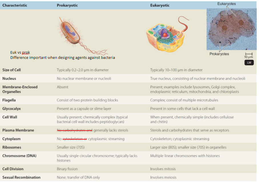
what are the main differences between Eukaryotes and Prokaryotes?
Eukaryotes often bigger in size than Prokaryotes
Prokaryotes don’t have a nuclear membrane or nuclei
DNA in Prokaryotes is free-floating - in plasmids as well
No membrane bound organelles in Prokaryotes e.g. mitochondria, EPR, golgi, chloroplast
Flagella can be present in both
Ribosome is smaller in Prokaryotes ~ 70s vs 80s
prokaryotes often have single circular chromosome with no histones
Prokaroytes divide by Binary fission
Eukaroytes divide by mitosis
Prokaryotes dont have sexual reproduction (they can transfer DNA) whereas Eukaryotes do by meiosis
Prokaryotic cell wall contains peptidoglycan, fungi has chitin
Prokaryotic plasma cell membrane lacks sterols unlike in eukaryotic
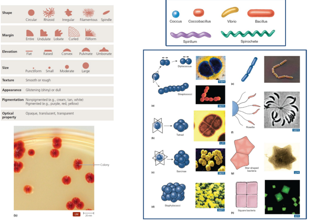
what are common shapes of bactera?
coccus - spherical
coccobaciullus - cylinder type
vibrio - thin and longand circular
bacillus - longer than coccobacillus
spirillium - spiral
spirochete - higher frewuency of swirls
e.g streptococci is 7 spheres connected as strept=7
What method do you use to distinguish between type of bacteria?
Gram staining
What are the main types of bacteria?
Gram positive
Gram negative
Acid-fast bacteria - not a type, basically a type of stain for specific bacteria
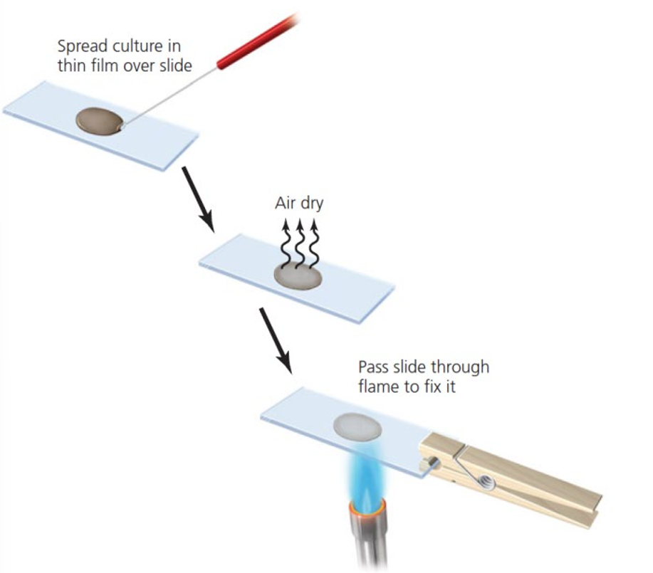
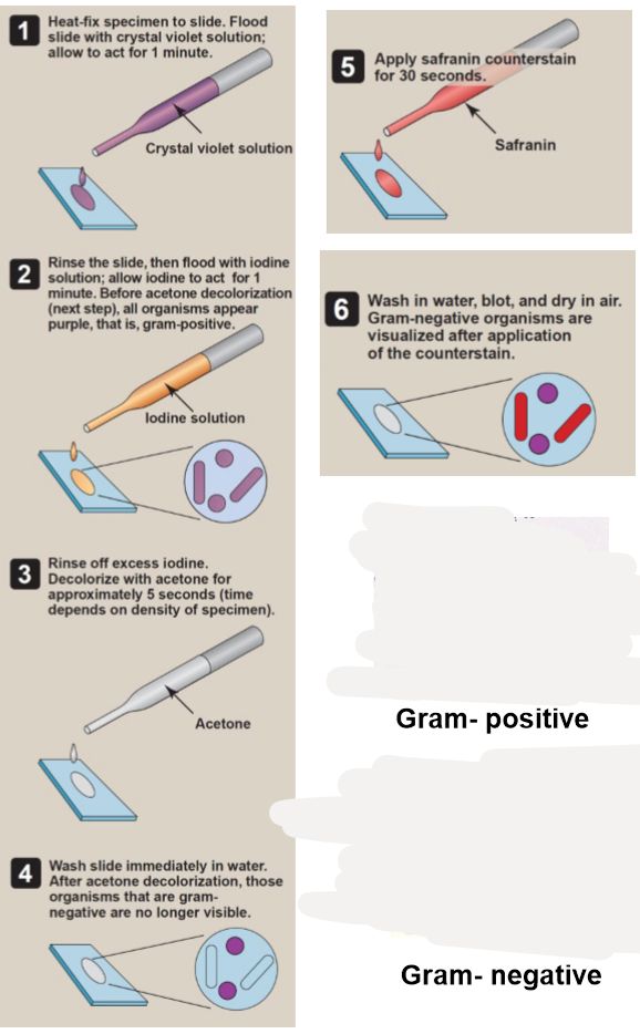
Explain the process of gram staining
Heat fix specimen to slide
flood slide with crystal violet solution and leave for 1 minute (purple)
rinse the slide then flood with iodine solution for 1 minute
Specimen will appear purple
Rinse of excess iodine (orange)
Declolourise with acetone for 5 seconds (depends on specimen) (clear)
wash slide immediatley in water
after washing, gram negative bacteria are not visible
apply safranin counterstain for 30 seconds (red)
wash in water, blot, and dry in air
gram negative bacteria will be visible after adding this counterstain
Describe the result for Gram (+) during staining
Staining depends on the ability of the bacteria to retain the dye
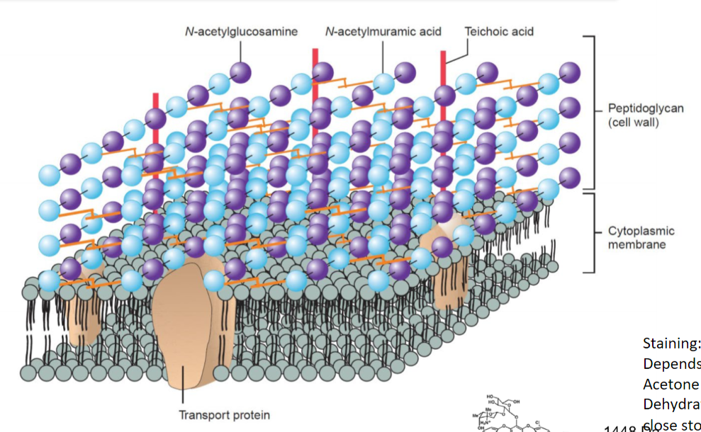
Gram (+) have thick peptidoglycam cell wall
Acetone dehydrates the cell wall and extracts the stain
dehydration collapses the cell wall, closes the pores, (causing it to intertwine) trapping the stain within the cells
so dye remains in the cell = purple appearance
so Gram (+) retains large crystal violet/iodine complex molecules upon acetone treatment
in pic, cell wall is thicker than cell membrane
Why can antimicrobial agents enter gram (+) bacteria?
Cell wall is porous, enzymes and antimicrobial agents enter the cell through here
max size entry is 50,000 daltons
most antimicrobial agents are <50,000 daltons
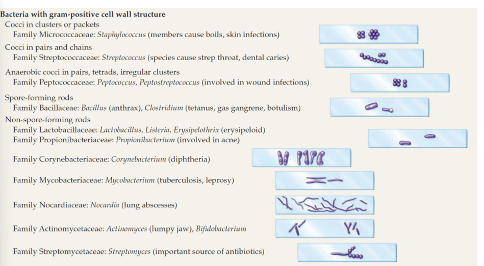
Give examples of Gram positive bacteria
Staphylococcus - skin infections
streptococcus - sore throat, dental caries
streptomyces - source of antibodies
Clostridium - wound infections
bacillus
lactobacillus
listeria
mycobacterium - leprosy, tuberculosis
often cocci shape
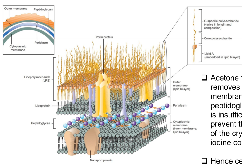
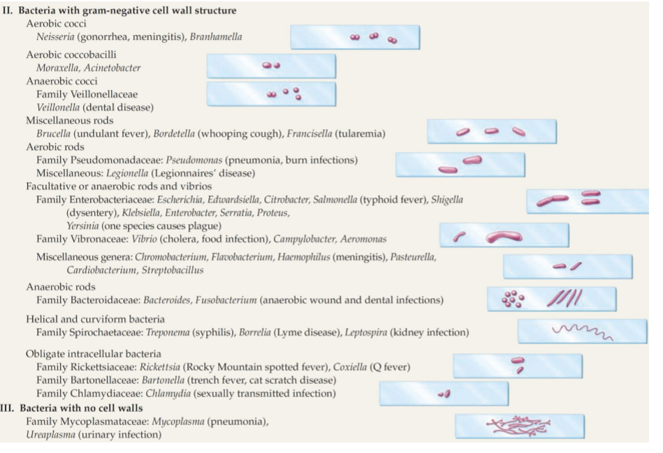
Describe Gram (-) bacteria cell wall
Thin peptidoglycan cell wall
an additional outer membrane
outer membrane contains porin proteins
higher content of sterols/lipids in cell wall
Describe gram (-) staining result
Acetone causes dehydration
Acetone removes the outer membrane
The thin cell wall peptidoglycan layer cannot prevent the removal of the crystal violet iodine complex
As, in the thin cell wall, there is insufficient cell wall layer
crystal violet/iodine complex washed away
counterstain safranin stains it red as a lighter colour than purple
What do Porin channels do?
allow diffusion of nutrients into the cell, into cytoplasm through membrane
water channels - hydrophilic
could be a drug target
Give examples of Gram (-) bacteria
Neisseria Gonnorhoea & meningitis
Brucella, Bordetella coughs and fevers
Pseudomonas
E.coli
Salmonella
shigella
enterobacter
V.cholera
chlamidya
campylobacter
fusobacterium (anaerobic wound and dental infection)
ureaplasma (urinary infections)
What do you use to stain acid-fast bacteria and why?
Carbolfuschin (red)
unlike gram (+) contains 60& lipid (mycolic acid) and less peptidoglycan - waxy prevents them from being stained
so they are hard to stain, but don’t get discoulored by alcohol
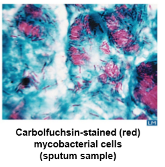
Describe Mycobacteria (acid-fast) structure and growth
thick cell wall
additional capsule-like-material ontop
unlike gram (+) contains 60& lipid (mycolic acid) and less peptidoglycan - waxy prevents them from being stained
grow slowly as lipids restrict flow of nutrients and other agents into the cell
lots of energy required to construct cell wall
this is why Tuberculosis takes months to grow
give examples of acid fast bacteria
mycobacterium tuberculosis (tuberculosis)
mycobacterium leprae (leprosy)
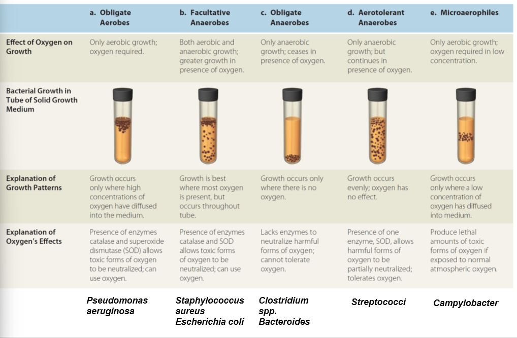
what are the types of bacteria growth
obligate aerobes
facultative anaerobes
obligate anaerobes e.g metronidazole in wounds- low oxygen
aerotolerant anaerobes
microaeophiles
SOD breaks down toxic oxygen
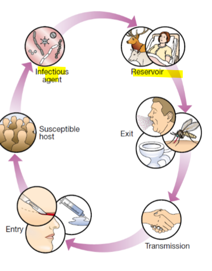
what are sources of microorganisms?
food
water
other humans
environment
animals, birds, insects, vectors
human self microflora
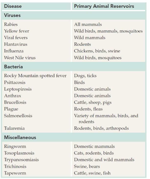
examples of zoonotic infections
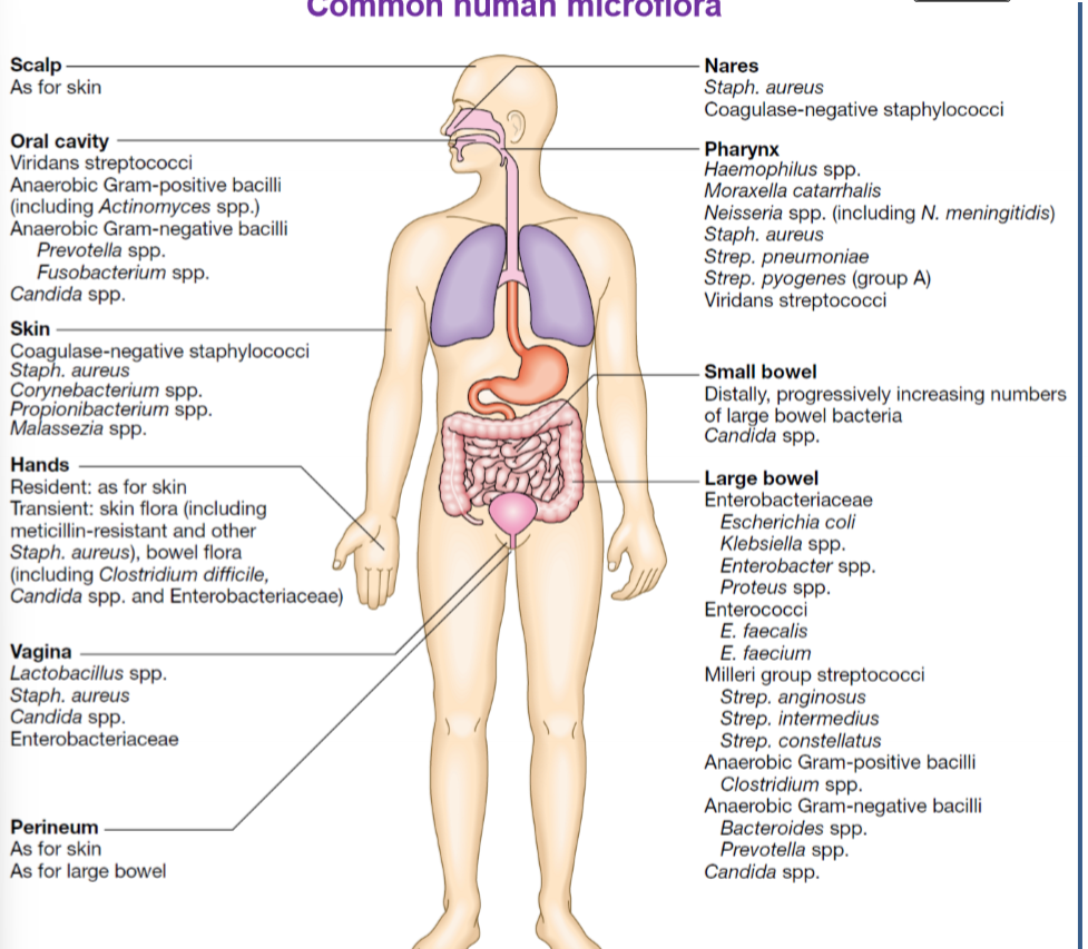
give examples of common human microflora
mostly in bowels as we know their used to break down bile acid, increase calcium absorption, makes vitamin B12, K, iron, calcium
what is a pathogen?
an organism capable of causing disease
what is a true pathogen?
causes disease in a healthy immunocompetent patient
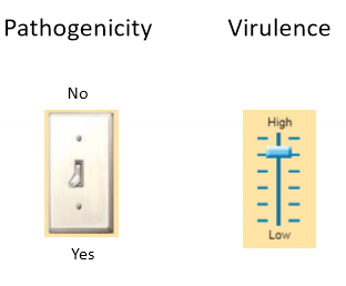
what is pathogenicity
ability to cause disease
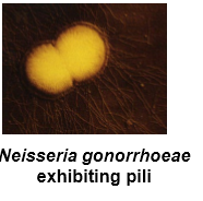
for example, neisseria gonorrhoeae requires pilli to attach to urethral epithelium
strains with no pilli are non-pathogenic as they do not causes disease
as washed away by urine
what is Virulences?
an organisms relative power to causes disease
illness can be asymptomatic, mild or very severe
so virulence factors effect extent of disease
a factor could be a human- how their immune system responds - human factors
give an example of pathogenicity vs virulence for understanding
streptococcus pneumoniae does not causes disease if it lacks a capsule so isnon-pathogenic
so the capsule determines the virulence of the organism
both pneumococci capsular type 3 and 30 produce capsules so are both pathogenic, however 3 causes severe disease whereas 30 rarely causes severe disease
why? as capsule protects it against immune system
how do you identify that a bacterium is a pathogen?
Koch’s Postulates
the suspected pathogen must be present in all cases of the disease and absent from healthy animals - how? - by using microscope
the suspected pathogen must be grown in pure culture - how? agar plate
cells from a pure culture of the suspected pathogen must cause disease in a healthy animal - how? inoculate healthy animal
the suspected pathogen must be re-isolated and shown to be the same as the original - how? - remove it from animal and observe under microscope
antibodies to the organism appear during course of the disease
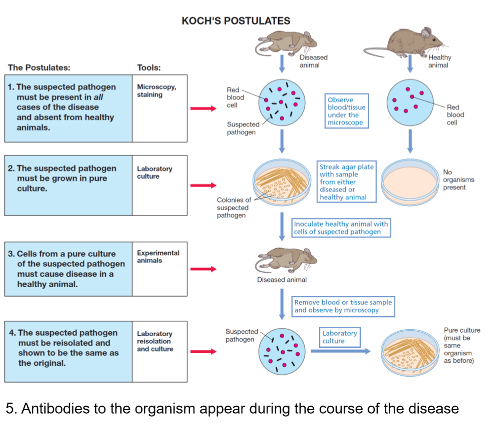
What are limitations to Koch’s Postulates
it works well for many bacteria, but does not work for all infectious disease
not all bacteria can be cultured in labs e.g. mycobacterium leprae
ethics prohibit attempts to transfer from person to person so we use animals instead
you need a pure sample, disease of possible polymicrobial origin wont be pure
immunosupression may lessen the antibody response and render the host very susceptible to the disease - which may not be a good reflection
genetic predisposition of individuals e.g if you have sickle cell, more likely to have certain infections
hosts own flora - how do we know its not your own flora e.g. e.coli, candida spp.
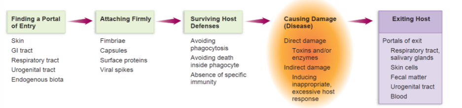
what are the 5 characteristics of a successful pathogen?
survival and transmission in the environment
attachment to the surface of the host
overcoming the body defences against infection
ability to damage the host, directly or indirectly (disease)
ability to replicate in the host, producing progency able to infect others (exit)
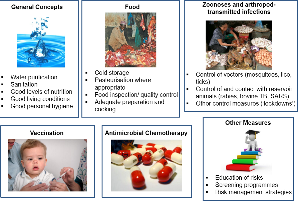
Give examples of strategies for controlling infectious disease
water purification
sanitation
good levels of nutrition
good living conditions (overcrowding)
good personal hygiene
food- cold storage
pasteurization
food inspection and quality control
adequate preparation and cooking e.g. meat
control of vectors e.g. mosquitoes, rats, lices, ticks
control of and contact with reservoir animals (rabies, bovine, TB, SARS)
control measures e.g. lockdown
vaccination
antimicrobial chemotherapy
education of risk
screening programmes
risk management strategies e.g. for covid or flu outbreak
what is antimicrobial chemotherapy?
antimicrobial medicine e.g antibiotics
depends on selectivity, so it must be selective
what must antimicrobials be, in order to be effective and what led to this discovery?
must be toxic in vivo to the microbe and not the host
use of dyes that differentially stained tryanosomes (protozoa) (stained the infective cells but not the actual human tissue) led to the concept of selective toxicity
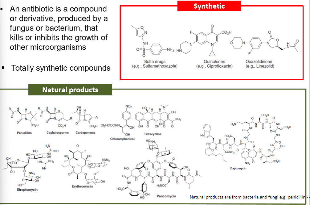
what is an antibiotic, and what is it made of?
is a compound or derivative produced by a fungus or bacterium that kills or inhibits the growth of other microorganisms
can be synthetic
can be natural e.g. by bacteria or fungi (penicllin)
how do we classify antibacterial agents?
bacteriocidal or bacteriostatic - inhibits growth
depends on the bacterial species e.g. chloramphenicol inhibits e.coli but kills haemophilus influenzae
by target site
by chemical structure
why does bacteriostatic effect work even after medicine is complete?
host immune system prevents the bacteriostatic bacteria from growing after
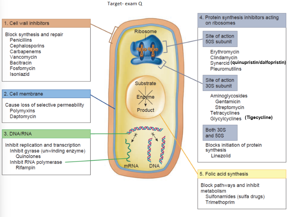
What are the antibacterial targets? EQ
Cell wall
cell membrane
DNA/RNA
folic acid synthesis
protein synthesis on ribosomes
what do you need to know about antibacterial agents?
what it is - chemical structure and synthesis
what it does - mechanism, target
where does it go - ADME
when its used
limitations - e.g. toxicit, efficacy, SE, CI
cost - more expensive, less? - just an idea
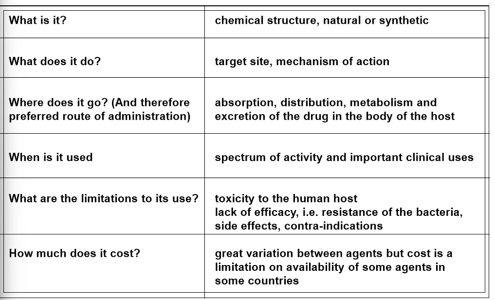
examples of common antibacterial agents
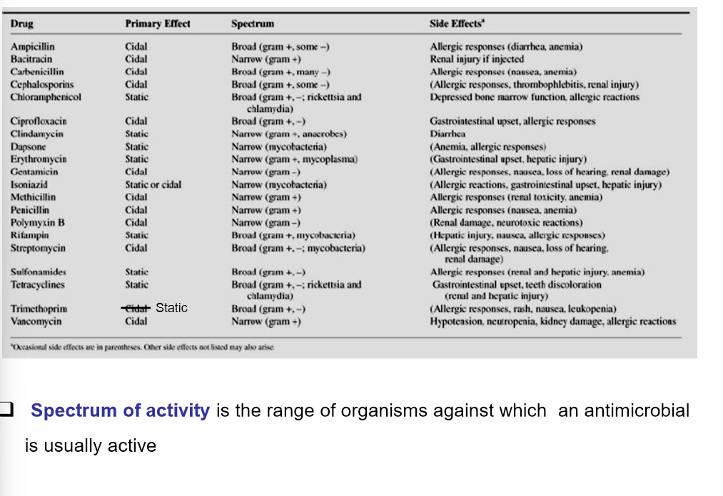
what is spectrum of activity?
which organisms are antimicrobials active against , so could be broad
How are antibiotics different from other drugs?
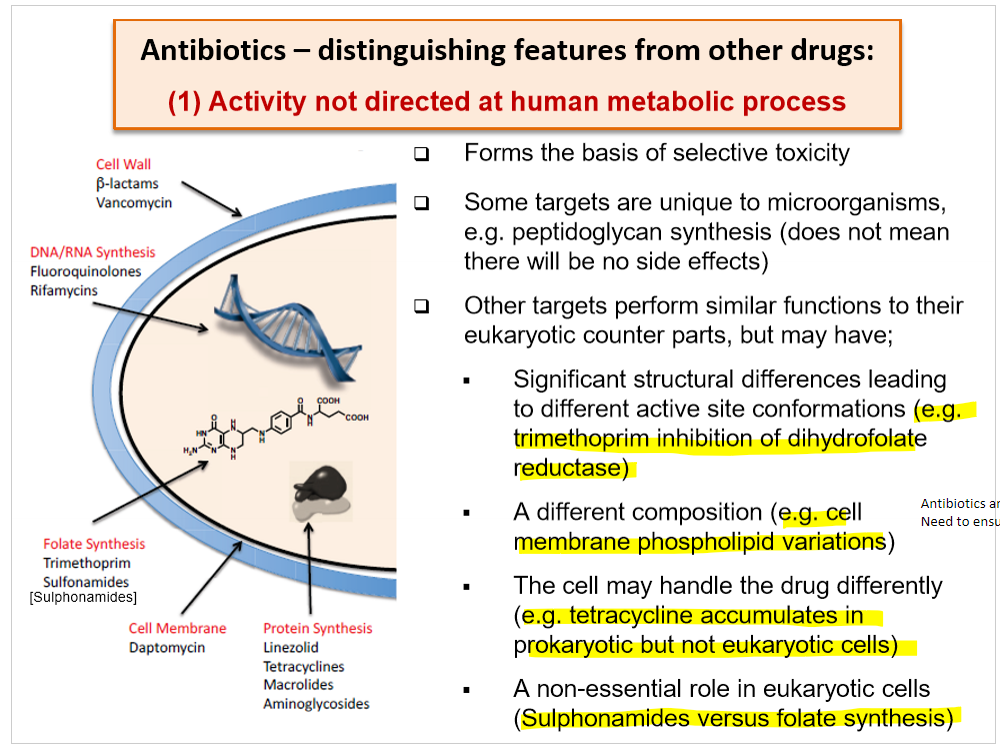
activity is not directed at human metabolic process - target is the microorganism
activity is needed against several body sites
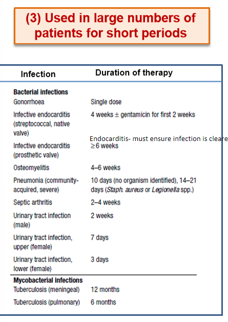
used in large numbers of patients for a short period
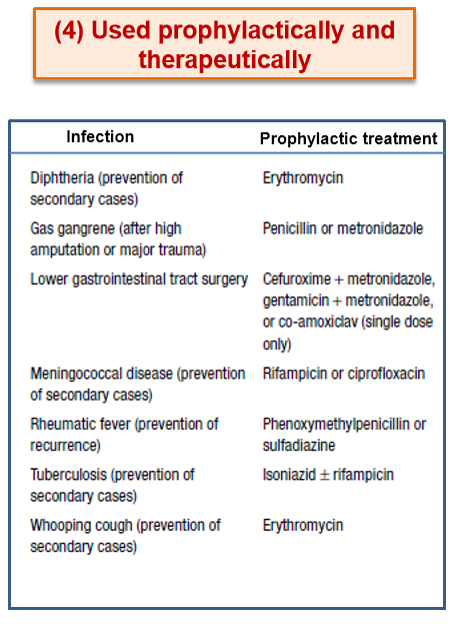
used prophylactically and therapeutically
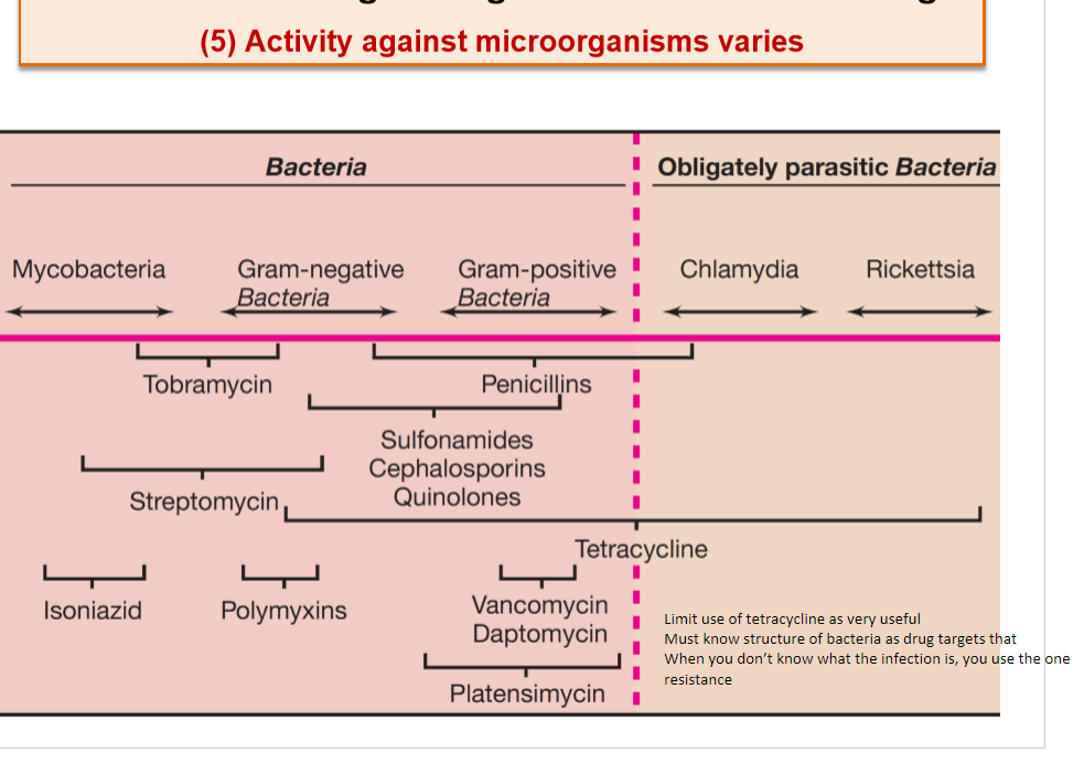
activity against microorganisms varies
drug resistance may transfer to other bacteria
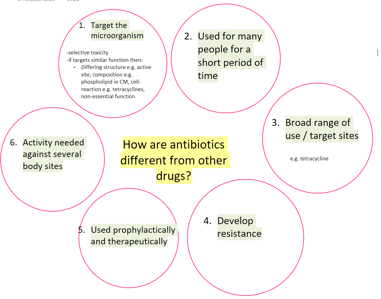
describe- activity is not directed at human metabolic process - target is the microorganism
it has selective toxicity
attacks the mircroorganism
some targets are unique to the microorganism e.g. peptidoglycan synthesis
other targets perform similar functions as those in eukaryotes but may have: significant structural differences leading to different active site conformations, different composition e.g. phospholipid, the cell may handle the drug differently, it may have a non-essential role in eukaryotic cells
this does not mean that there will not be side effects to the patient
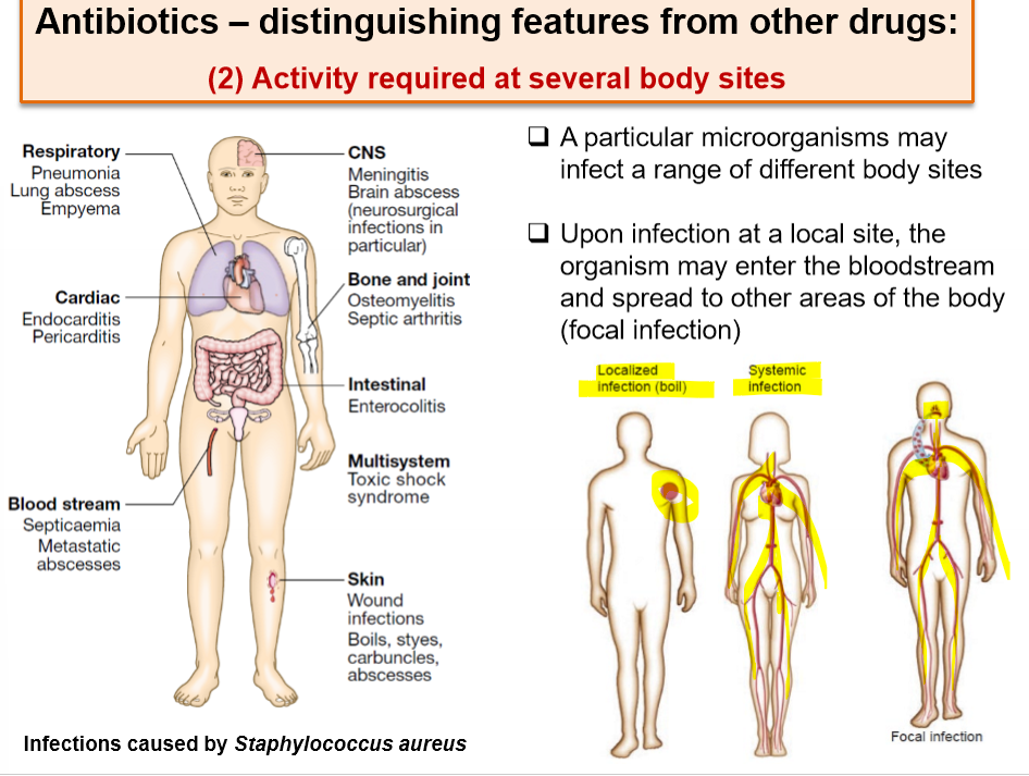
describe- activity is needed against several body sites
one microorganism may infect a range of different body sites - why? travel in blood
this is known as a focal infection- enters one part, then spreads by bloodstream
describe - used in large numbers of patients for a short period
e.g. 3-5-7 days only
for endocarditis, its used for a long period to ensure the infection has cleared
describe - used prophylactically and therapeutically
used to prevent infection
used in infection
describe - activity against microorganisms varies
one antibiotic can have multiple targets
to do this, the drug target must be known
when you don’t know what infection somebody has, you use an antibiotic with broad targeting e.g. tetracycline
Describe - drug resistance may transfer to other bacteria
drug resistance may affect the recipient and may also disseminate clonally or by gene transfer to other bacteria
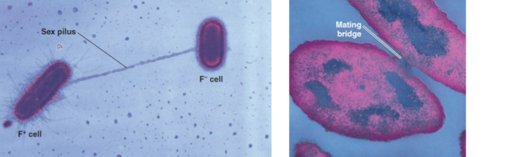
plasmids are extracellular loops of DNA
some plasmids (conjugative plasmids) are capable of being passed between different bacterial species and even genera
so plasmids can spread antibiotic resistance e.g. mating bridge
bacteria can be spread by individuals (next card) & movement of people/travelling
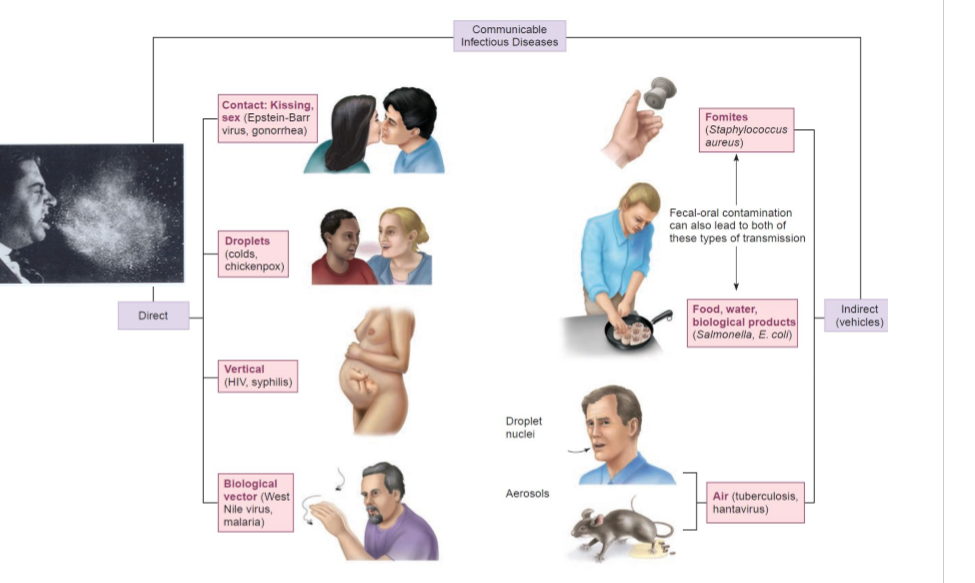
how can disease be spread between individuals?
communicable disease
movement of people, travelling leading to resistant organisms
for example, you carry your microflora with you
what are parts that make up spreading disease? like a cycle