Parasites, Viruses, and Bacteria of GIT
1/52
There's no tags or description
Looks like no tags are added yet.
Name | Mastery | Learn | Test | Matching | Spaced | Call with Kai |
|---|
No analytics yet
Send a link to your students to track their progress
53 Terms
Spirocerca lupi
an esophageal parasite in dogs that migrated from the stomach and form a granuloma between aorta + esophagus
This parasite can cause diffuse hypertrophic gastritis in horses
trichostrongylus (nematode -roundworm)
Helicobacter
A gram-negative spiral bacteria with flagella. Most common bacteria in monogastrics. Found in healthy and unhealthy animals. Common cause of gastritis
Ollulanus tricuspis
Nematode. Direct life cycle. Gastric parasite of cats, usually asymptomatic.
Physaloptera
Nematode that infects GIT. Indirect life cycle. Arthropod IH.
Transmission of ollulanus tricuspis
leave the host in vomitus
Diagnosis and treatment of ollulanus tricuspis
Induction of vomiting then baermann technique. Fecal floats are NOT reliable. Treat with ivermectin or fenbendazole
Diagnosis and treatment of physaloptera
Gastroscopy or visualization in vomitus.
Fibrobacter succinogenes
A cellulolytic bacteria of the rumen that breaks down lignin
Entodinium
A ciliate protozoa that accounts for most of the protozoa population in the rumen
Methanogens
Archaea in rumen that are responsible for producing methane from hydrogen
Siphoviridae
most dominant virus of the rumen that is important in genetic exchange with other rumen microbes
lactobacillus (monogastric)
a bacteria that ferments carbs (lactose) into lactic acid, creating an acidic environment
E. coli (monogastric)
A bacteria that helps with synthesis of vitamin K and B-complex vitamins
Enterotoxigenic E. coli
Causes hypersecretory diarrhea via enterotoxins (cause increase in cAMP, leading to CFTR releasing more Cl). Gram negative, LPS.
Common effect of enterotoxigenic E. coli
Neonatal colibacillosis in piglets (watery diarrhea, enteritis, septicemia, bacteremia)
Heat labile enterotoxigenic E. coli toxin
Affects cAMP leading to hypersecretion of fluids
Shiga toxin-producing E. coli
Causes edema and bleeding in GIT of weaned pigs. Attach to enterocytes and produce shiga toxins that can enter the bloodstream.
Significance of transmissible R plasmids
They harbor antibiotic resistant genes in E. coli, creating need for antimicrobial sensitivity testing
Lawsonia intracellularis
Seen in foals and weaned pigs. Inflammatory. Malabsorptive diarrhea. Gram negative.
Salmonella
Uses a Type 3 secretory system (a needle like structure that projects toxins into host cells) to activate secretory pathways, causing diarrhea. Gram negative: LPS.
Feline panleukopenia
Targets undifferentiated crypt cells. Malabsorption diarrhea
Rotavirus
Destroys small intestine enterocytes on the tip of the villi causing malabsorption. Has a toxin called nsp4 that causes secretory diarrhea
Coronavirus
Causes death of microvilli and blunting of villi (disorder of absorptive enterocytes). Effects both small and large intestine. Malabsorption diarrhea
Mycobacterium paratuberculosis (Johne’s disease)
Malabsorptive diarrhea. Causes submandibular edema. Effects cattle. Causes lesions of the lamina propria (M cells take bacteria into peyers patches and infect macrophages) leading to villous blunting and fusion.
Yersinia enterocolitica
Cause secretory diarrhea. Gram negative, LPS. Enter mucosa through M cells and use Type 3 secretory system to inject toxins. . Produce Yersinia outer proteins (Yops) that interfere with phagocytosis and produce ROS)
Brachyspira hyodysenteriae
seen in weaned pigs. Causes muco-hemorrhagic diarrhea (invade goblet cells and have hemolysins). Gram negative, LPS
Clostridium perfringens
Gram positive bacteria that produce 4 kinds of toxins (alpha, beta, epsilon, iota)
Clostridium perfringens type A
Causes canine hemorrhagic gastroenteritis and necrotic enteritis in chickens
Clostridium perfringens type C
Produces alpha (hemolysis) and beta toxins (vascular necrosis) and causes hemorrhagic enteritis in pigs
Canine Parvovirus
Systemic virus that infects and destroys rapidly dividing cells in crypts of lieberkuhn, lymph tissue, and bone marrow. Causes malabsorption and disrupted gut barrier leading to exudative diarrhea.
What do rotavirus, coronavirus, and norovirus have in common
They are all enteric viruses
Qualities of enteric viruses:
Enter through the fecal-oral route and are usually naked viruses so can persist in the environment
Most common viruses causing milk scours/viral diarrhea in calves:
rotavirus and coronavirus
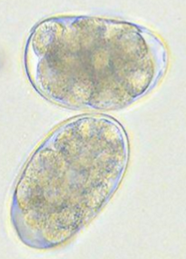
Cystoisospora
A coccidia found in the small intestine that infects dogs/cats (host specific). Causes watery diarrhea
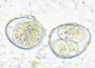
cystoisospora: right is sporulated/infective stage
eimeria
coccidia of small intestine that infects ruminants causing hemorrhagic diarrhea and CNS signs
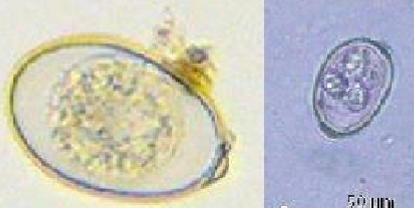
eimeria: right is sporulated
giardia duodenalis
Protozoa of small intestine. Causes pale mucus diarrhea. trophozoite is infective stage.
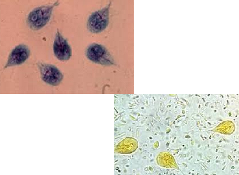
giardia: cyst is infective, trophozoites have flagellum
crytosporidium parvum
Causes yellow watery diarrhea in cattle. protozoa that infects small intestine, zoonotic.
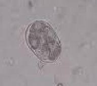
cryptosporidium
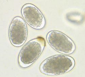
Ancylostoma caninum
Causes dark tarry diarrhea. Nematodes with 3 pairs of teeth; infect the small intestine; L3 is infective.
hookworm eggs (ancylostoma)
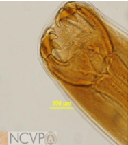
3 pairs of teeth
ancylostoma caninum
ancylostoma tubaeforme
1 pair of teeth
ancylostoma braziliense
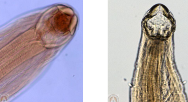
cutting plates
uncinaria stenocephala
bunostomum
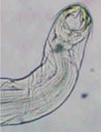
strongyloides stercoralis
nematode that infects small intestine, zoonotic. Causes bloody diarrhea
trichostrongylus colubriformis
ostertagia
cooperia
Nematodes that infect small intestine of ruminants. Ostertagia infect abomasum
looks the same as ancylostoma but only infects ruminants
trichostrongyles
tritrichomonas blagburni
infects large intestine of cats causing water diarrhea
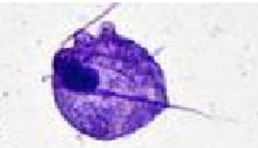
tritrichomonas
trichuris vulpis
whipworm that infects large intestine of dogs, infective stage is L1. Causes diarrhea with bright blood