Lecture 5: Gas Exchange
1/45
There's no tags or description
Looks like no tags are added yet.
Name | Mastery | Learn | Test | Matching | Spaced | Call with Kai |
|---|
No analytics yet
Send a link to your students to track their progress
46 Terms
need of oxygen
to generate ATP, which is needed for cellular function and metabolism. as a result of cellular activity, high levels of CO2 is generated, which must be expelled.
cellular respiration
oxidative processes within cells
external respiration
exchange of O2 and CO2 between an organism and its environment
factors for effective gas-exchange surfaces
moist
thin
relatively large surface area
vascularization
formation/presence of blood vessels; helps enhance the effectiveness of diffusion
Types of Respiratory Organs
cutaneous respiration
tracheal systems
gills or brachia
lungs
cutaneous respiration
respiratory organ that refers to direct diffusion of respiratory gasses to skin
paramecium, marine sponge, jellyfish, flat worms, amphibians
tracheal systems
respiratory organ referring to a branching system of tubes, with openings called spiracles (usually no well-defined circulatory system)
in insects
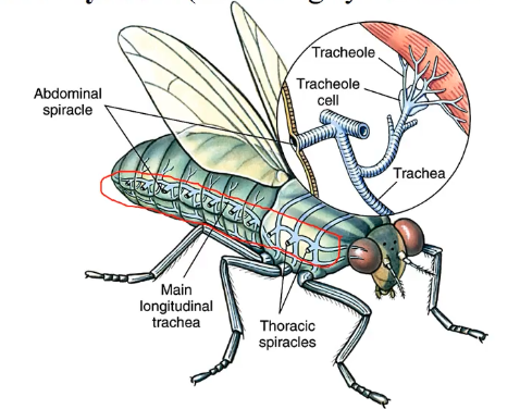
gills or brachia
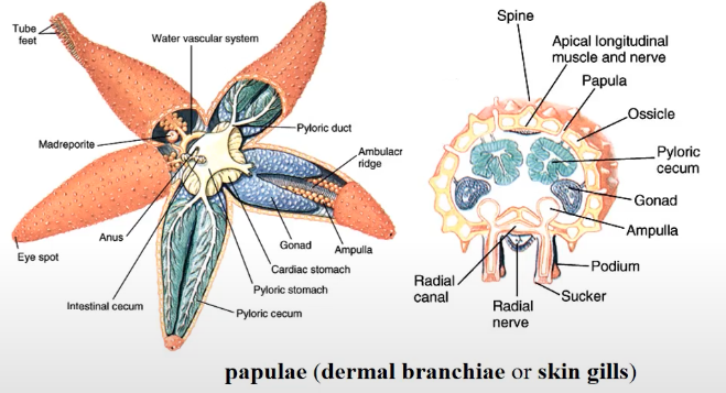
respiratory organ that retain its shape in the presence of water, and possess papulae (extension of the fluid-filled coelom)
in fish, starfish, axolotl, ragworms, etc.
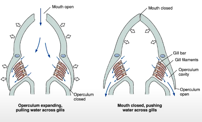
way in which a fish ventilates its gills
gills are given its shape from counterflow of water
when mouth open, water enters and a structure called the operculum is closed & water collects in gills
when mouth is closed, the operculum opens, pushing water across gills to exit
gill filaments
part of gill that help filter oxygen out of the water
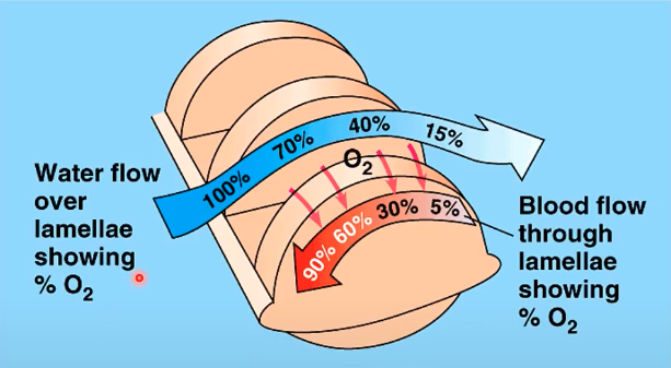
blood vessels in gills mechanism
countercurrent flow/exchange:
as oxygen-rich water enters the filaments, oxygen is filtered in and distributed to blood vessels in the gill for these to be oxygenated (arteries)
as water exits the gill filaments in the form of de-oxygenated water, this is where unoxygenated blood residues (veins)
lungs
respiratory organ that are invaginations, and are more suited for terrestrial animals
air gives lungs its structure
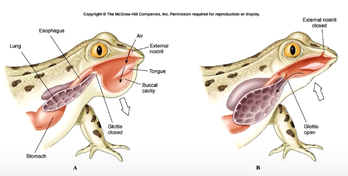
lung mechanism in frogs
positive-pressure breathing
pressure in the buccal cavity increases (more air) = inflation
lung mechanism of birds
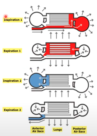
possess air sacs attached to the lung proper, since birds are suited for flight
inspiration 1: oxygen-rich air goes to posterior air sac
the pressure pushes air to the more thoracic component, once it reaches the cervical component, it becomes deoxygenated
second inspiration: repeat steps 1-2
the second batch of air that enters the anterior air sac pushes the previous air outwards (cycle repeats)
component of lungs in mammals
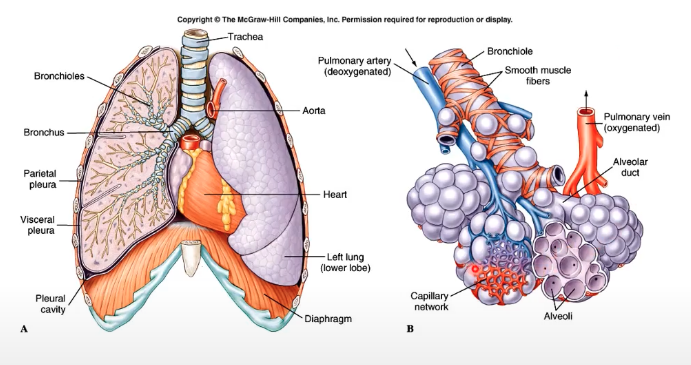
passage of air: trachea → bronchus → bronchioles → alveoli
associated with arteries and veins (capillary network in alveoli, where gas exchange occurs)
lung mechanism of mammals
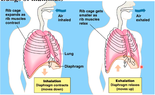
negative pressure breathing
upon inhale, diaphragm contracts (moves downward) = ribcage expands, but “stomach” area is sucked in
upon exhale, diaphragm relaxes (moves up)- rib cage smaller
altitude and oxygen content
higher altitude - less saturation of oxygen
alveoli open up more for more efficient exchange
forms of lung capacity/volume
tidal volume
vital capacity
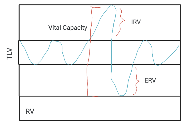
tidal capacity
refers to the volume of air that an animal inhales and exhales with each breath (normal breathing in and out)
~500 ml in resting humans
vital capacity
refers to the maximum tidal volume during forced breathing (total amount of air you can inhale)
~3.4L female, 4.8L male
residual volume
needed amount of air for lungs to retain shape; left-over air when you force exhale
inspirational reserved volume capacity (IRV)
maximum volume of air that can be inhaled
expiratory reserve volume capacity (ERV)
maximum volume of air that can be exhaled
when you exhale, you don’t exhale all of air content of lungs (since without air, lungs will collapse)
respiratory pigments
proteins where oxygen is bound to for transport
types of respiratory pigments
hemacyanin
hemoglobin
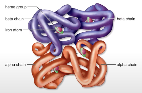
appearance of hemoglobin
reddish because of iron presence (where oxygen binds to)
excess CO2 in body
drop in pH = increased depth & rate of breathing = excess CO2 is eliminated in exhaled air
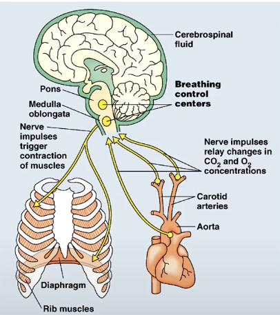
determinant of pH level in blood
gas concentration
more oxygen = basic (blood alkalosis)
more carbon dioxide = acid (blood acidosis)
pons and medulla
region in the brain that detects shifts in blood pH, which relays impulses to the brain and instructs the diaphragm and ribs accordingly
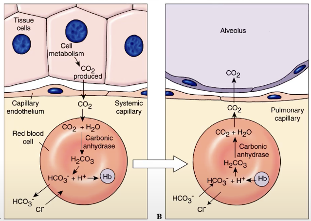
pulmonary capillaries
where oxygen diffuses into
where carbon dioxide diffuses out of
oxyhemoglobin
oxygen + hemoglobin (in RBCs)
carbaminohemoglobin
carbon dioxide + hemoglobin (in RBCs)
bicarbonate ion
most CO2 is transported in this form (not yet combined with hemoglobin)
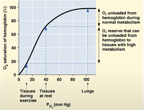
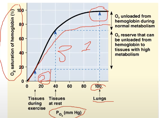
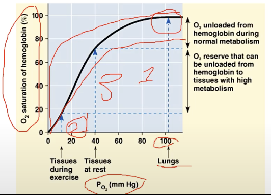
oxygen dissociation curve
a graph that tells us how fast any typical tissue can be saturated with oxygen, and reflective of how long a particular tissue can hold onto oxygen
factors of oxygen dissociation curve:
metabolic activity
temperature
altitude
thoughts
increase of the following: acidosis
decrease of the following: alkalosis
components of the oxygen dissociation curve
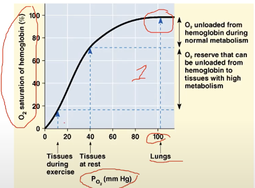
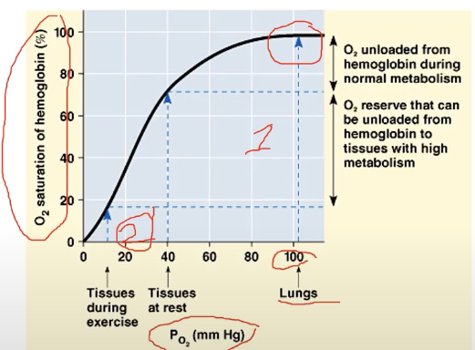
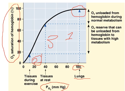
x axis → oxygen pressure
y axis → oxygen saturation of hemoglobin
more oxygen saturation = more oxygen pressure
high oxygen pressure & high saturation point
results in slow oxidation of oxygen (since full already)
usually takes place in the Lungs
low oxygen pressure and low saturation point
takes place when tissues are active (e.g. calf muscles)
because always active, saturation point will be a lot faster & oxygen in red blood vessel is detached a lot quickly because of how utilized this is (muscles needing O2)
average pressure and saturation of oxygen
takes place when tissues are at rest
red blood cells can hold on to oxygen longer
implication if dissociation curve flattens earlier (but same slope)
assume that saturation is faster, and more metabolic activity
implication if dissociation curve has a steeper slope (but same area where it flattens; longer area how its flattened)
assumes that body holds onto more oxygen
Bohr shift
refers to a drop in pH that lowers the affinity of hemoglobin for O2 (shift to the right)
RBC gives more oxygen to sustain metabolic activity going on
results in higher CO2 concentration
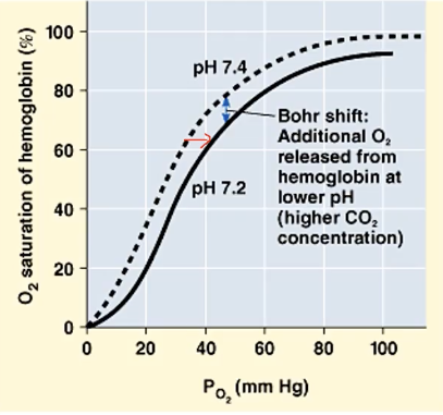
haldane effect
refers to increase in pH, when blood holds onto O2 more (shift to left)
implies you do not have good metabolism
relation of activity to O2/CO2 levels
increase activity = increase metabolism = increase respiration = faster oxygen tends to dissociate = faster saturation of oxygen = decrease oxygen = increase carbon dioxide
acidosis (shift of diss. curve to right)
decrease activity = decrease metabolism = decrease respiration = increase oxygen = decrease carbon dioxide
alkalosis (shift of diss. curve to left)
temperature and oxygen content
higher temperature favors faster dissociation of oxygen, thus more acidified blood