Immunology Chapter 10 Preventing Infection at Mucosal Surfaces
1/21
There's no tags or description
Looks like no tags are added yet.
Name | Mastery | Learn | Test | Matching | Spaced | Call with Kai |
|---|
No analytics yet
Send a link to your students to track their progress
22 Terms
________________ are all sites of communication, where material and information are passed between the body and its environment.
Because of their physiological functions of gas exchange (lungs), food absorption (gut), sensory activity (eyes, nose, mouth, and throat), and reproduction (uterus, vagina, and breast), the mucosal surfaces are by necessity dynamic, thin, permeable barriers to the interior of the body.
These properties make the mucosal tissues particularly __________________________________________.
Mucosal tissues; vulnerable to subversion and breach by pathogens
mucosa (plural mucosae)
mucus-secreting epithelium such as that lining the respiratory, intestinal, and urogenital tracts. The mammary glands and the conjunctiva of the eye are also placed in this category. Mucosal epithelium communicates with the external environment and is the route of entry for most pathogens.
mucus
slimy protective secretion composed of glycoproteins, proteoglycans, peptides, and enzymes that is produced by the goblet cells in many internal epithelia.
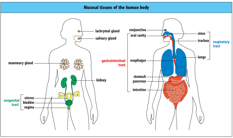
Distribution of mucosal tissues.
This diagram of a woman shows the mucosal tissues. The mammary glands are a mucosal tissue only after pregnancy, when the breast is lactating. Red, gastrointestinal tract; blue, respiratory tract and conjunctiva; green, urinary tract; yellow, genital tract; orange, secretory glands.
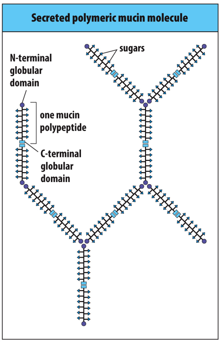
The structure of ______ gives mucus its characteristic protective properties. A major constituent of the mucin glycans is _________. Can bind defensins, other antimicrobial peptides and IgA. IgA traps bacteria and defensins kill the bacteria.
mucin; sialic acid
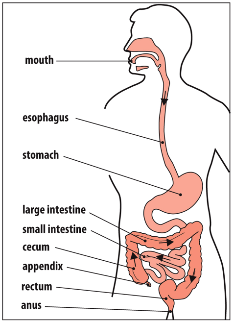
The ________________ extends from the mouth to the anus and is about 9 m in length in an adult human being. As food travels along the gastrointestinal tract and becomes increasingly degraded, it passes through environments with increasing numbers of ____________. Starting in the stomach at 1000 bacteria per milliliter of gut contents, numbers increase to 105 to 108 per milliliter in the small intestine and reach 1012 per milliliter in the colon.
gastrointestinal tract; resident bacteria
The population of commensal microorganisms, known as the _________, is an important and integral part of a health.
microbiota
Because the immune system is only _________________ at birth, its continuing development after birth is ___________ by the acquisition of the microbiota.
partially developed; strongly influenced
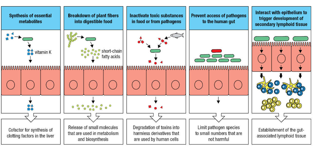
Five ways in which the commensal gut microbiota benefit their human hosts.
Synthesis of essential metabolites
Breakdown of plant fibers into digestible food
Inactivate toxic substances in food or from pathogens
Prevent access of pathogens to the human gut
Interact with epithelium to trigger development of secondary lymphoid tissue
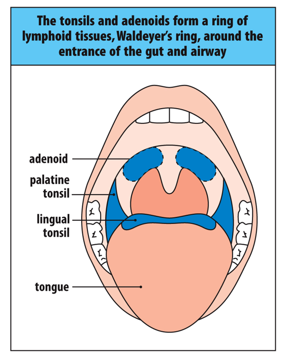
A ring of lymphoid organs guards the entrance to the gastrointestinal and respiratory tracts.
Called Waldeyer's ring,. Lymphoid tissues are shown in blue. The adenoids lie at either side of the base of the nose, and the palatine tonsils lie at either side of the palate at the back of the oral cavity. The lingual tonsils are on the base of the tongue.
Gut-associated Lymphoid Tissue and Lymphocytes (GALT)
the most extensive secondary lymphoid tissues in the human body
____________ are _______________ that underline gut epithelium and consist of a T-cell area (blue), B-cell follicles (yellow), and a ‘dome’ area (striped blue and yellow) blow the epithelium and populated by B cells, t cells and dendritic cells.
Peyer’s patches; secondary lymphoid organs

_________ enters a Peyer’s patch from the gut via the_________
Antigen; M cells
Peyer’s patches _________________ but their efferent lymphatics connect with the lymphatics ______________ to the ___________________.
have no afferent lymphatics; carrying lymph; mesenteric lymph nodes
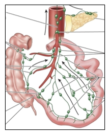
Mesenteric Lymph Nodes
One of a chain of lymph nodes in the mesentery,
the membrane that holds the gut in place. These
lymph nodes connect by lymphatics to the
lymphoid tissues of the gut, and pathogen-
bearing dendritic cells are transported there to
initiate additional adaptive immune responses
against gut pathogens.
Microfold cells have characteristic membrane ruffles.
This scanning electron micrograph of intestinal epithelium has a microfold cell(or M cell) in the center. It appears as a sunken area of the epithelium that has characteristic microfolds or ruffles on the surface. M cells capture microorganisms from the gut lumen and deliver them to Peyer's patches and the lymphoid follicles that underlie the M cells on the basolateral side of the epithelium. Magnification ×23,000. Electron micrograph courtesy of Allan Mowat.
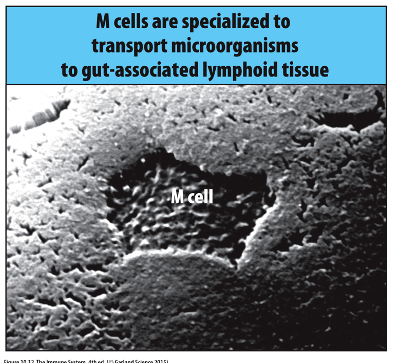
Uptake and transport of antigens by M cells.
Adaptive immune responses in the gut are initiated and maintained by M cells that sample the gut's contents and deliver this material to the intraepithelial pockets on the basolateral side of the M cell. Here, dendritic cells and B cells take up antigen and stimulate the proliferation and differentiation of antigen-specific T cells and B cells.

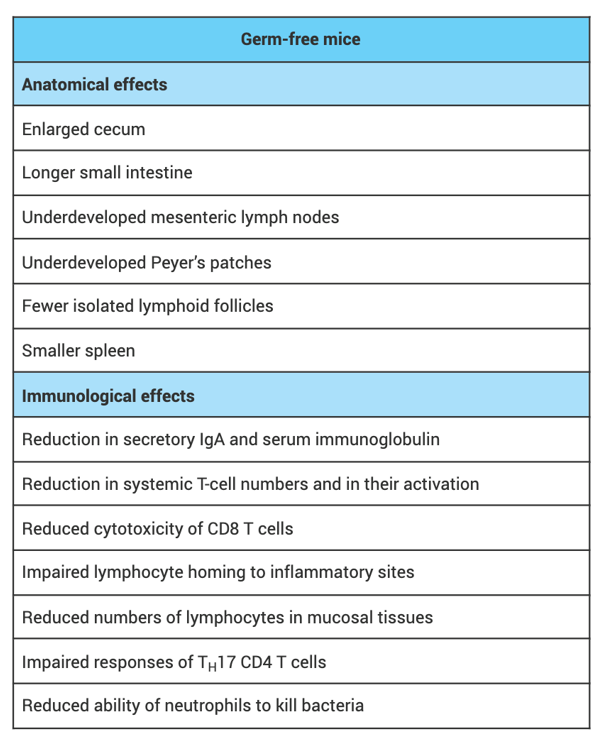
In the absence of a microbiota, the immune system develops abnormally and functions poorly.
Listed here are the differences distinguishing mice born and raised under sterile conditions from those raised under nonsterile conditions. The former have no microbiota; the latter have normal gut microbiota.
Germ-free mice are mice that are completely devoid of any _____________ including bacteria, viruses, fungi and parasites
microorganisms
Purpose of Germ-Free Mice
Used in research to study the role of the gut microbiome in various health conditions such as: Digestive disorders, immune system function, obesity, diabetes, and cancer
Generation of Germ-Free Mice
Typically generated by cesarean section from mothers that have been sterilized. The pups are then placed in sterile isolators, where they are raised in a germ-free environment
Characteristics of Germ-Free Mice
Lack of gut microbiota, altered immune system, Increased susceptibility to certain infections, altered metabolism, and different behavioral patterns.