Muscular System I
1/123
There's no tags or description
Looks like no tags are added yet.
Name | Mastery | Learn | Test | Matching | Spaced | Call with Kai |
|---|
No analytics yet
Send a link to your students to track their progress
124 Terms
Thoracic Wall
ENTIRE STRUCTURE that is the muscles of the thoracic cage
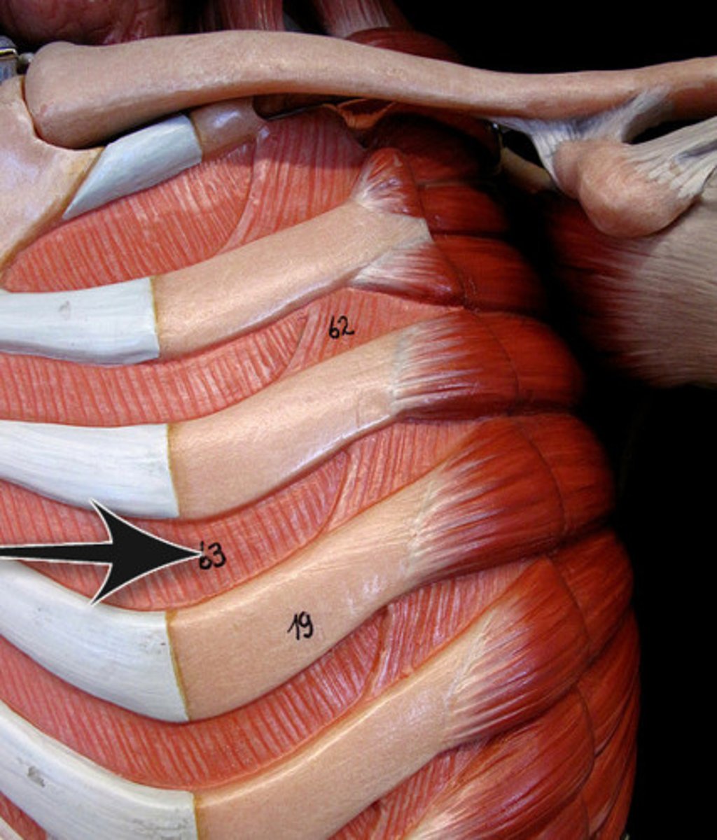
External Intercostal Muscles
STRUCTURES that are part of the thoracic muscles; muscle striations are slanted diagonally down and in (lateral to medial) over the ribs
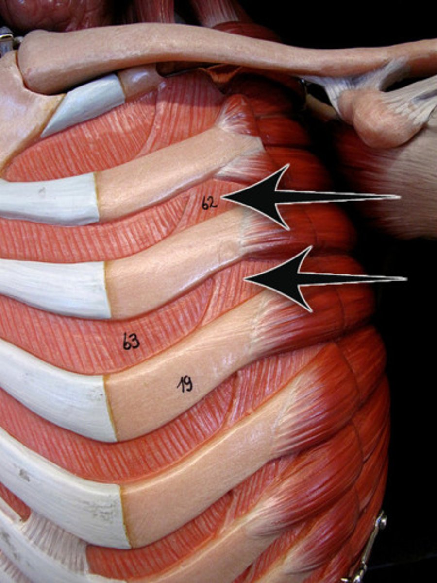
Internal Intercostal Muscles
STRUCTURES that are part of the thoracic muscles; muscle striations are slanted diagonally down and out (medial to lateral) over the ribs
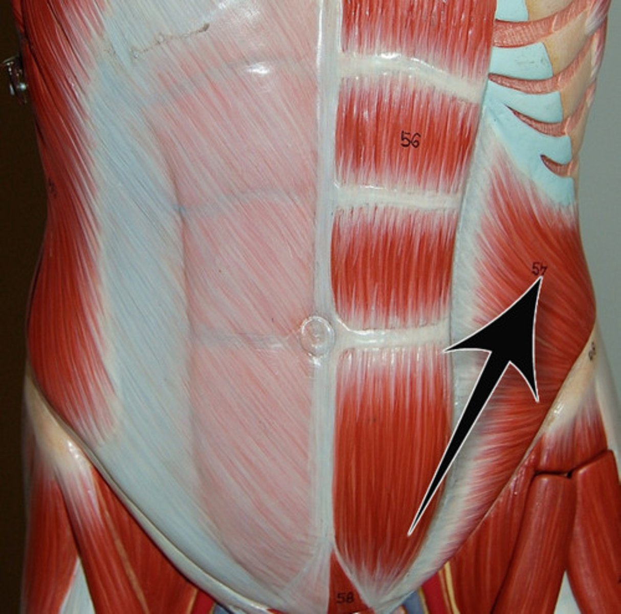
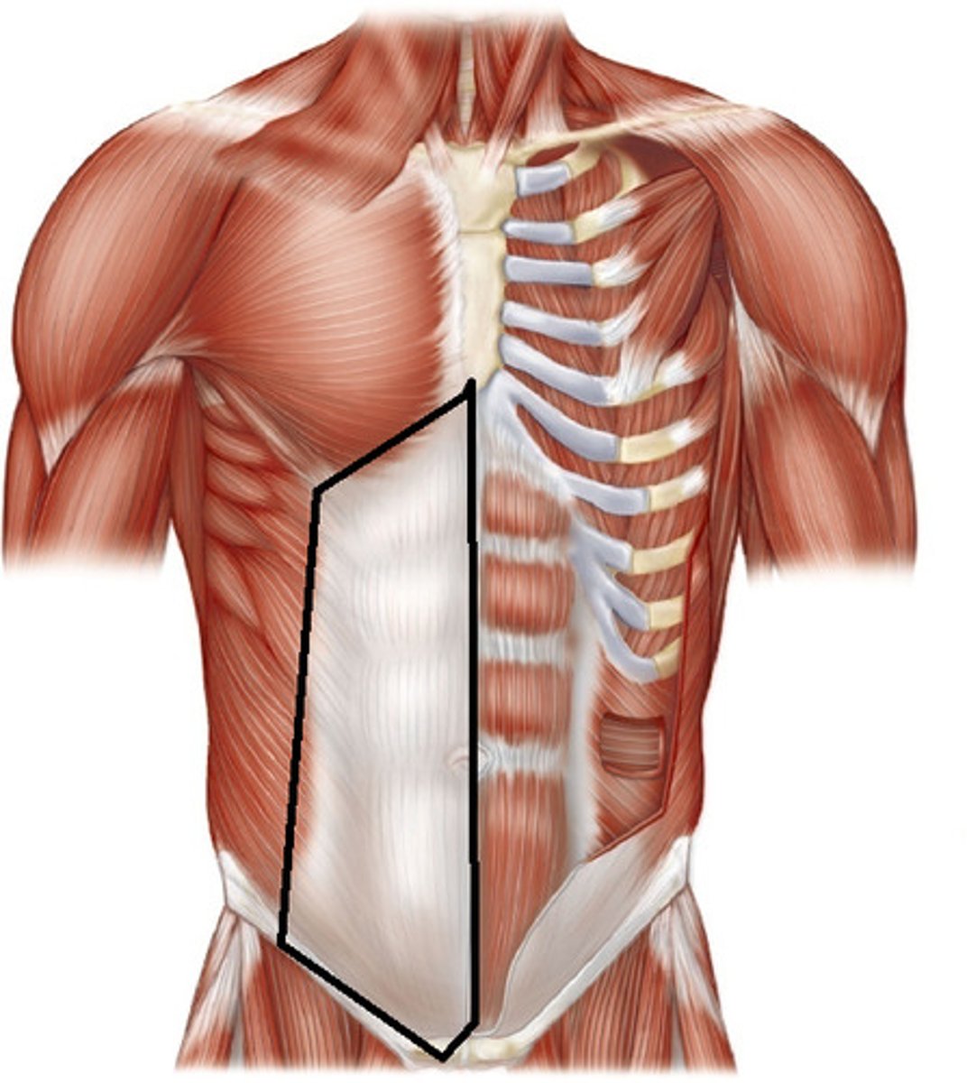
External Oblique Muscles
STRUCTURE that is the (exterior) outside flap just below the rib cage
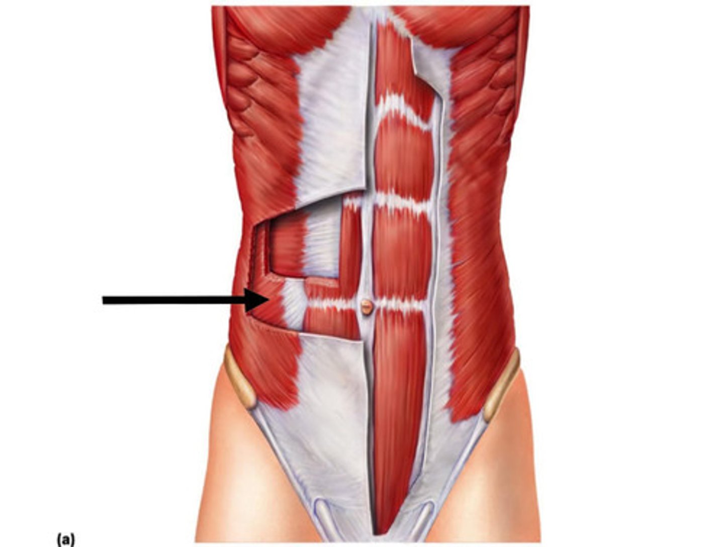
Internal Oblique Muscles
STRUCTURE that is the flab just (middle) under the external oblique muscles; located below the rib cage
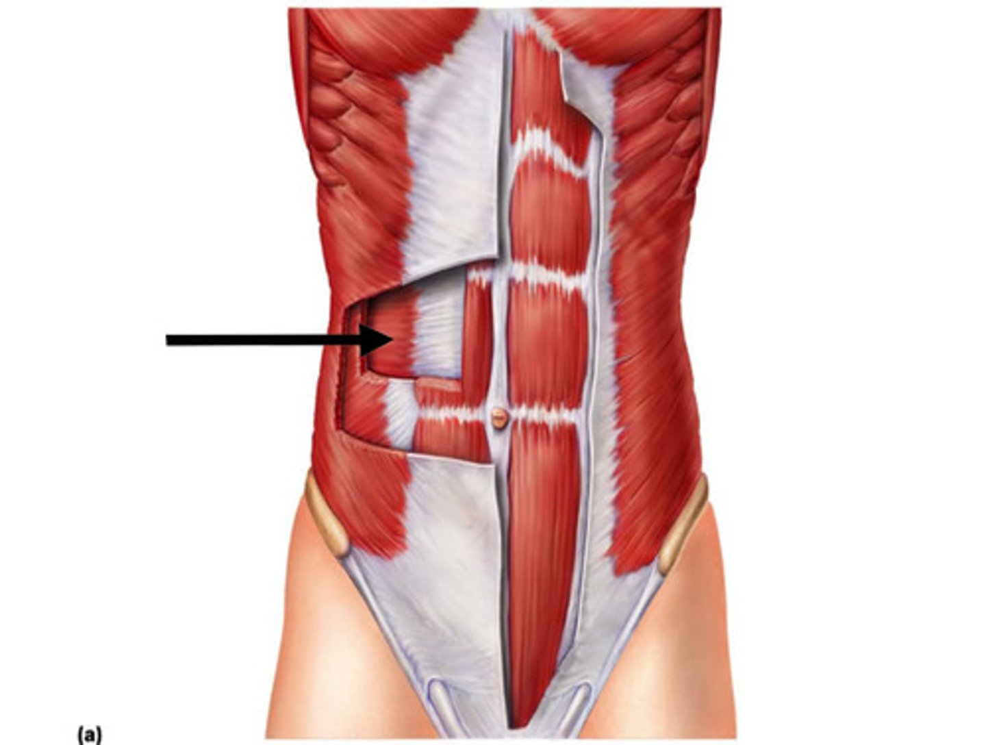
Transverse Abdominis Muscles
STRUCTURE that is the (interior) deepest flap just under the internal oblique muscles; right over intestines
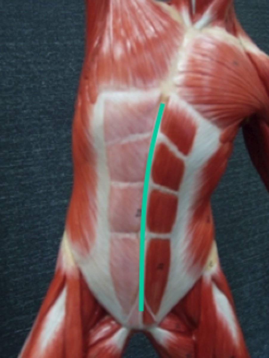
Linea Alba
DIVISION that is the center ab line
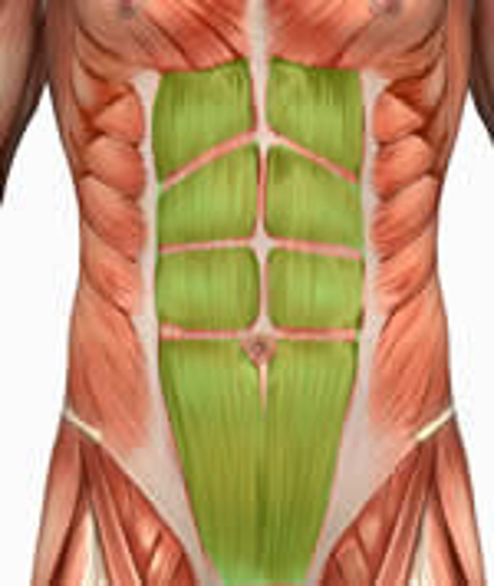
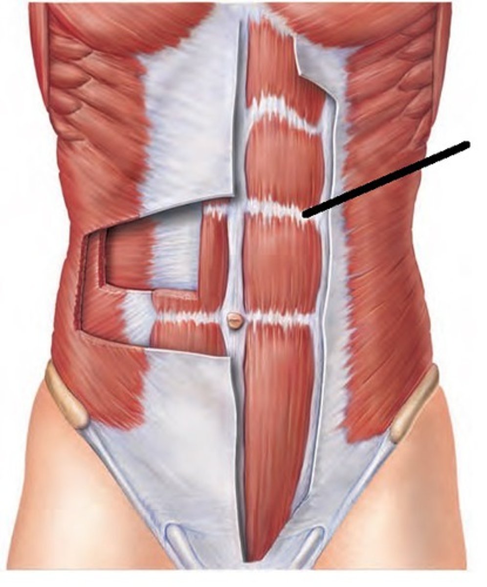
Rectus Abdominis Muscles
STRUCTURE that is the individual sections of muscles that make up the abs (green)
Tendinous Intersections
DIVISIONS that are the horizontal lines that make up ab pack; seen as a white line over rectus abdominis muscles flap
Rectus Sheath
COVERING that is the white thin flap that covers the rectus abdominis muscle flap
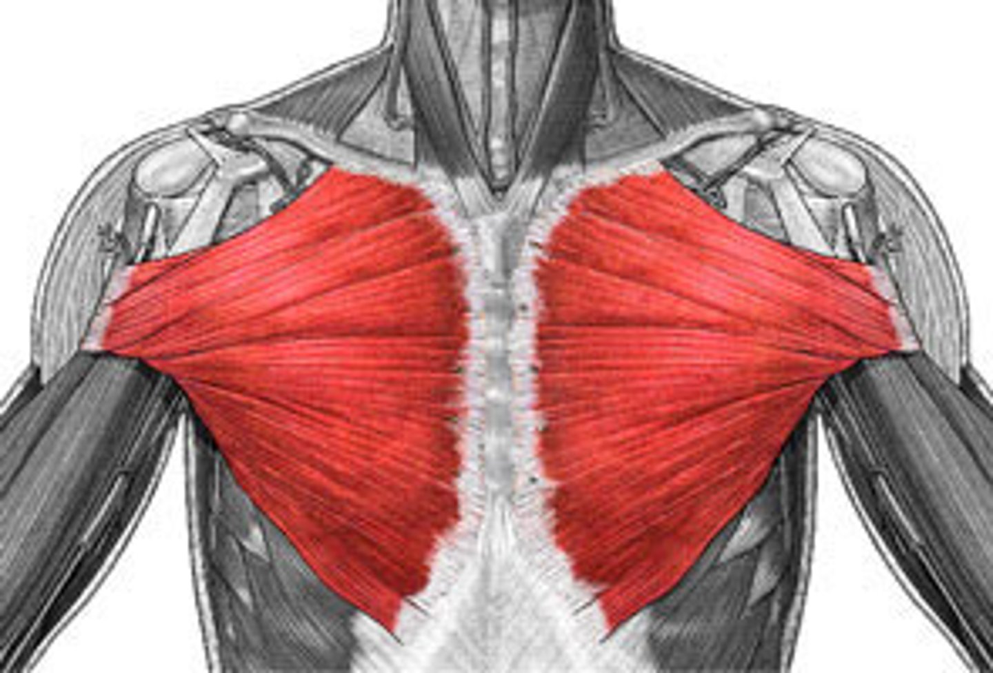
Pectoralis Major Muscle
STRUCTURE that is the big pectoral flap
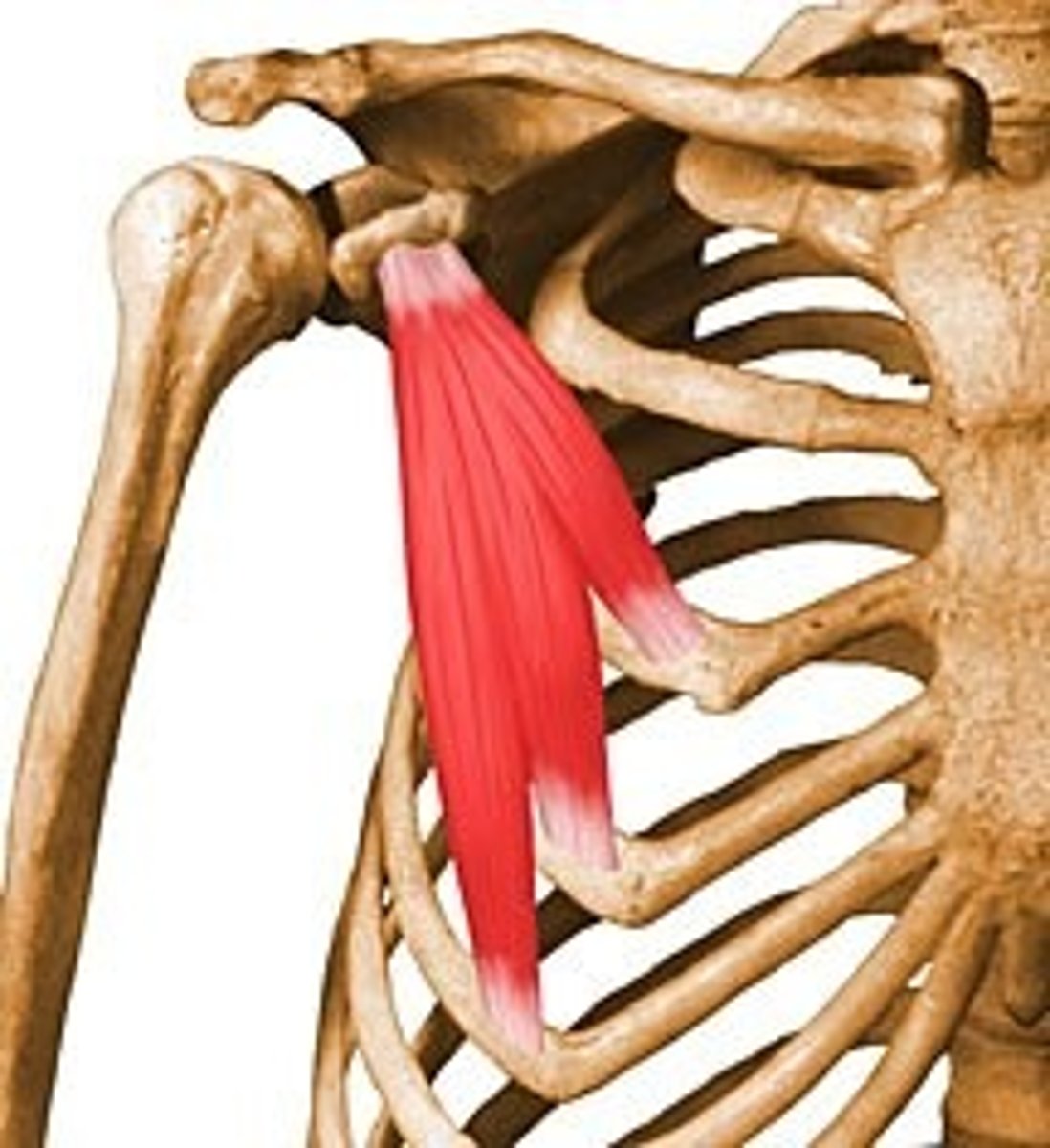
Pectoralis Minor Muscle
STRUCTURE that is the small flap attached to the back of the pectoralis major muscle near the shoulder
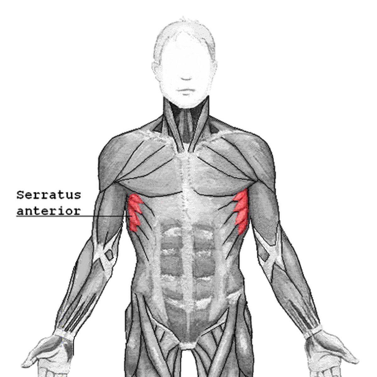
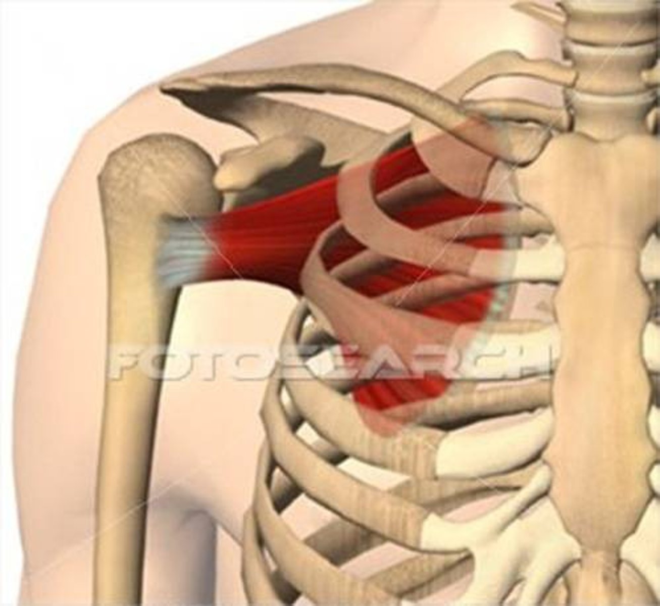
Serratus Anterior Muscle
STRUCTURE that makes a scalloped edge on both sides of upper ribs
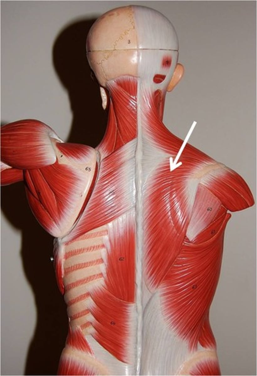
Trapezius Muscle
STRUCTURE that are flaps that go from the tops of the shoulders down the back
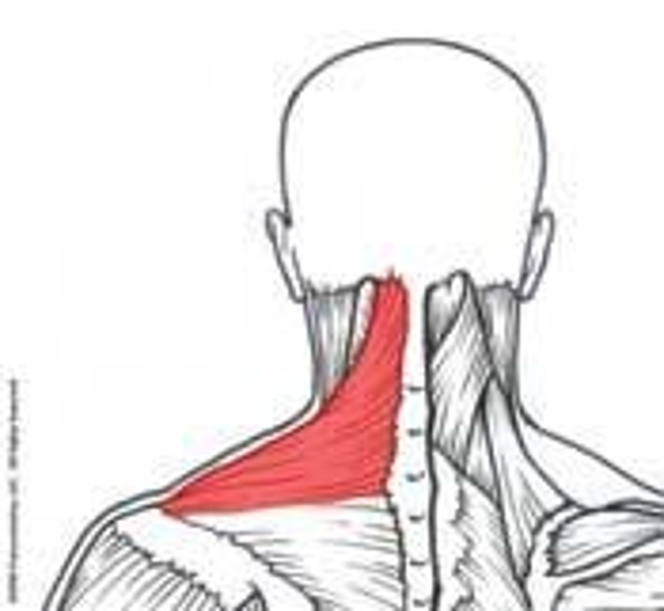
Descending Part of the Trapezius Muscle
PORTION of the Trapezius muscle that is at the top with muscle striations that run in a swooping horizontal into the neck
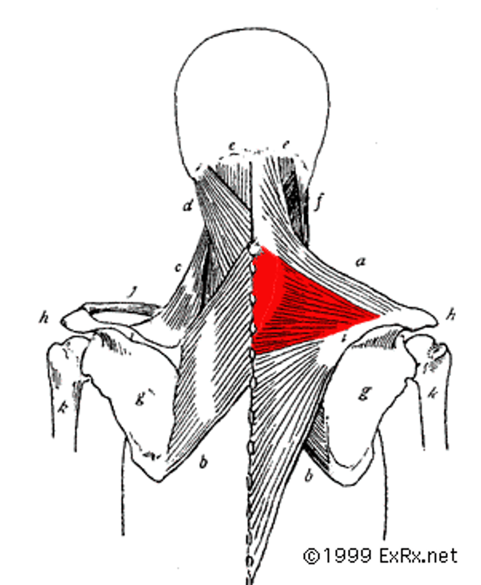
Transverse Part of the Trapezius Muscle
PORTION of the trapezius muscle that is in the middle and has horizontal striation
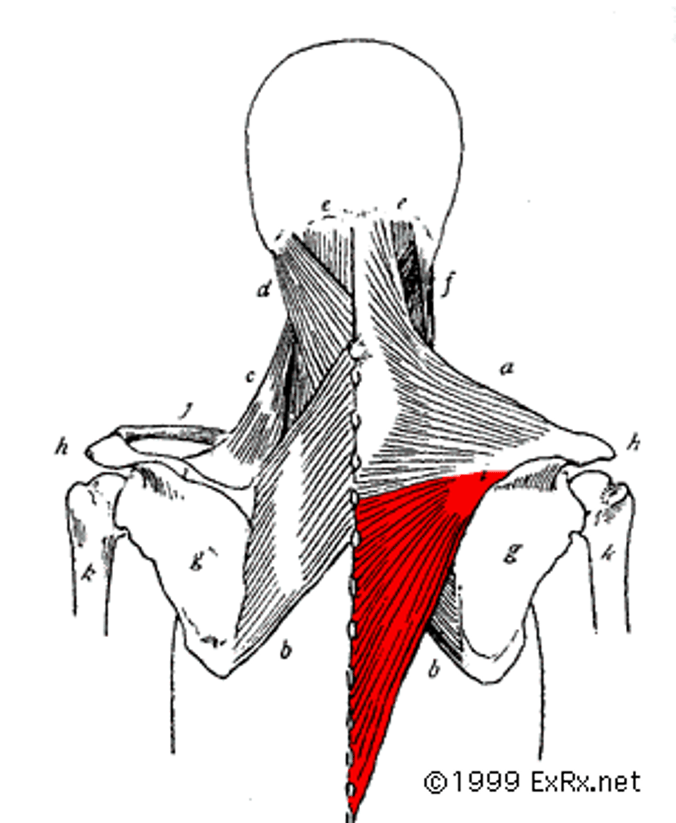
Ascending Part of the Trapezius Muscle
PORTION of the trapezius muscle that is at the bottom and has diagonal striations running medial into the spine
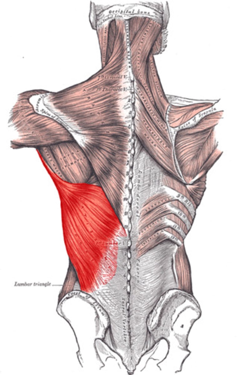
Latissimus Dorsi Muscle
STRUCTURE that is the "wings" on the back/sides of the back ribs
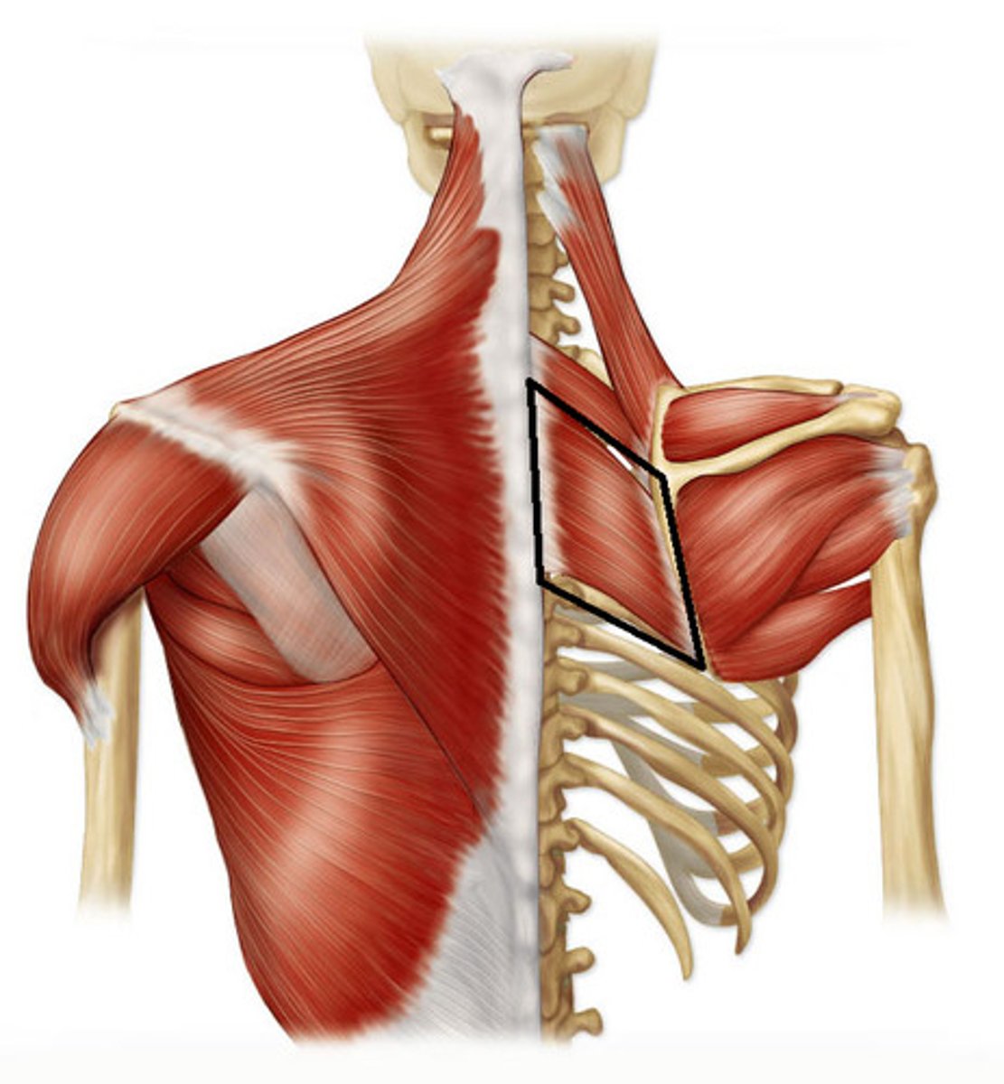
Rhomboids Major Muscle
STRUCTURE that is the flap just over the scapula, under the trapezius muscle flap
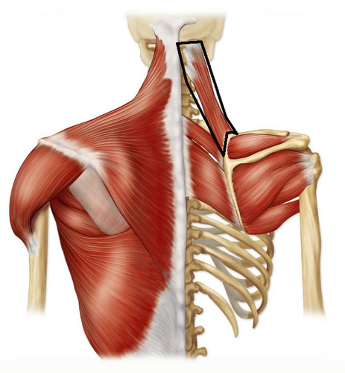
Rhomboids Minor Muscle
STRUCTURE that is the thin shred pointing back up to the neck at the top of the rhomboids major muscle; forms a "V" with the levator scapulae muscle

Levator Scapulae Muscles
STRUCTURE that is right next to the rhomboids minor and leads up to the neck; forms the outside edge of the "V"
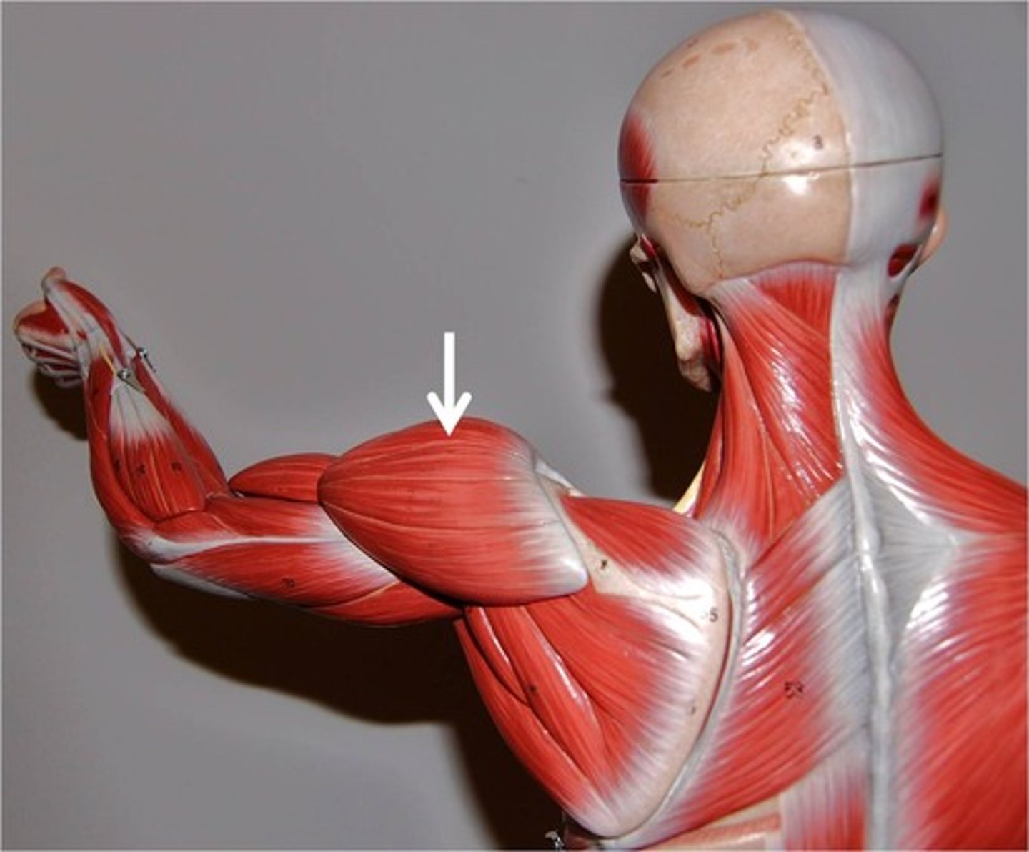
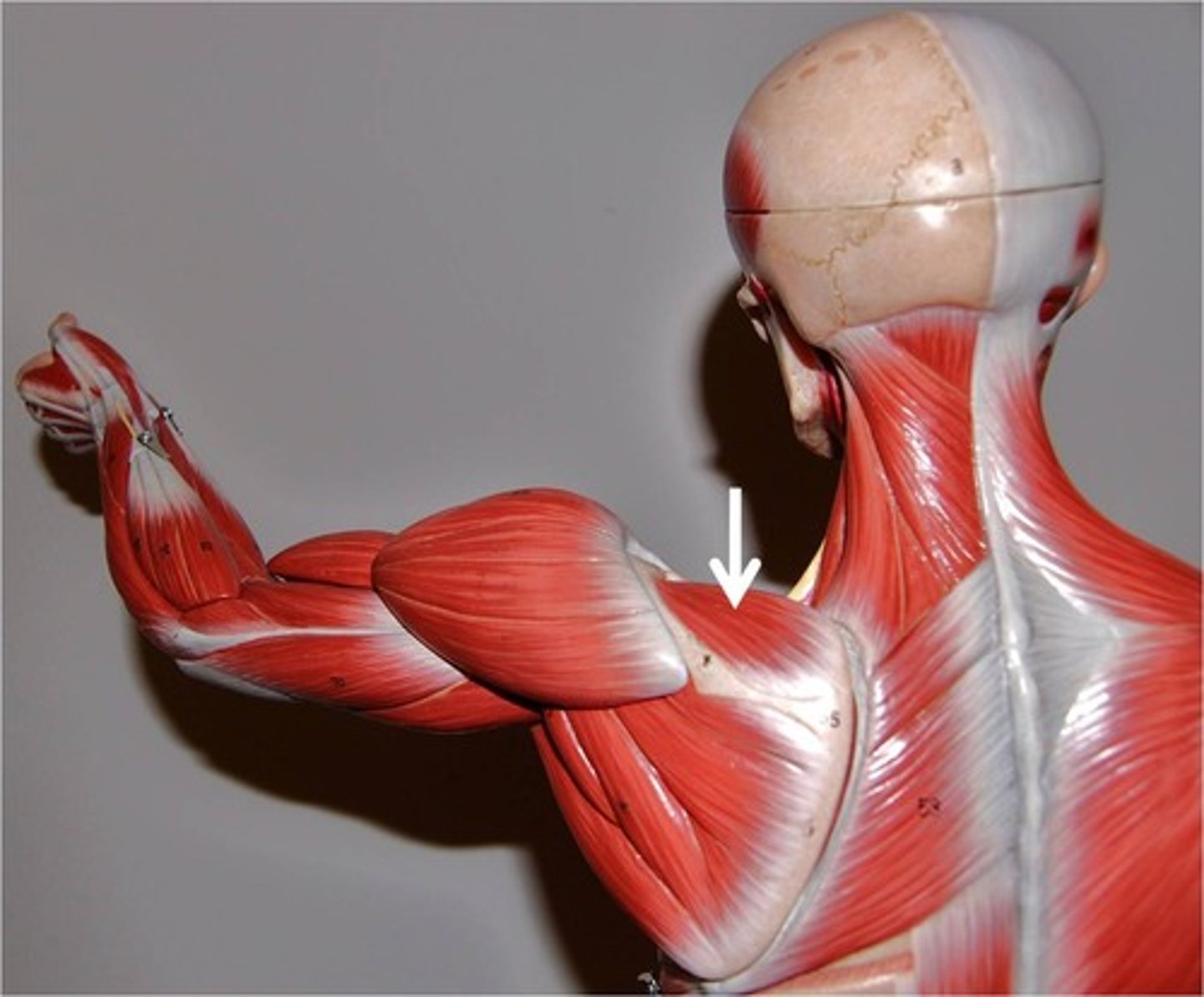
Deltoid Muscle
STRUCTURE that is a thick band at the shoulder
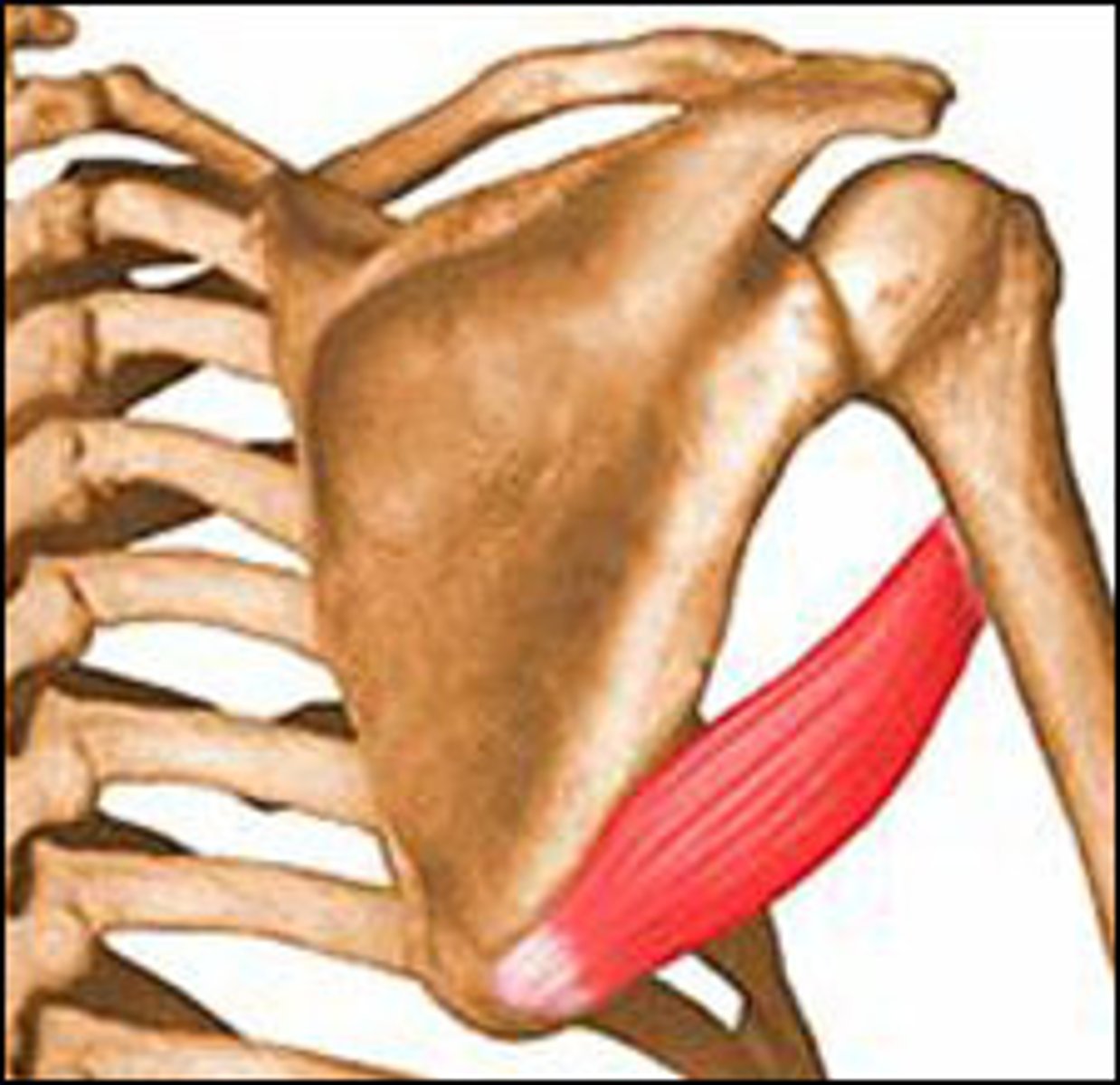
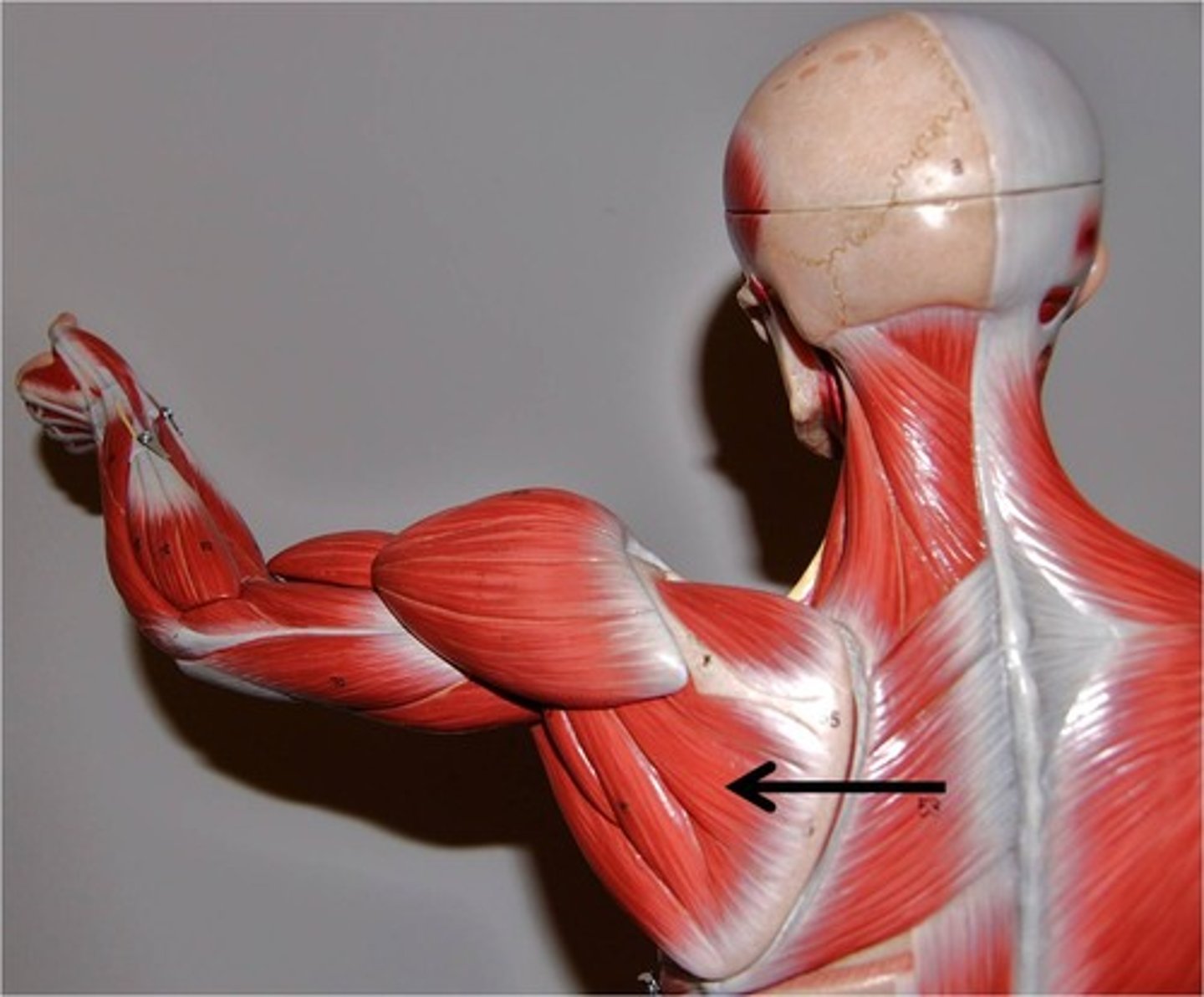
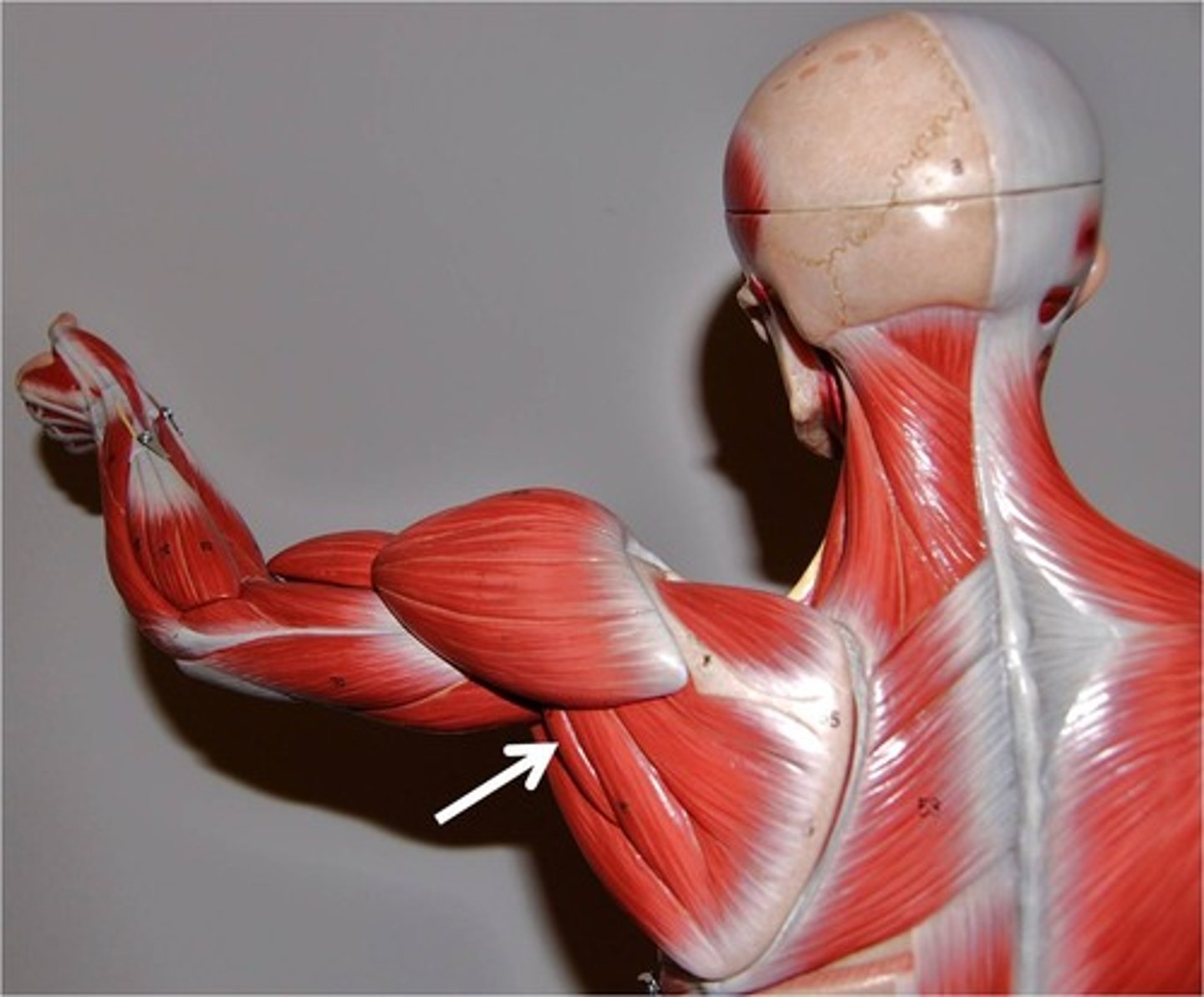
Teres Major Muscle
STRUCTURE that is the thick, attached band under the deltoid
Supraspinatus Muscle
STRUCTURE that is above the spine of the scapula bone; medial to deltoid muscle
Infraspinatus Muscle
STRUCTURE that is just below the scapula bone spine; just below the deltoid muscle
Teres Minor Muscle
STRUCTURE that is a horizontal band that runs under the deltoid, and just above the Teres Major M
Subscapularis Muscle
STRUCTURE that is on the inside of the scapula; we can't see it on the cadaver, so the TA will put hand behind scapula
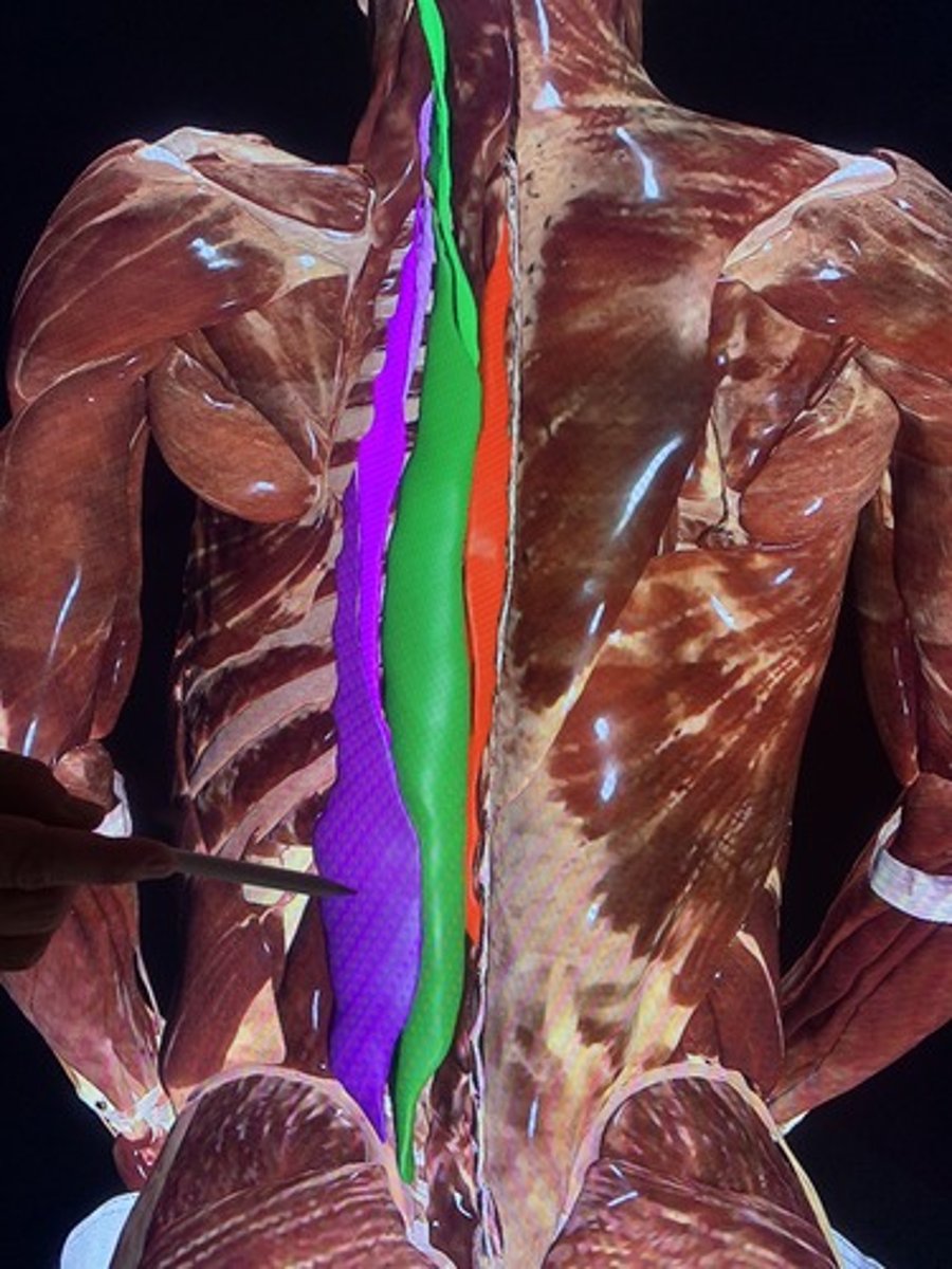
Spinalis Muscle
STRUCTURE that is in the back (part of erector spinae) and runs most medial to the spine; little loose band of muscle
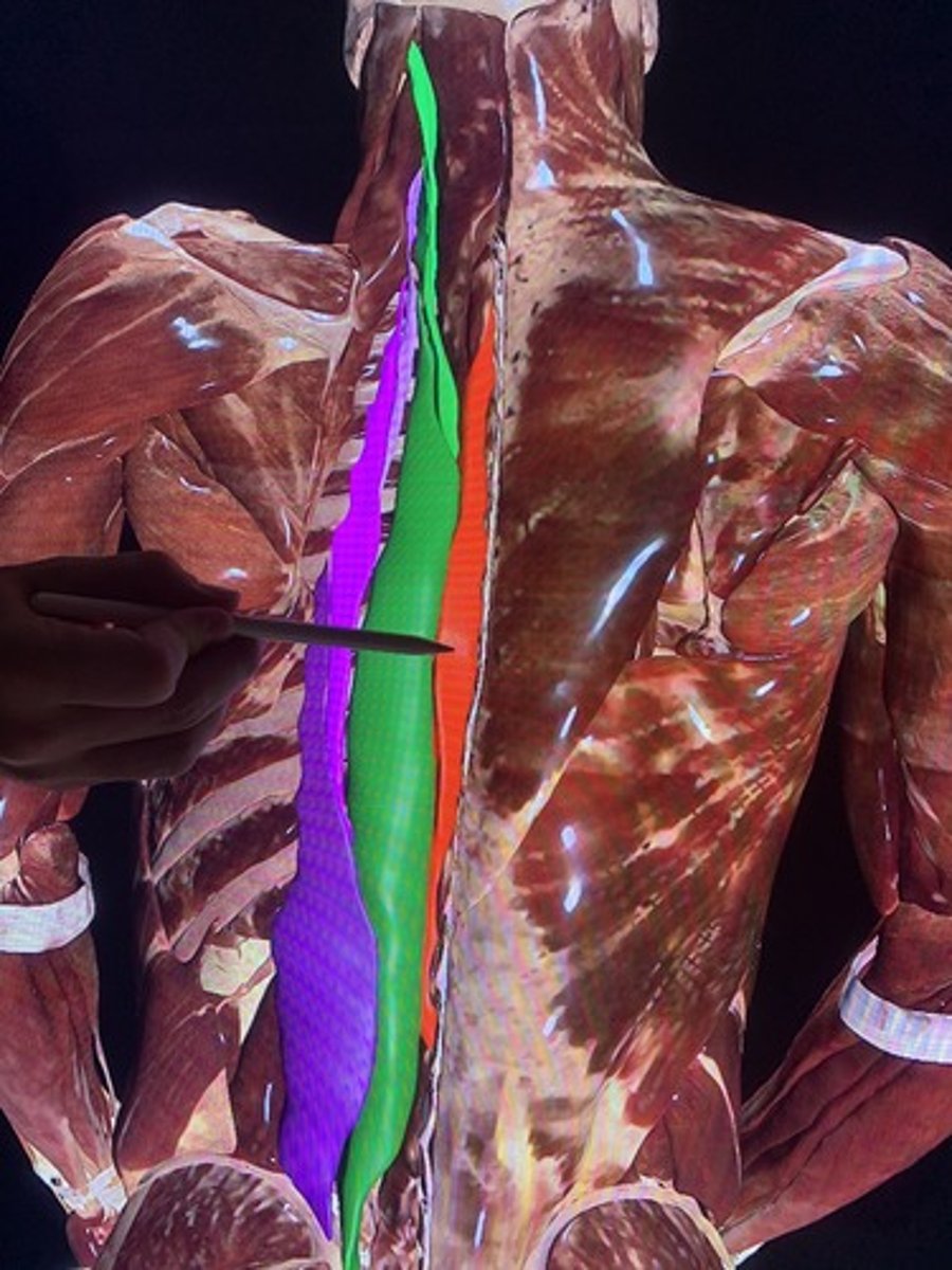
Longissimus Muscle
STRUCTURE that is the thick middle band of muscles of the erector spinae
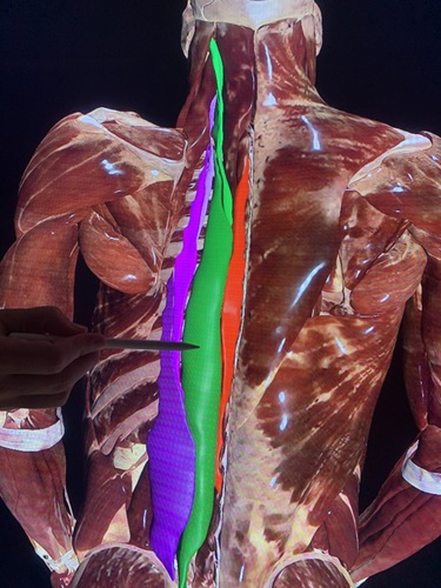
Iliocostalis Muscle
STRUCTURE that is the most lateral muscle of the erector spinae; loose long band
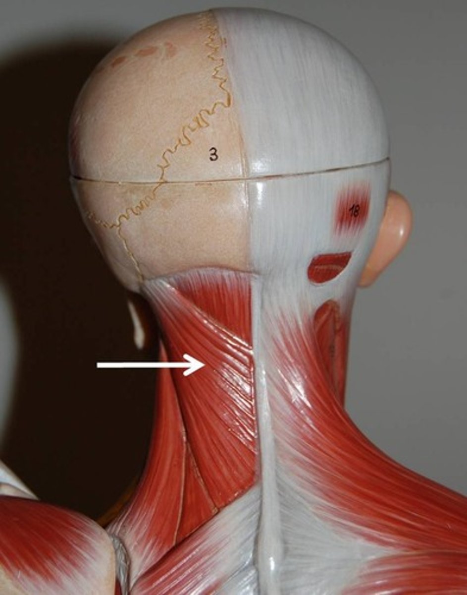
Splenius Capitis Muscle
STRUCTURE that is a downward pointing triangle on the back of neck leading to the sides of the spine; loose on cadaver
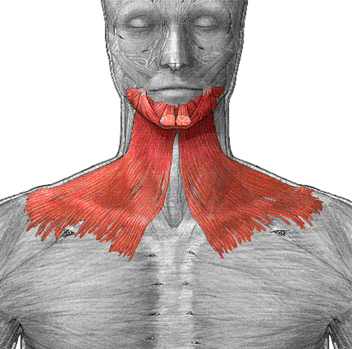
Platysma Muscle
STRUCTURE that is the thin sheath on the front of the neck
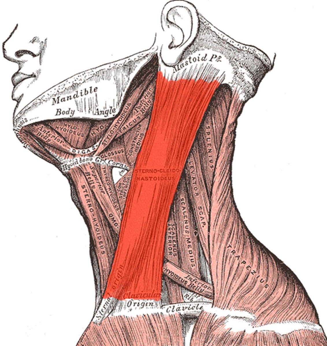
Sternocleidomastoid Muscle
STRUCTURE that is a loose band that goes from the ear to the center of lower neck
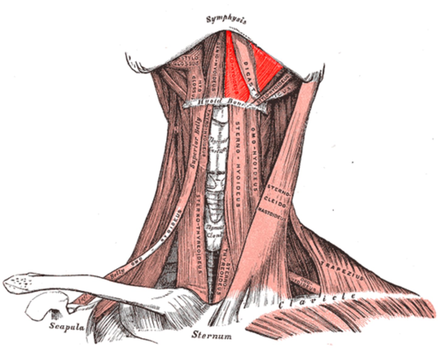
Digastric Muscles
STRUCTUREs that are just under the chin on both sides
Mylohyoid Muscle
STRUCTURE that is in between the two digastric muscles on the underside of the chin
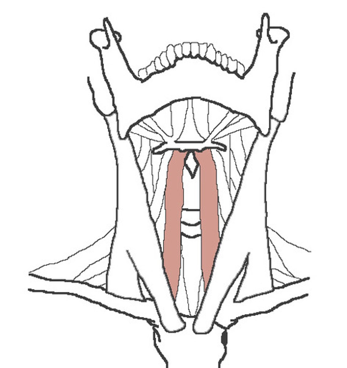
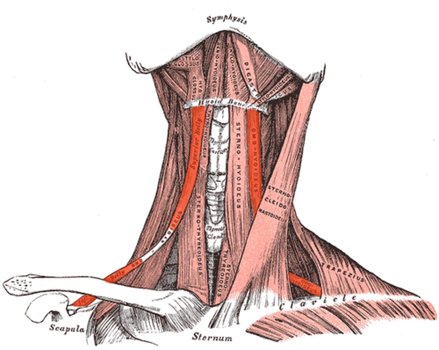
Sternohyoid Muscles
STRUCTUREs that runs from hyoid down the center neck; loose flap
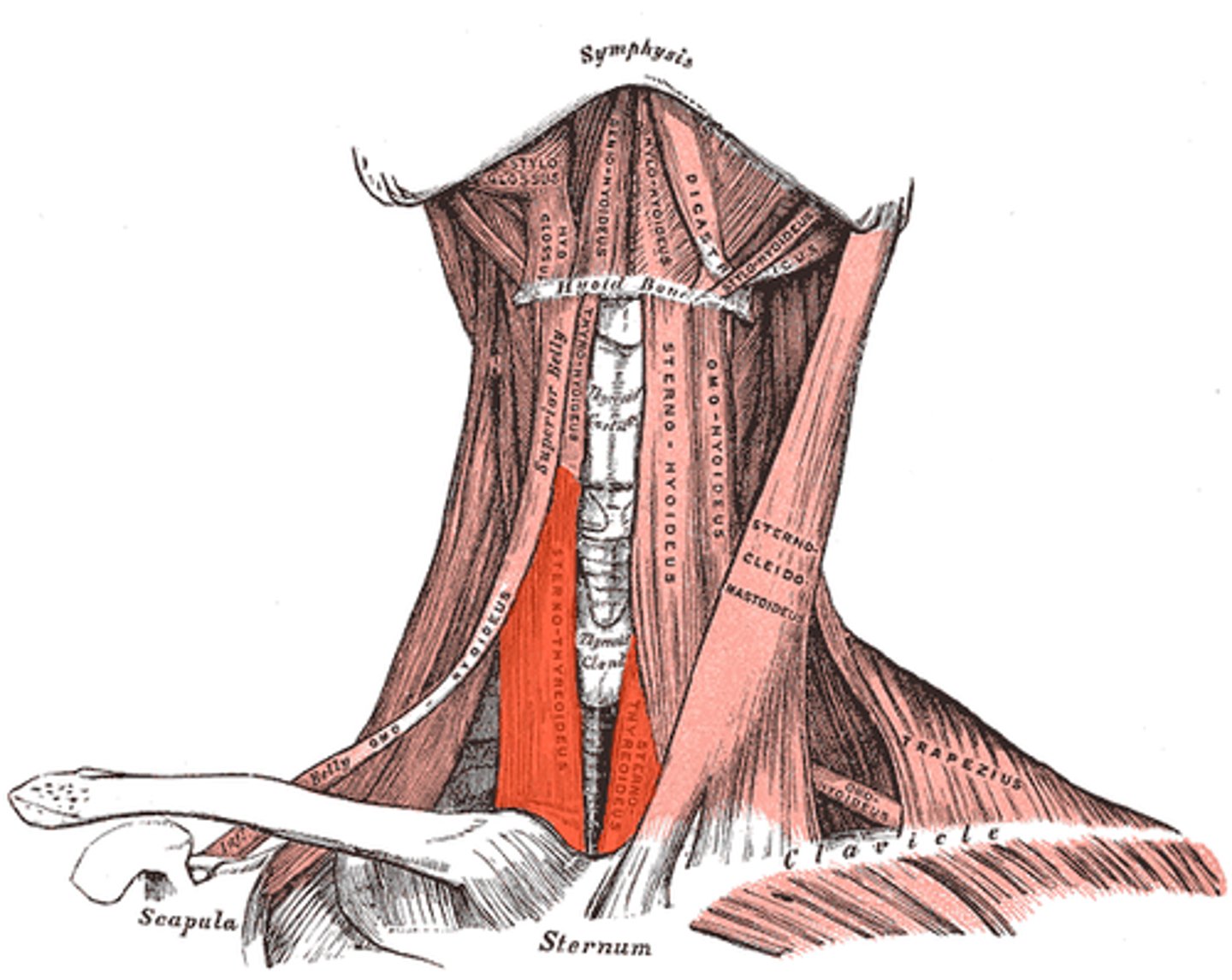
Sternothyroid Muscle
STRUCTUREs that run down the sides of front neck, under sternohyoid flap (seen on left)
Thyrohyoid Muscle
STRUCTURE that is a round muscle right above sternothyroid muscle
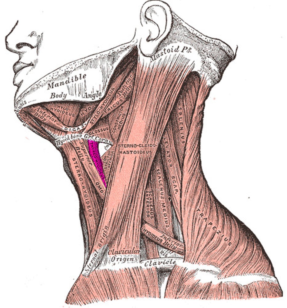
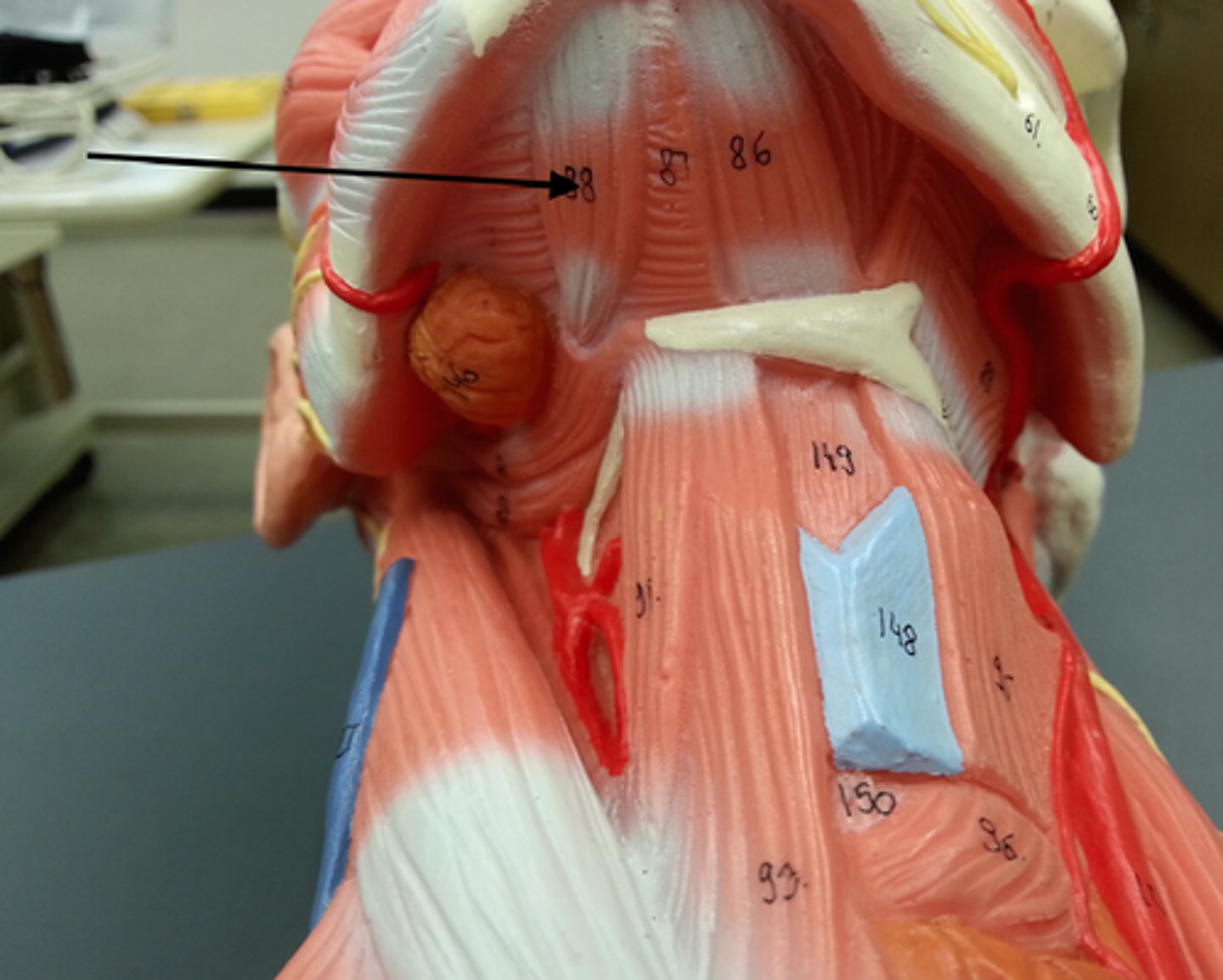
Omohyoid Muscle
STRUCTURE that is a thin loose band that runs from sternohyoid to right base of neck
Superior Belly of the Omohyoid Muscle
PORTION that is the thick part of the loose rope that attaches to the sternohyoid muscle
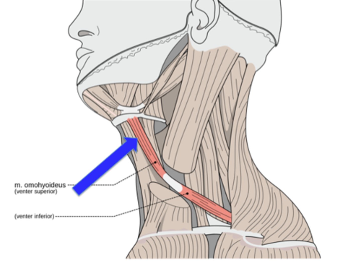
Inferior Belly of the Omohyoid Muscle
PORTION that is the thin part of the loose rope that attaches to the side of the base of the neck (bottom part)

Occipitofrontalis Muscle
ENTIRE STRUCTURE that is the top/front to back of the head

Frontal Belly of the Occipitofrontalis Muscle
PORTION that is the forehead
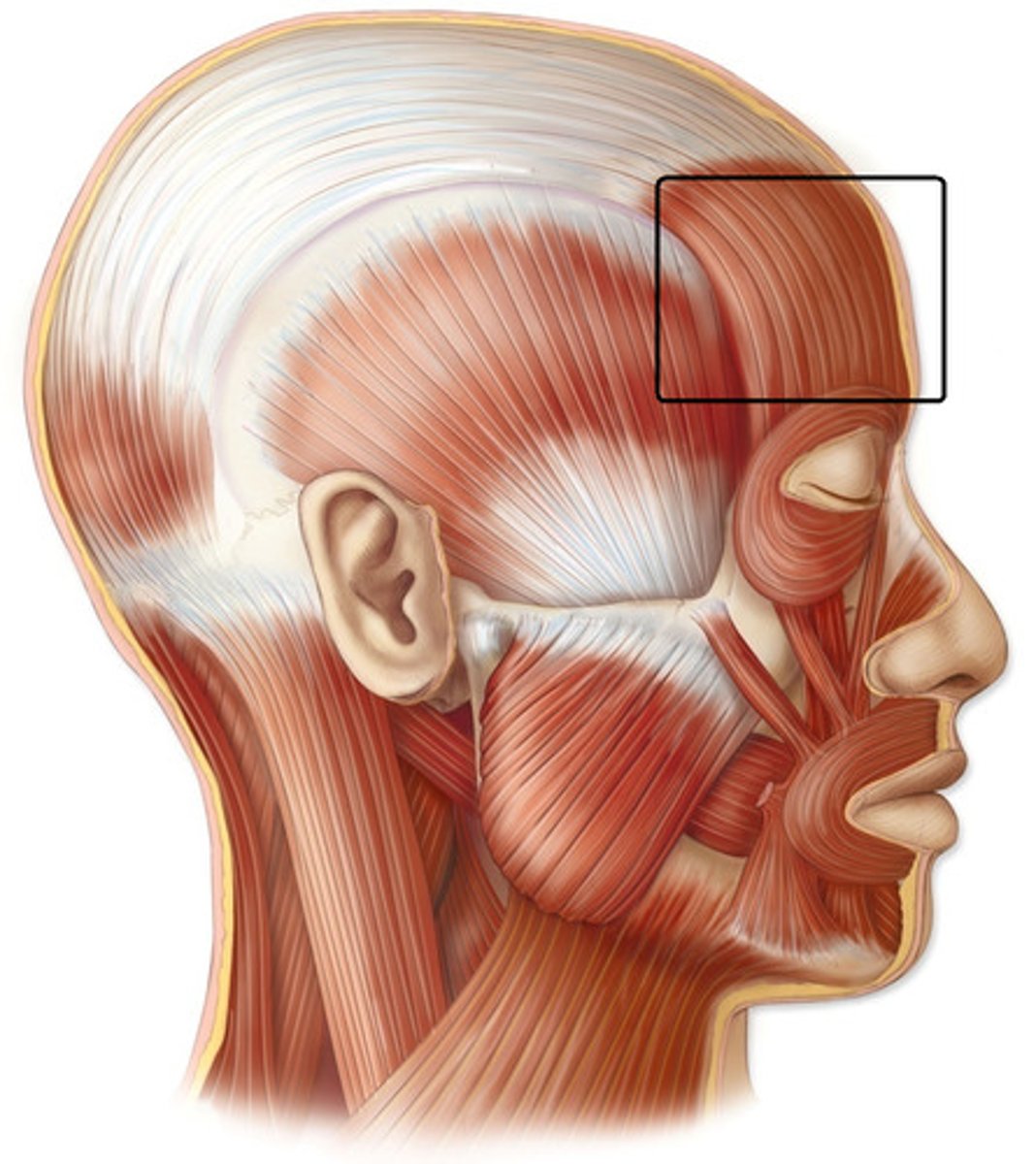
Occipital Belly of the Occipitofrontalis Muscle
PORTION that is the base of the head (over the occipital bone)

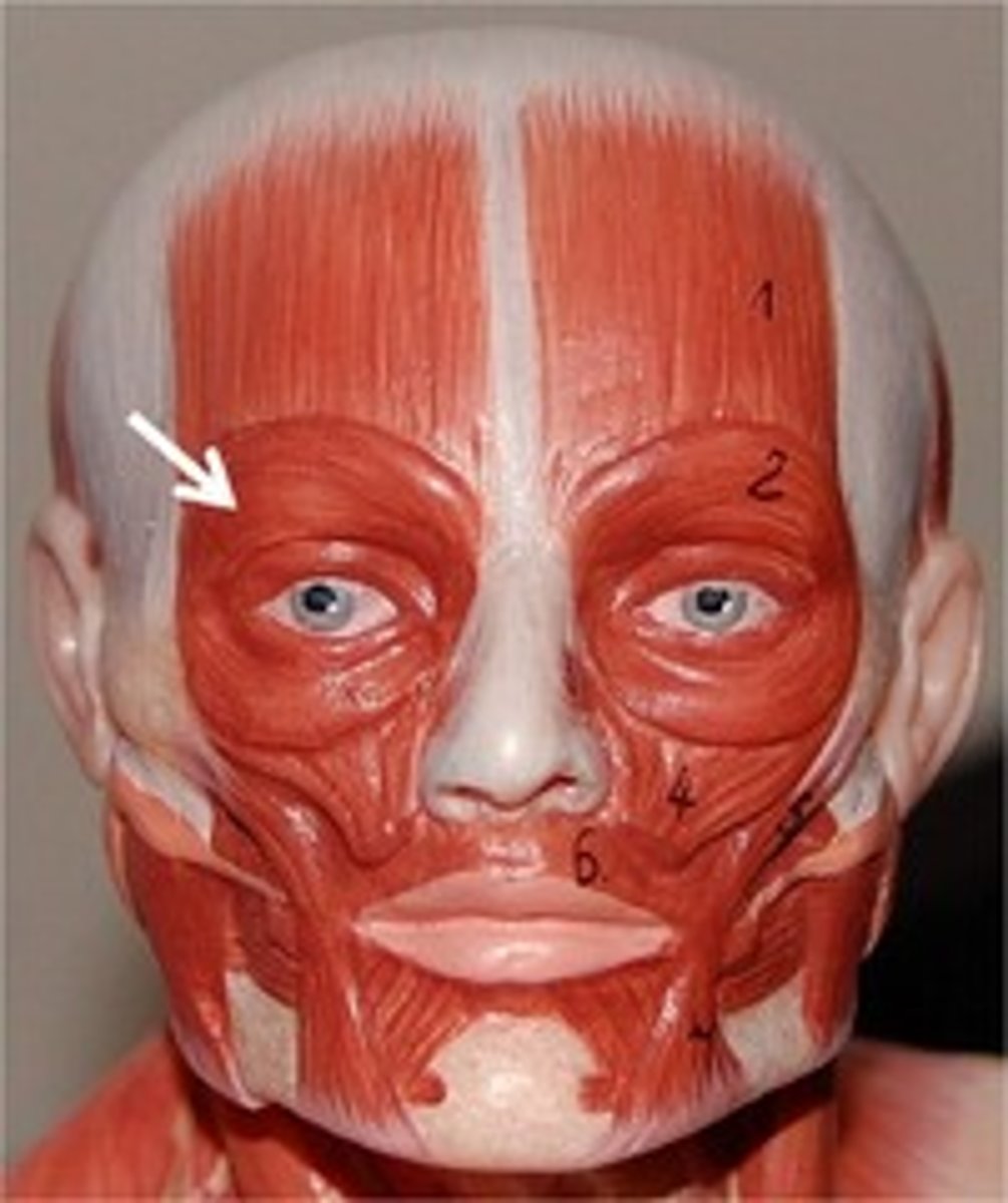
Orbicularis Oculi Muscle
STRUCTURE that is the muscles around the eyelids (probe will circle the eyes)
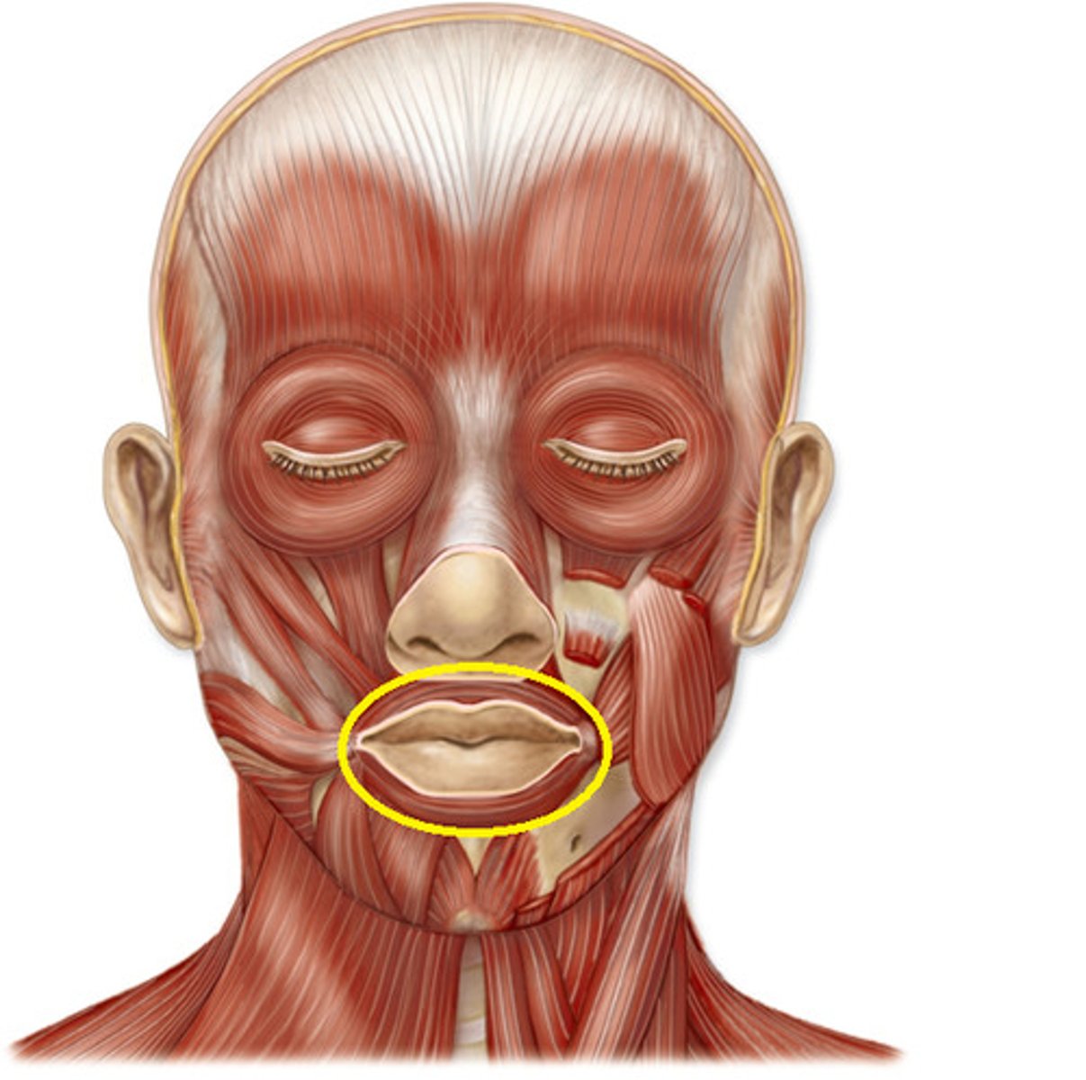
Orbicularis Oris Muscle
STRUCTURE that is the muscle around the lips (probe will circle lips)
Buccinator Muscle
STRUCTURE that is the triangle flap on the side of the cheek, attached at the cheekbone
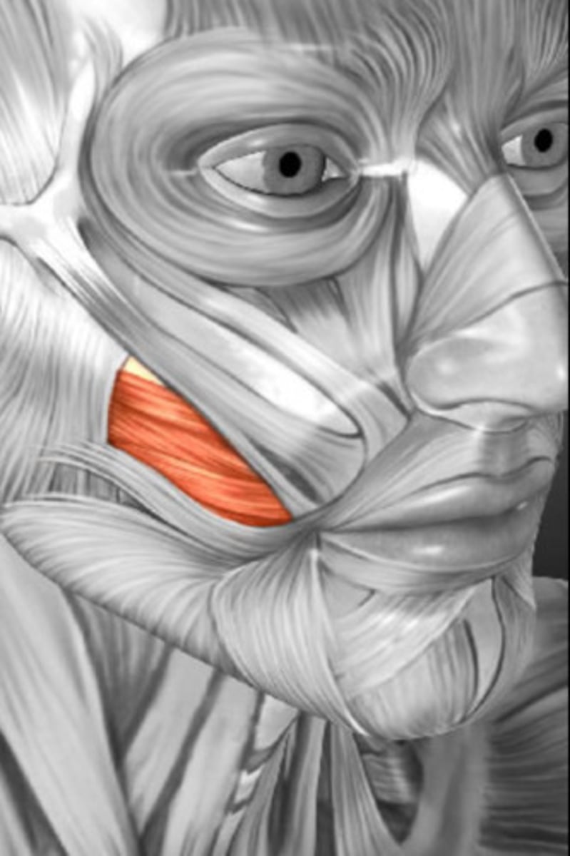
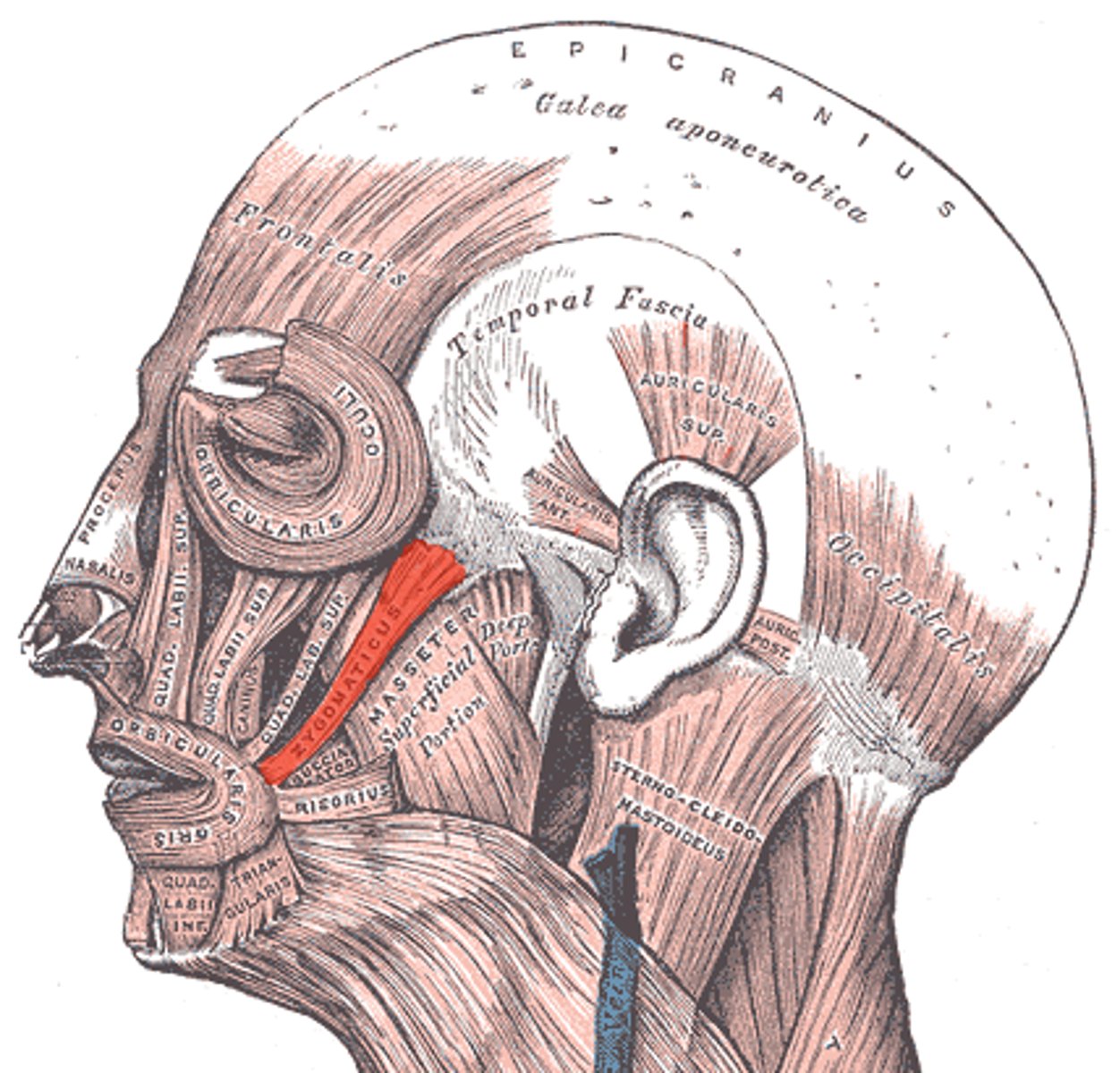
Zygomaticus Major Muscle
STRUCTURE that attaches by corner of mouth to cheekbone
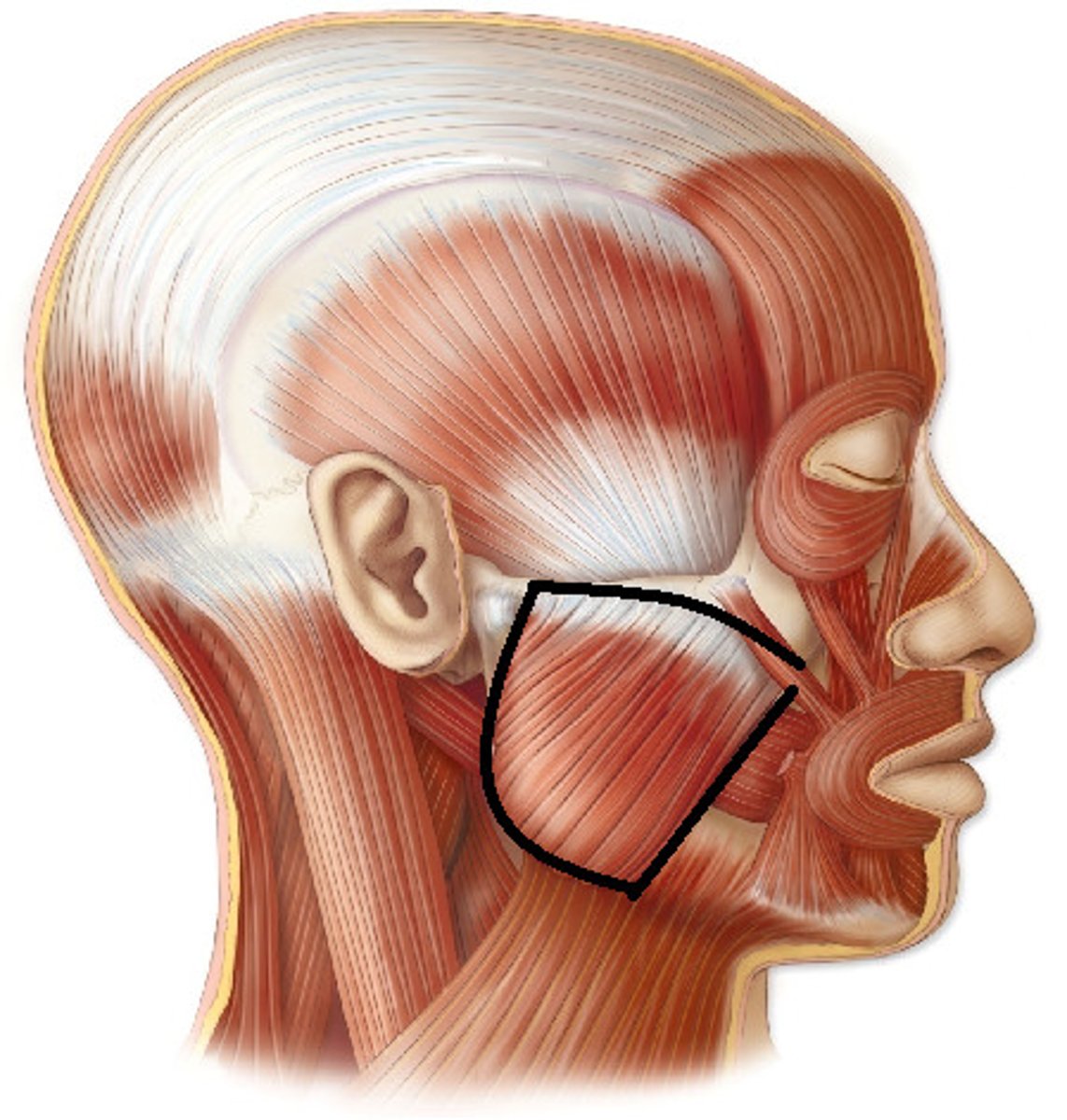
Masseter Muscle
STRUCTURE that is the large flap of the cheek (attached at jaw line)
Temporalis Muscle
STRUCTURE that is where the temple is

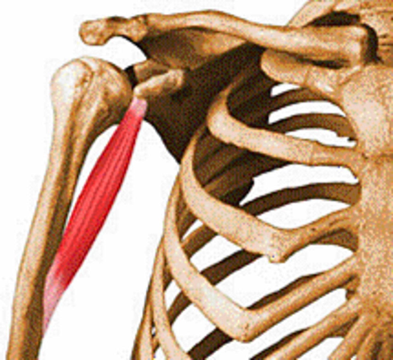
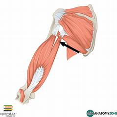
Coracobrachialis Muscle
STRUCTURE that attaches right at the pectoralis muscle
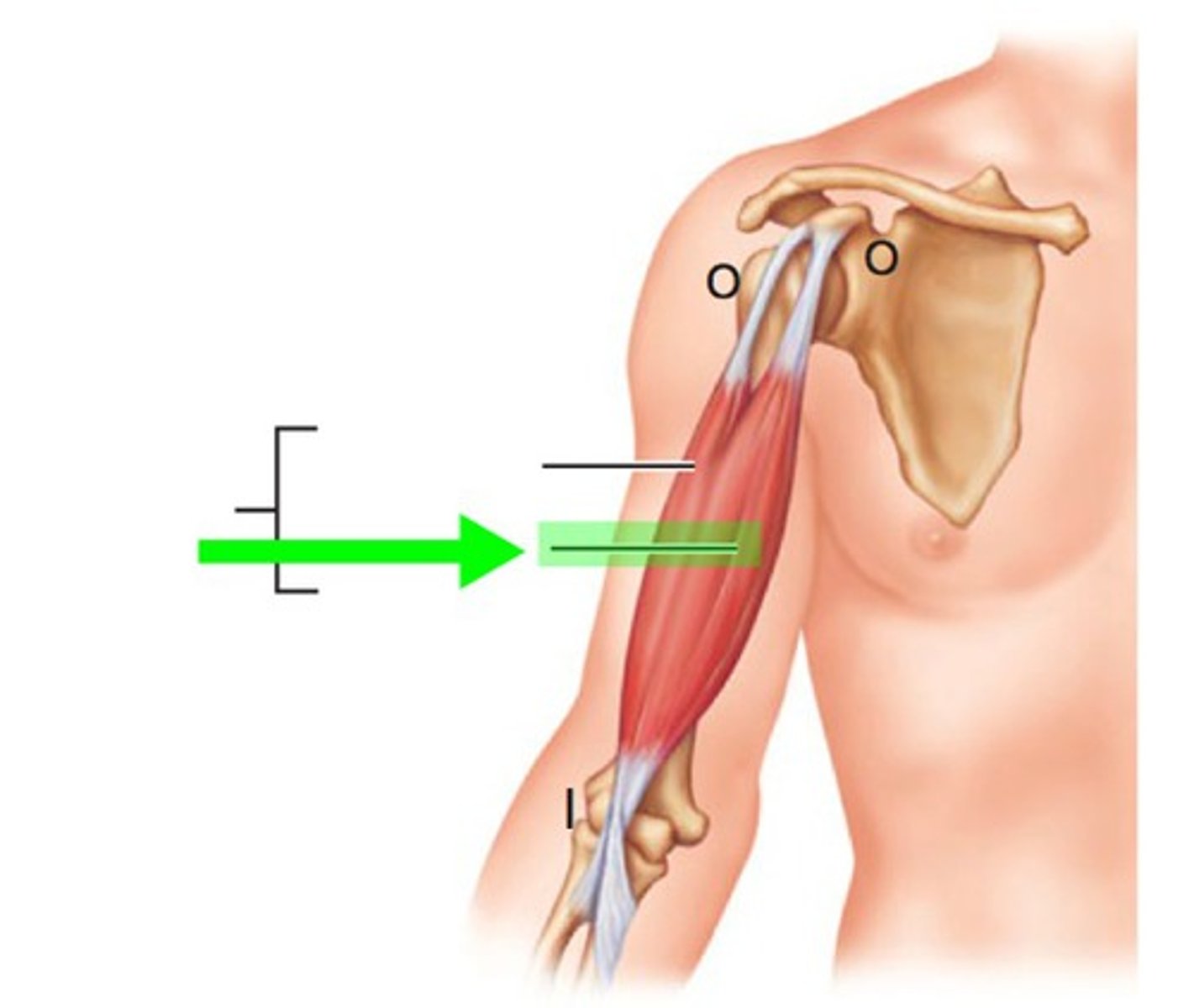
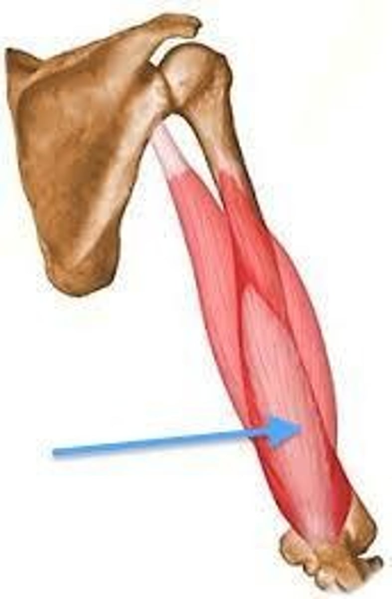
Biceps Brachii Muscle
STRUCTURE that is the thick rope of muscles on the inside of the arm
Long Head of the Biceps Brachii Muscle
STRUCTURE that is the back of the biceps brachii muscle, nearest the bone
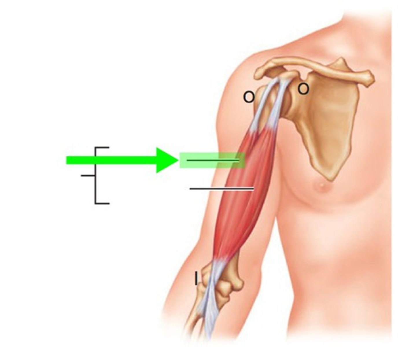
Short Head of the Biceps Brachii Muscle
STRUCTURE that is the "peak" of the biceps brachii muscle
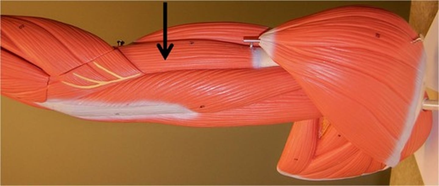
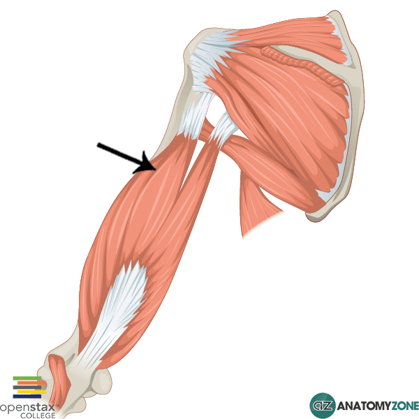
Brachialis Muscle
STRUCTURE that runs next to the biceps brachii muscle; lateral to bicep
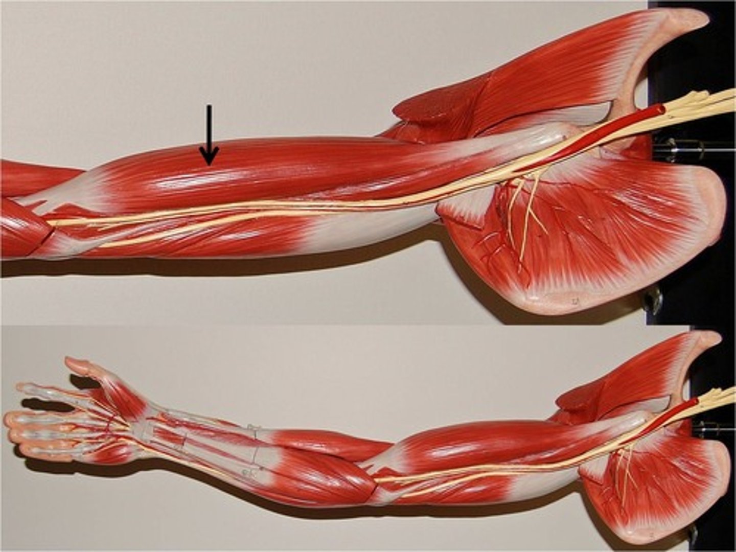
Triceps Brachii Muscle
ENTIRE STRUCTURE that is on the elbow/back side of the arm

Long Head of the Triceps Brachii Muscle
STRUCTURE that is the largest part of the triceps brachii muscle; hangs off the back of the arm, near the shoulder
Medial Head of the Triceps Brachii Muscle
STRUCTURE that is the portion just below the long head; along the length of the triceps brachii muscle
Lateral Head of the Triceps Brachii Muscle
STRUCTURE that is just medial to the medial head of the medial head of the triceps brachii
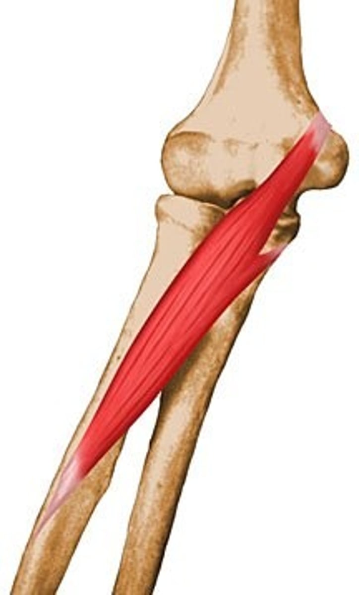
Pronator Teres Muscle
STRUCTURE that starts at the crook of the elbow and runs from the middle of elbow down half of forearm
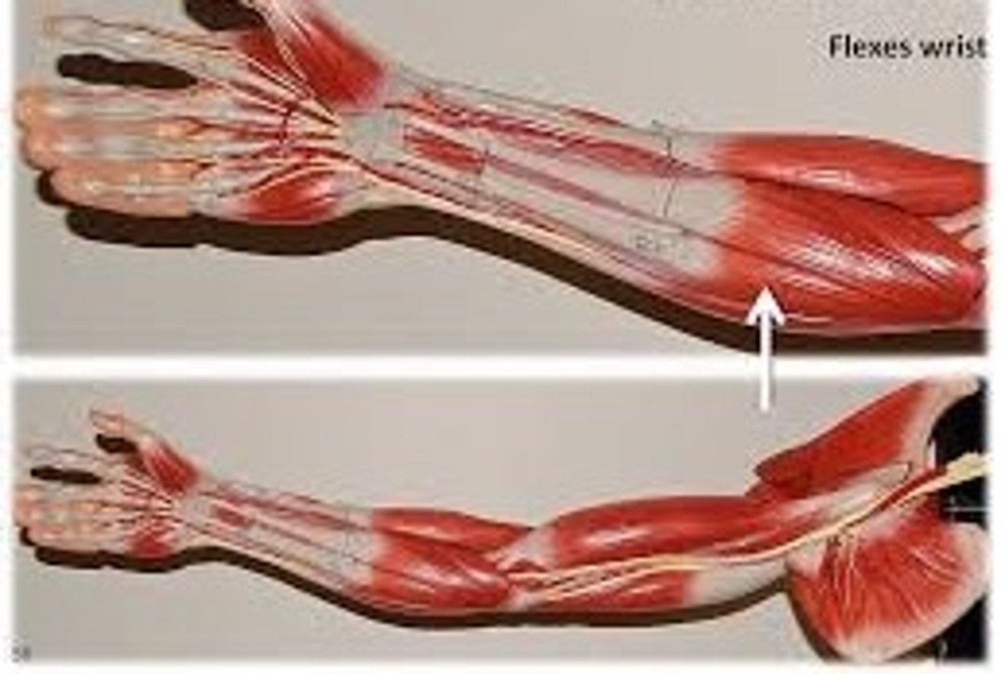
Flexor Carpi Radialis Muscle
STRUCTURE that is medial to the pronator teres muscle; thick attached bands that runs down the length of the forearm
Palmaris Longis Muscle
STRUCTURE that has a very long, thin ligament that attaches at wrist/hand; muscle is very small and at the top of the inside forearm
Flexor Carpi Ulnaris Muscle
STRUCTURE that is the long portion on the pinky side; it has not been dissected fully
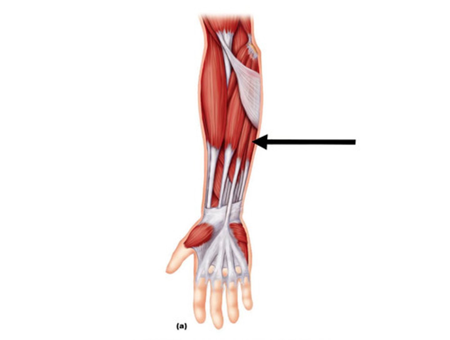
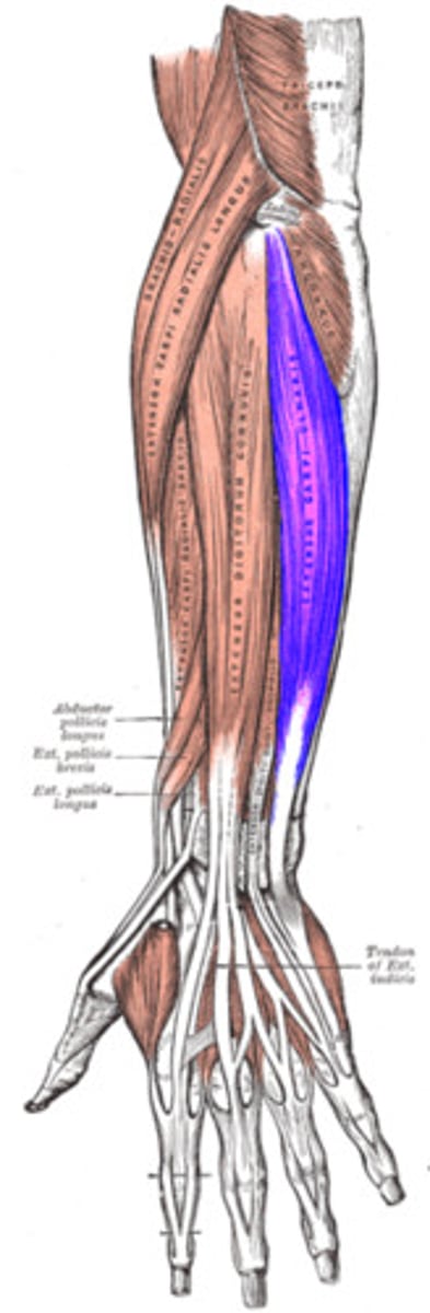
Flexor Digitorum Superficialis Muscle
STRUCTURE that is a big muscle slab under all to-layer forearm muscles; runs down center of forearm
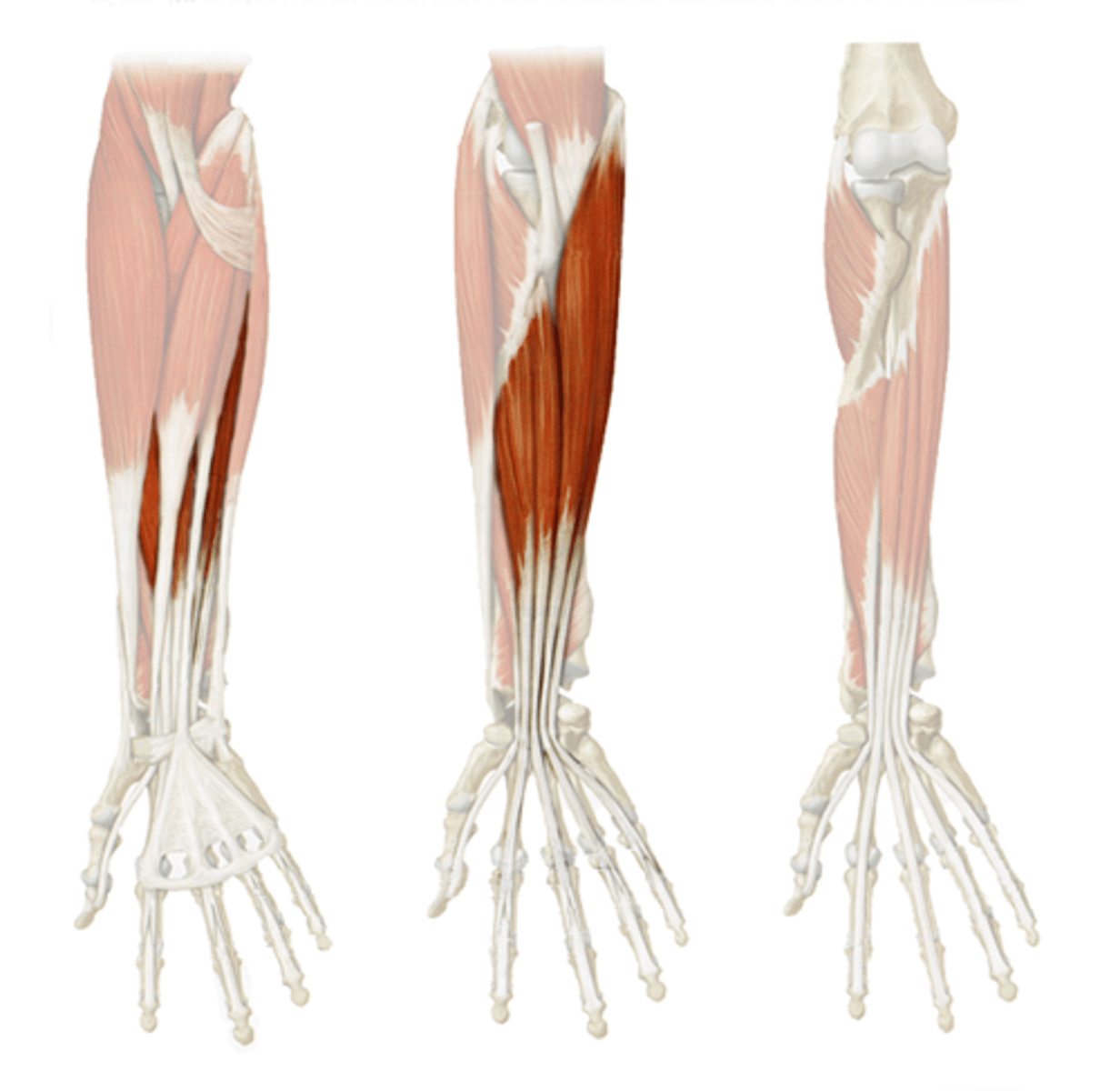
Flexor Digitorum Profundus Muscle
STRUCTURE that is two thick bundles of white in the under cavity of the forearm; on pinky side
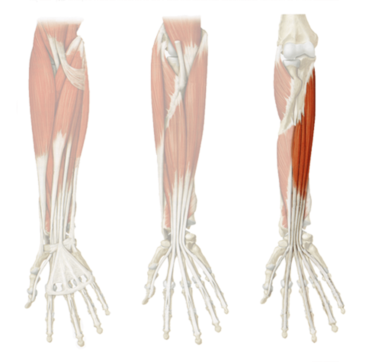
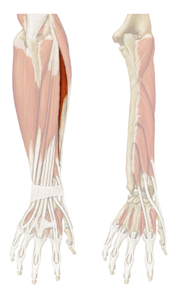
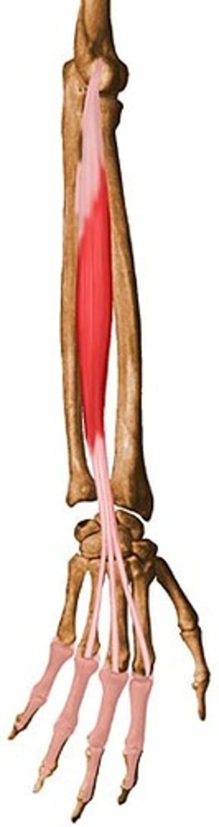
Flexor Pollicis Longus Muscle
STRUCTURE that is deep in the forearm cavity; white line back towards the thumb
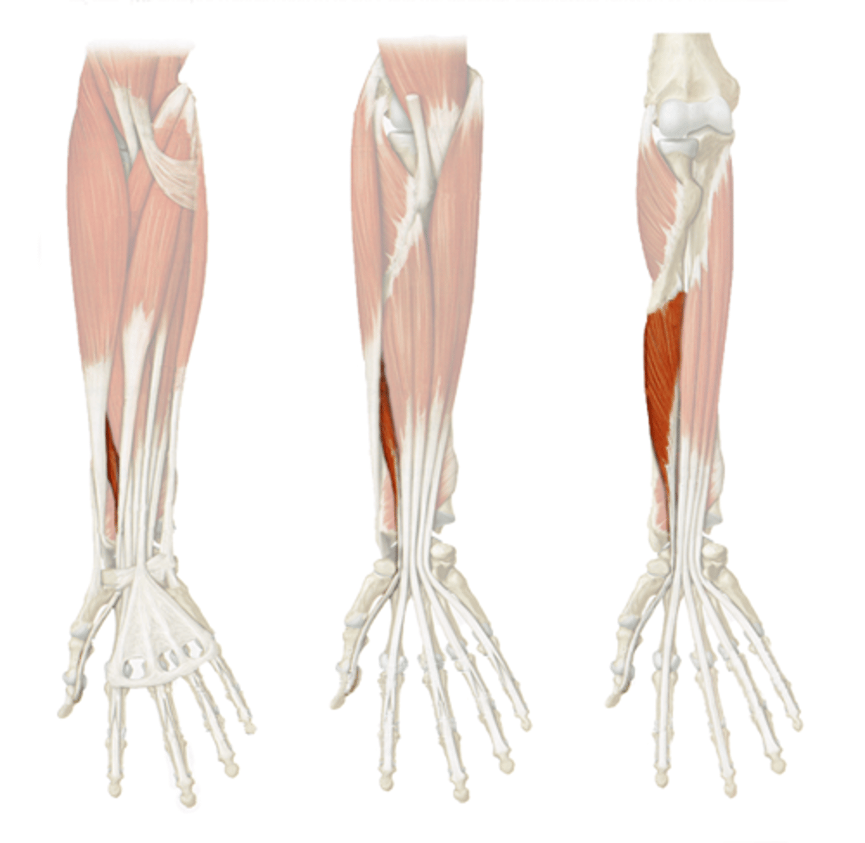
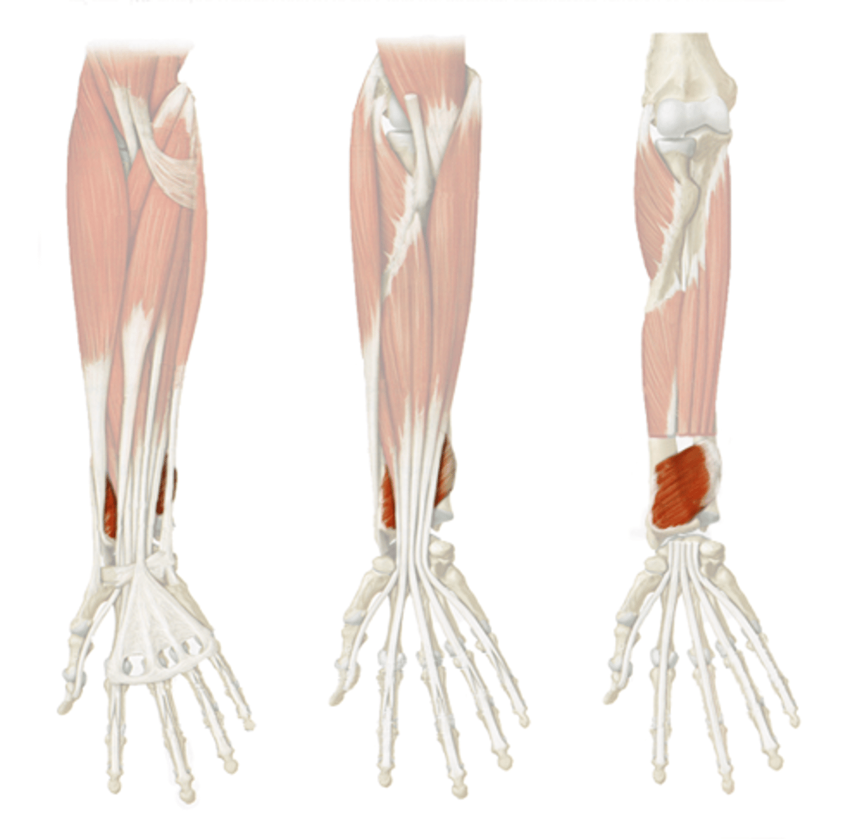
Pronator Quadratus Muscle
STRUCTURE that is the white muscle wall deepest in the forearm cavity
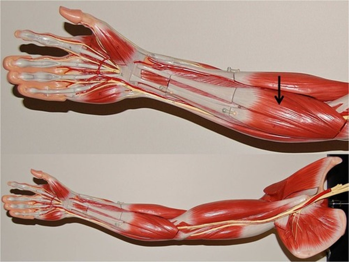
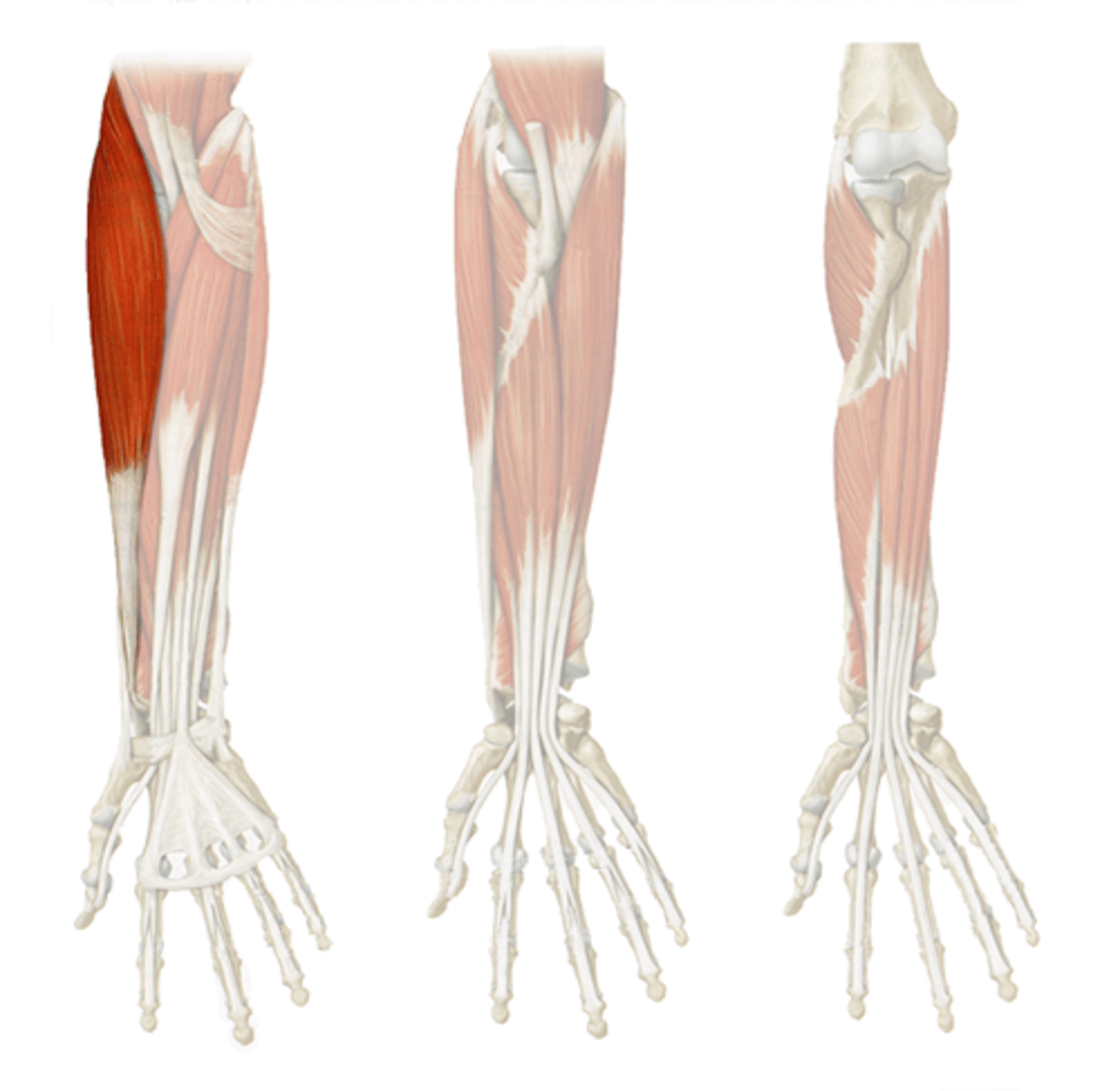
Brachioradialis Muscle
STRUCTURE that is on the lateral edge of the forearm; runs all the way from wrist to bicep
Extensor Carpi Radialis Longus Muscle
STRUCTURE that is on the back of the forearm; lateral to brachioradialis muscle; runs from wrist to elbow only
Extensor Carpi Radialis Brevis Muscle
STRUCTURE just under and medial to the extensor carpi radialis longus muscle; on back of forearm; runs from just below elbow to just above the wrist
Extensor Digitorum Muscle
STRUCTURE that is a bundle that connects to the back of the hand and the fingers
Extensor Carpi Ulnaris Muscle
STRUCTURE that is undissected and on the pinky side of the back forearm
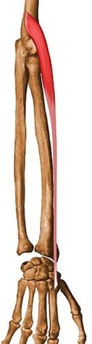
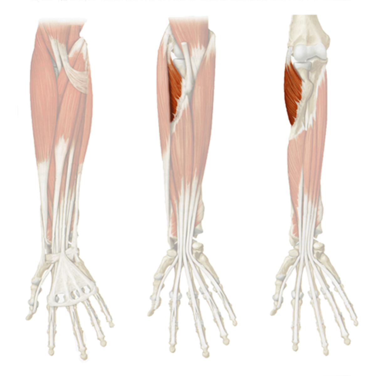
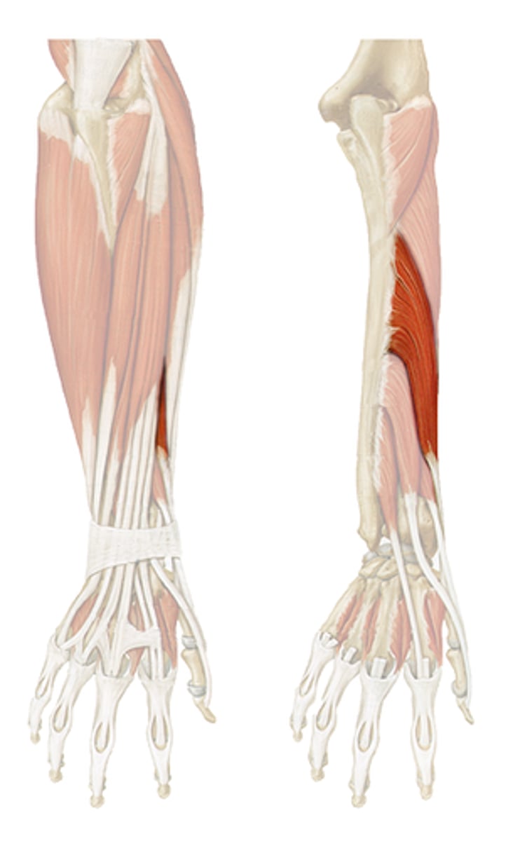
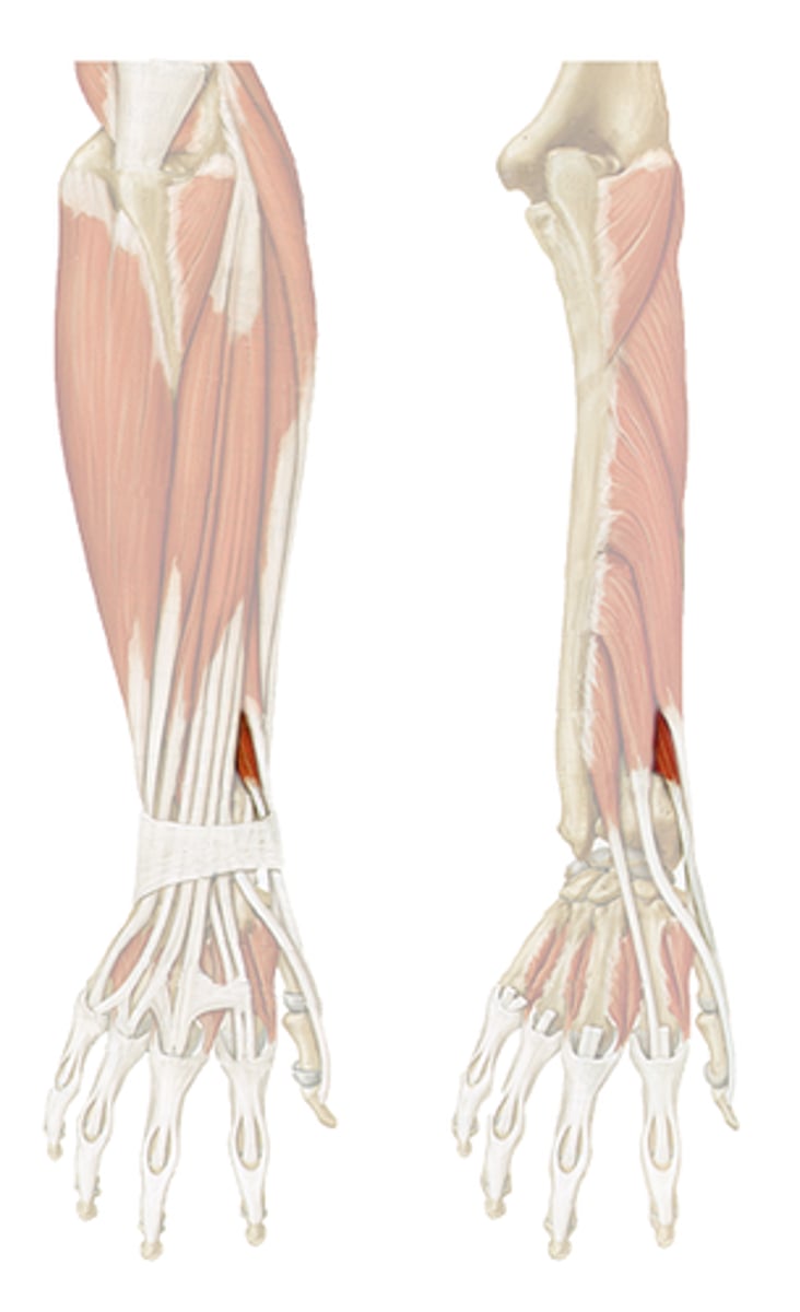
Supinator Muscle
STRUCTURE that is a seen as a small string under the brachioradialis muscle; only muscle under the brachioradialis we will look at
Abductor Pollicis Longus Muscle (1)
STRUCTURE that is closes to the thumb side on the back forearm; we only see a small portion that is tucked between two long muscles
Extensor Pollicis Longus Muscle
STRUCTURE directly on the pinky side of the abductor pollicis longus muscle; very short and tucked between surrounding long muscles
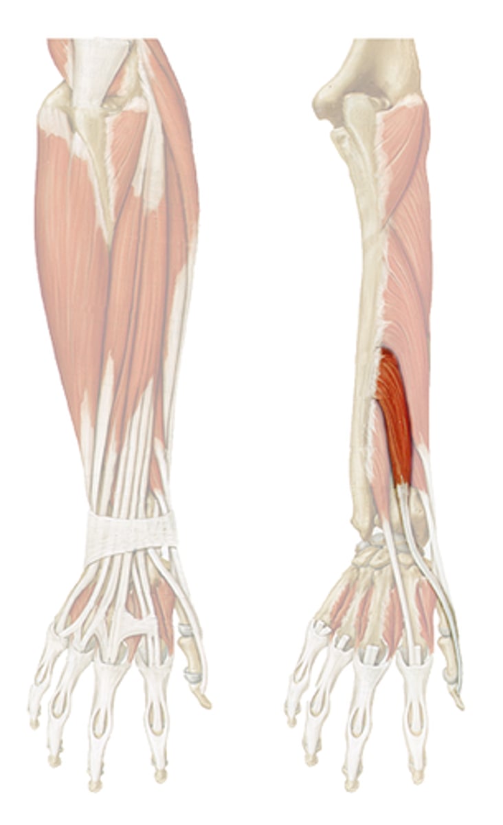
Extensor Pollicis Brevis Muscle
STRUCTURE that is very short (only about 1 inch visible) and tucked between the extensor pollicis longus m. and abductor pollicis longus m.
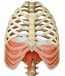
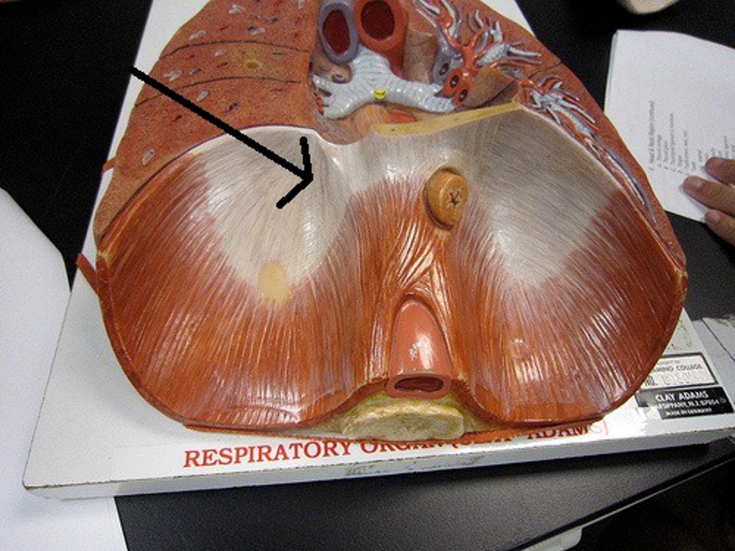
Diaphragm Muscle
COLLECTIVE STRUCTURE that is a sheet-like muscle perpendicular to the bottom of the lungs; covers the inferior thoracic aperture (attaches at the red outline in the diagram)
Sternocostal Part of the Diaphragm Muscle
PORTION that is from the shiny purple line and outward; the main part/majority of the diaphragm muscle
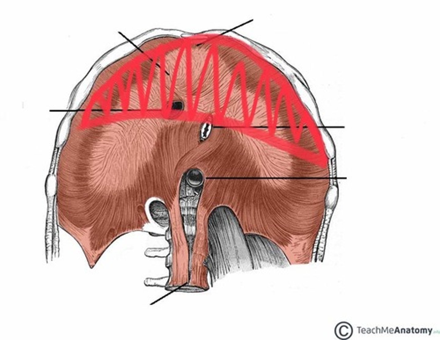
Lumbar Part of the Diaphragm Muscle
PORTION on the inside of the shiny purple line; the part of the diaphragm muscle that is closest to the spine
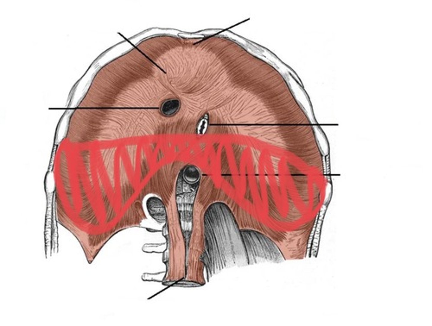
Central Tendon of the Diaphragm Muscle
STRUCTURE that is the shiny purple line that divides the sternocostal and lumbar parts of the diaphragm muscle
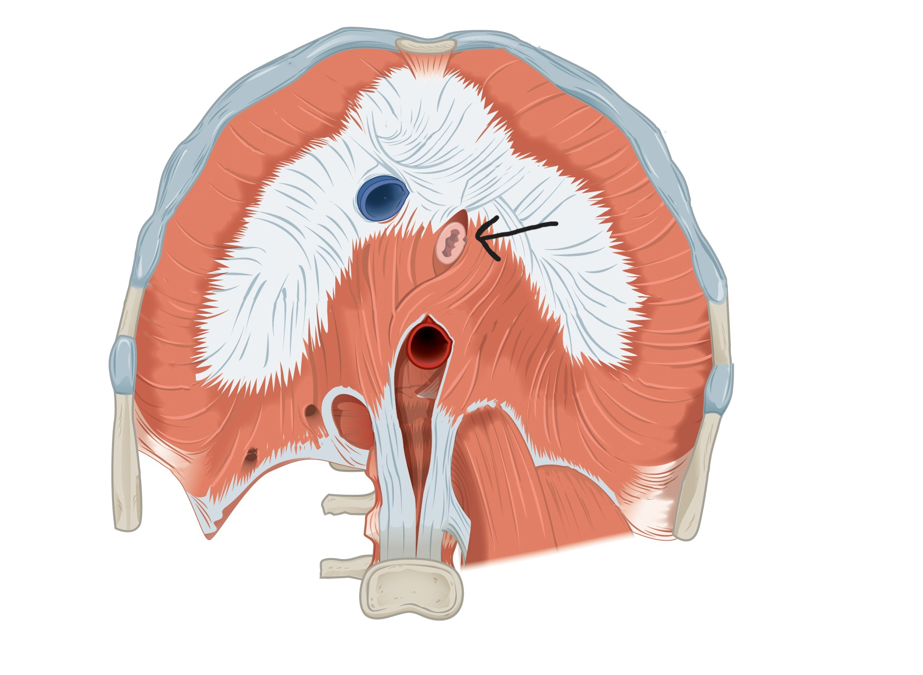
Aortic Hiatus of the Diaphragm Muscle
OPENING in the diaphragm muscle; probe will go under the esophagus, along the aorta, and then through the diaphragm muscle from the top

Esophageal Hiatus of the Diaphragm Muscle
OPENING in the diaphragm muscle; probe will go up the shredded-looking esophagus and through the diaphragm muscle from the bottom
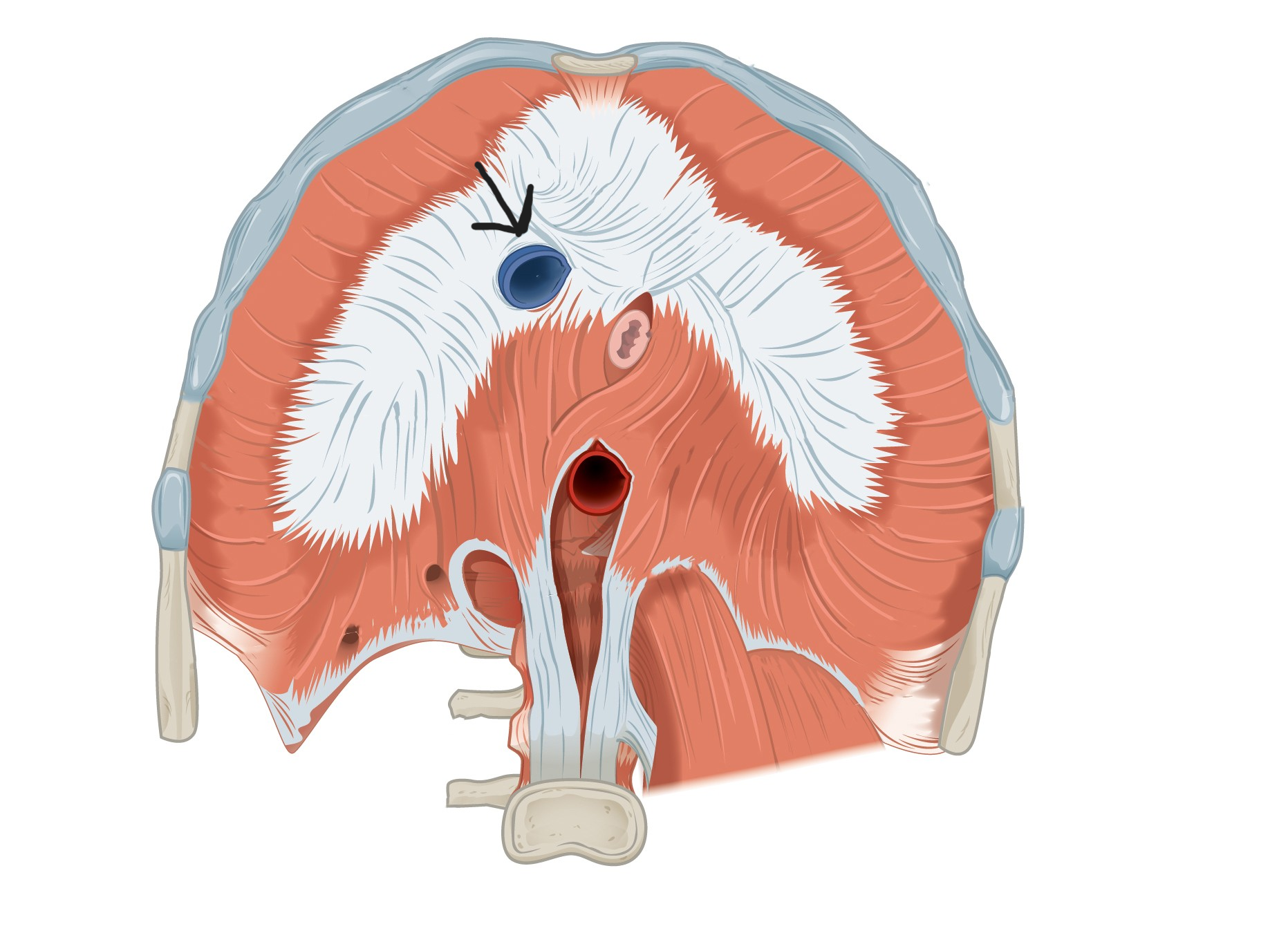
Caval Opening of the Diaphragm Muscle
OPENING in the diaphragm muscle; the probe will go in through the giant hole (from the top) and then through the diaphragm muscle, down into the vena cava
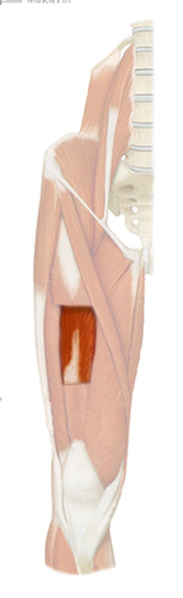
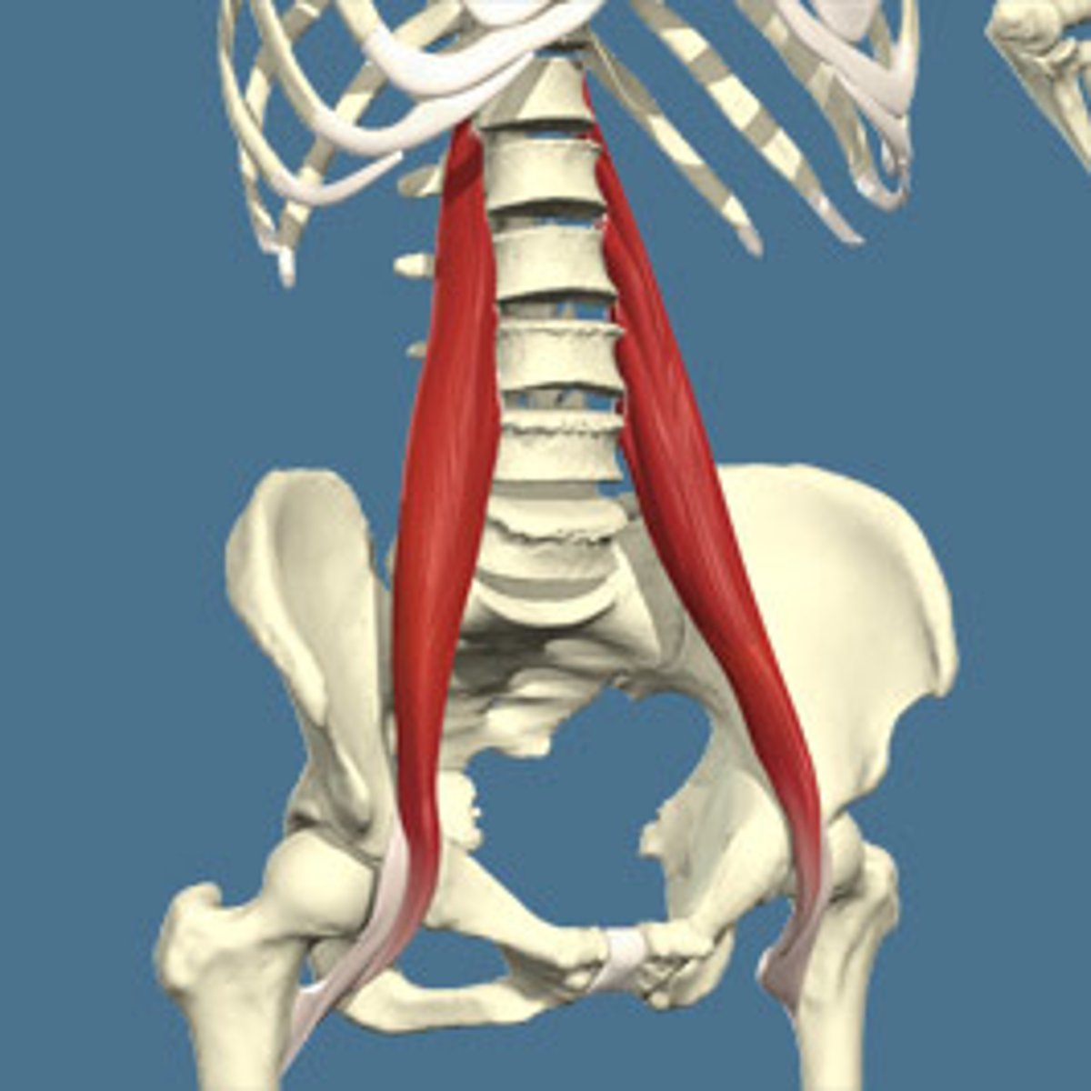
Psoas Major Muscle
STRUCTURE that is the large muscle of the pelvic region that is loose; attaches near head of femur and up along sides of lumbar vertebrae
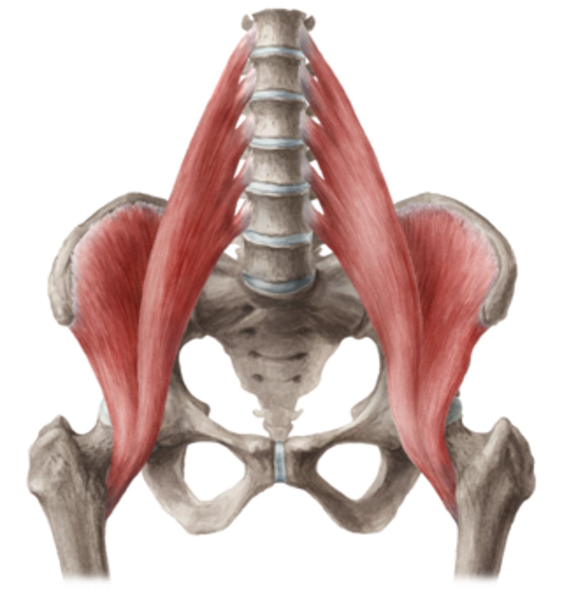
Iliacus Muscle
STRUCTURE that fills the fossa of the illiac bone (seen on the right leg)
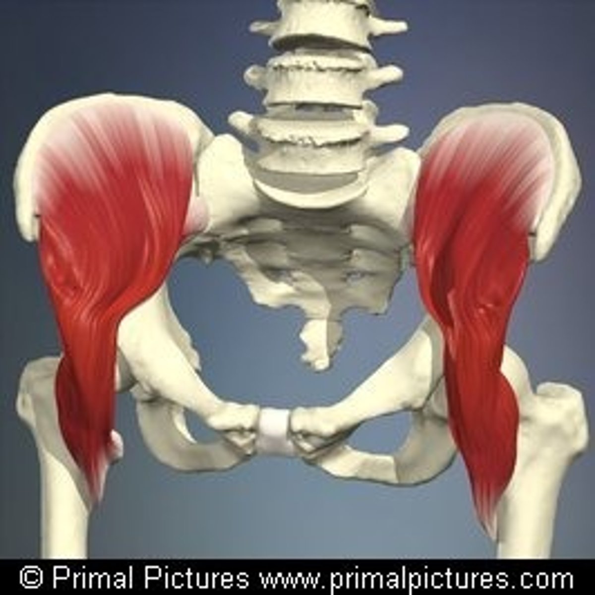
Iliopsoas Muscle
STRUCTURE that is in line with humerus head, where the iliacus and psoas major muscles meet
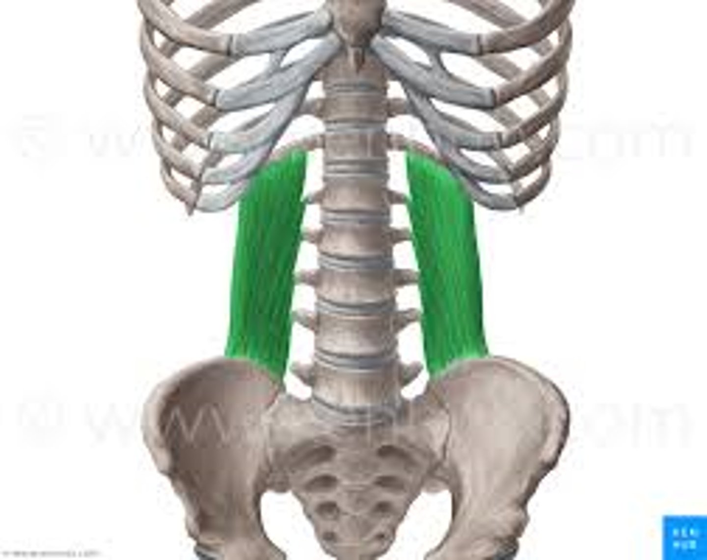
Quadratus Lumborum Muscle
STRUCTURE that forms a smooth cup under the kidney (left kidney will be moved to view)
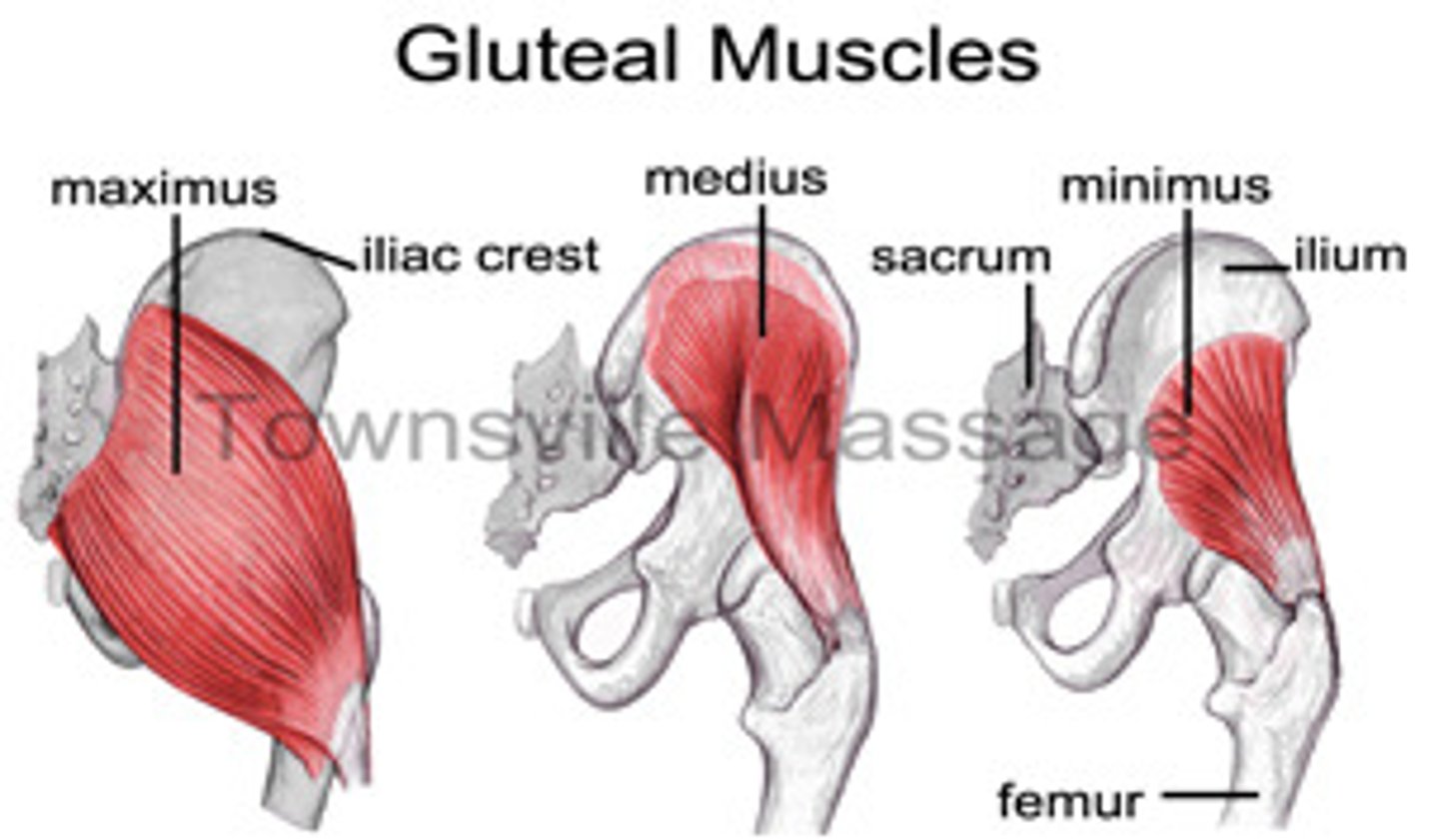
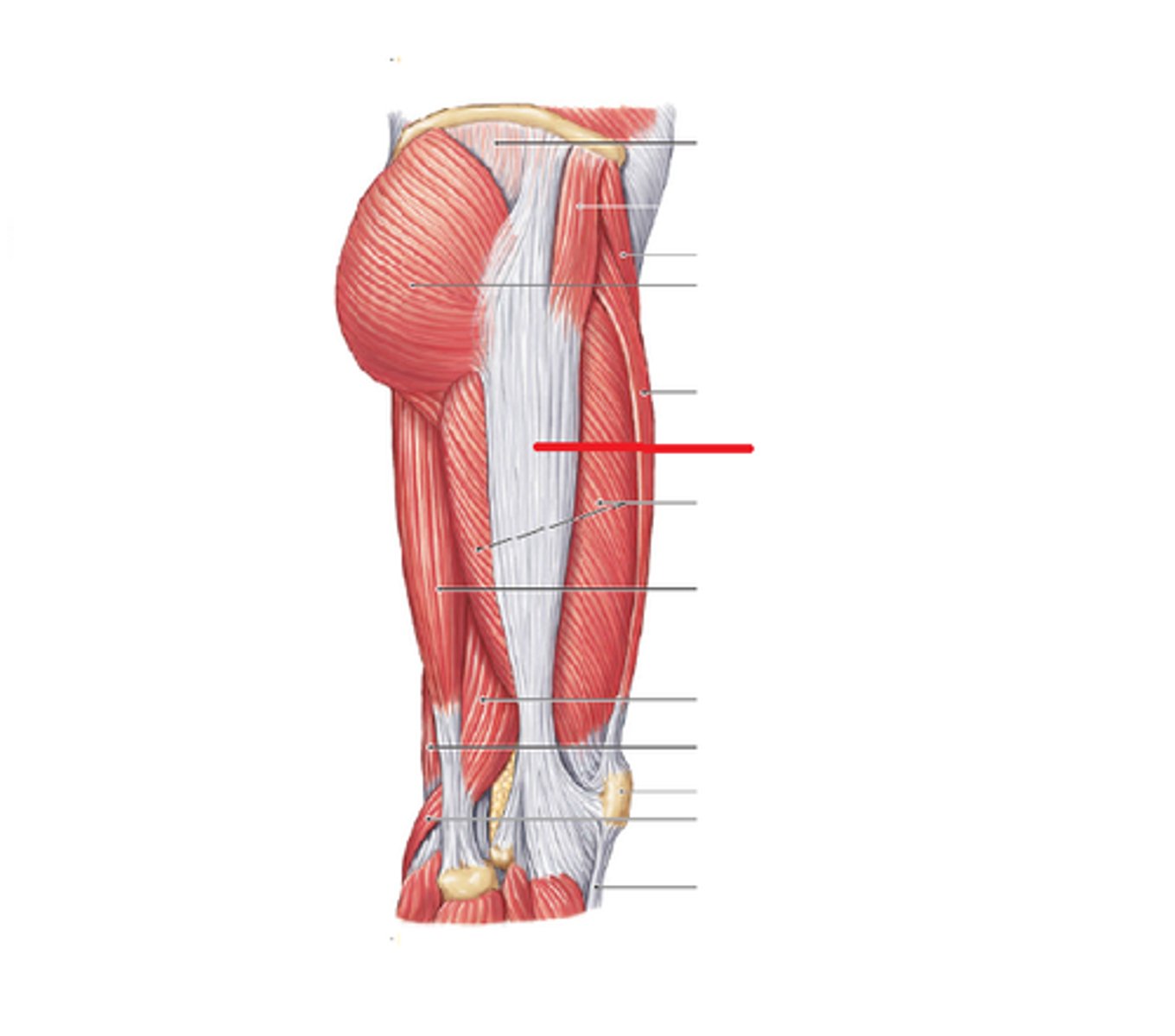
Gluteus Maximus Muscle
STRUCTURE that is the outermost and biggest flap covering the butt
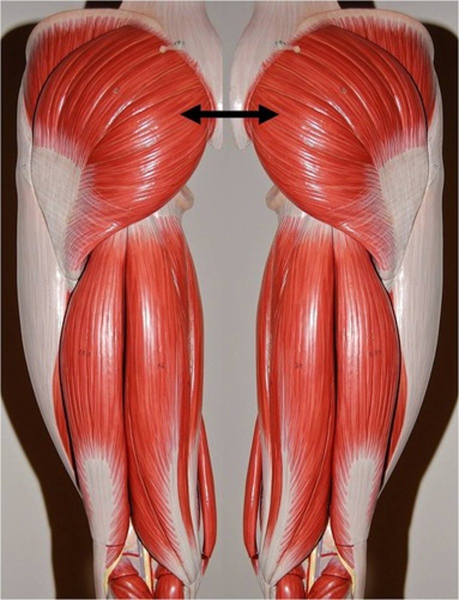
Gluteus Medius Muscle
STRUCTURE that is the hand-sized flap at the top and outer-side of the butt
Gluteus Minimus Muscle
STRUCTURE that is smooth and attached just under the gluteus medius muscle
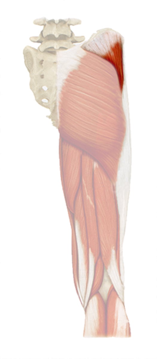
Tensor Fasciae Latae Muscle
STRUCTURE that is just under the IT band; short and covers the lateral edge of hip
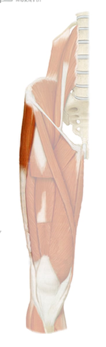
Iliotibial Tract (Band)
STRUCTURE that is the papery-looking "skin" on the outside/lateral edge of the thigh
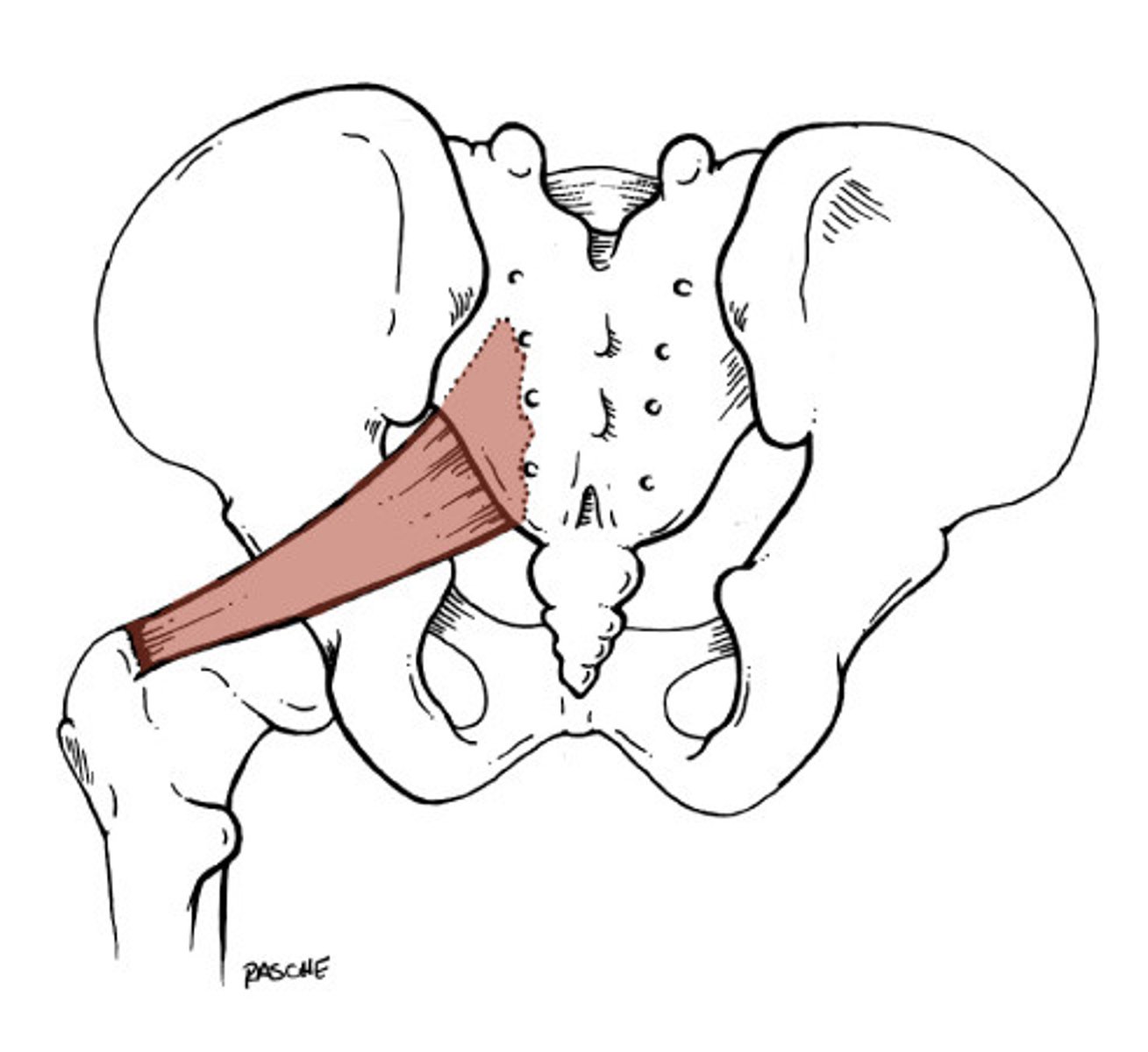
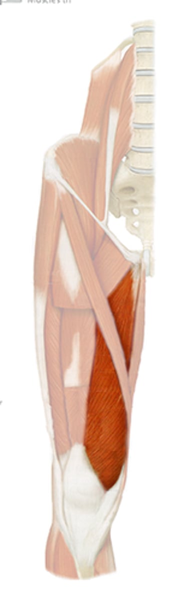
Piriformis Muscle
STRUCTURE that runs horizontal just inferior to the gluteus medius
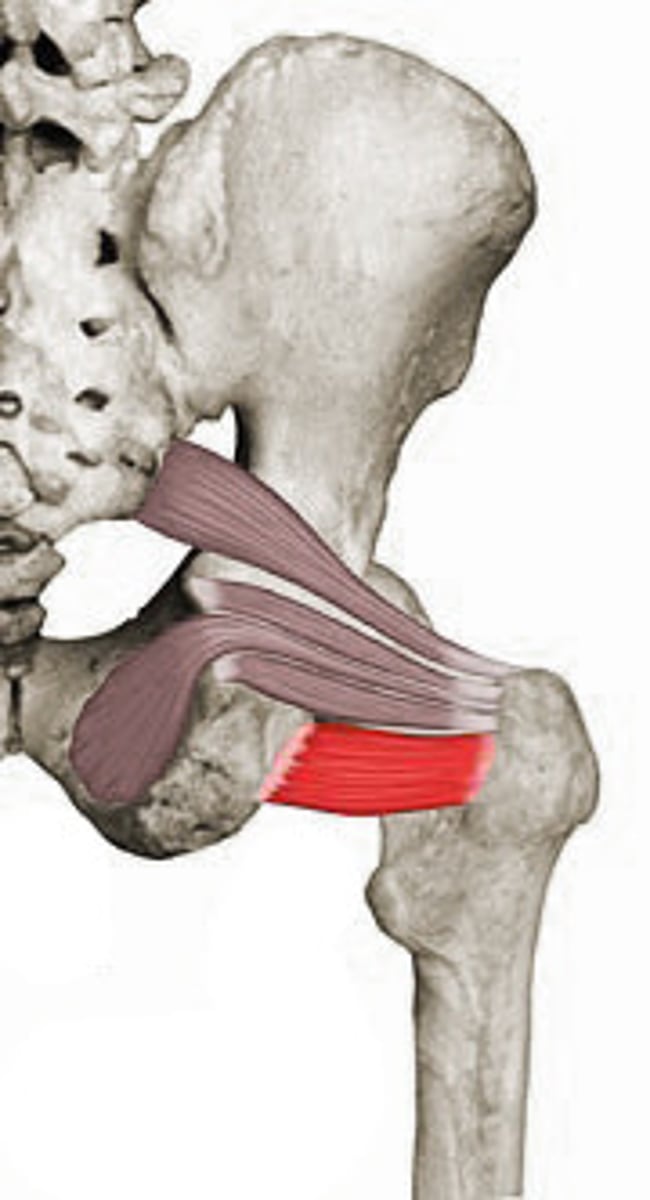
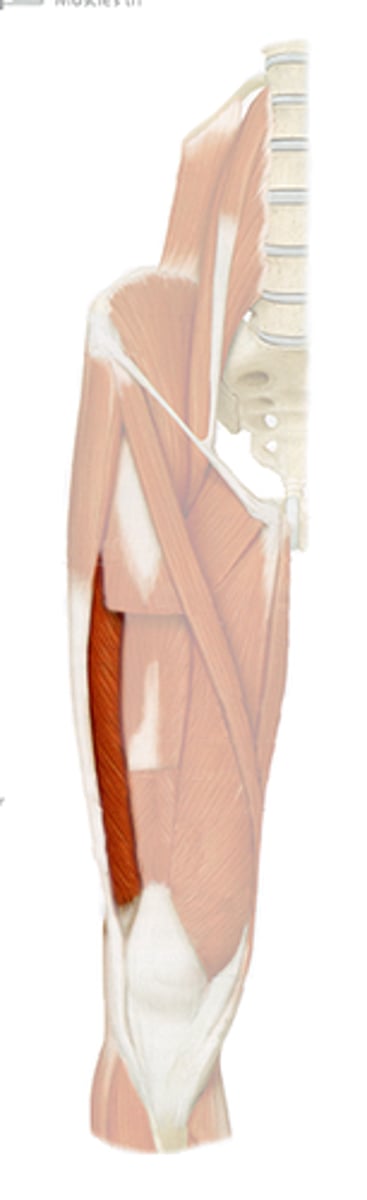
Quadratus Femoris Muscle
STRUCTURE that is the horizontal grey rectangle at the top of the femur bone
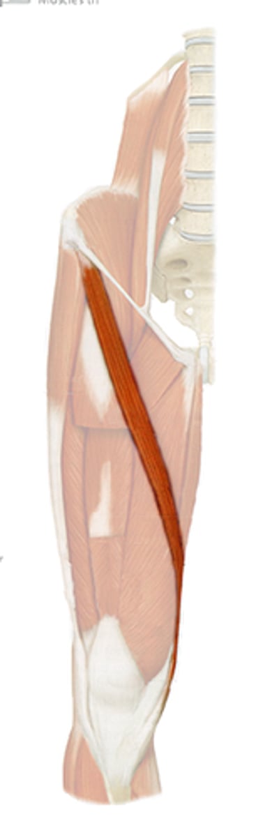
Sartorius Muscle
STRUCTURE that runs from outside of hip to inside of knee; about an 1 1/2 inches in diameter
Quadriceps Femoris Muscle
COLLECTIVE STRUCTURE that includes the 4 muscles of the anterior leg; TA will tap all 4 muscles near knee
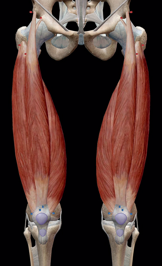
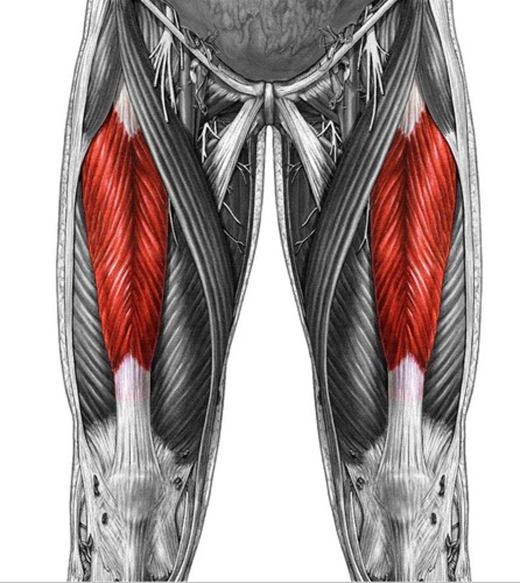
Rectus Femoris Muscle
STRUCTURE that is the muscle that runs down middle of thigh to the top of the knee; can be lifted by the probe
Vastus Medialis Muscle
STRUCTURE that is the thick, attached band on the medial edge of the thigh
Vastus Lateralis Muscle
STRUCTURE that is the thick band just lateral to the rectus femoris muscle
Vastus Intermedius Muscle
STRUCTURE that is directly underneath the rectus femoris; shiny and attached