8. Radiographic Interpretation Part 3
1/43
There's no tags or description
Looks like no tags are added yet.
Name | Mastery | Learn | Test | Matching | Spaced |
|---|
No study sessions yet.
44 Terms
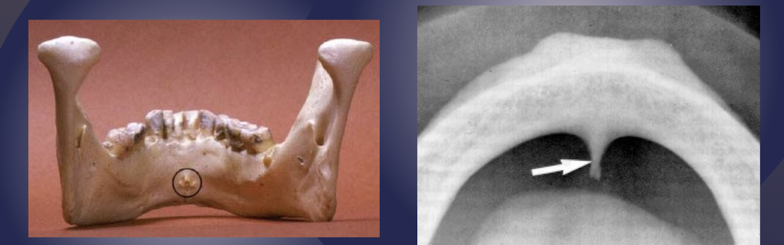
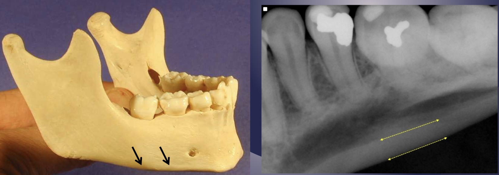
what are the landmarks of the mandible?
genial tubercles
lingual foramen
metal triangle/ridge
mental fossa
vascular/nutrient channels
IAN canal
mental foramen
external oblique ridge
internal oblique ridge/mylohyoid ridge
submandibular gland fossa
coronoid process
inferior border of mandible
genial tubercles
which muscles attach to superior tubercles?
genioglossus
which muscles attach to inferior tubercles?
geniohyoid
radiopaque mass in midline, below the incisors
genial tubercles
genial tubercles
lingual foramen
lingual foramen, saggital
which landmark of the mandible?
Lingual aspect of midline of mandible
Region of genial tubercles
Neurovascular bundles from lingual arteries and nerves
Sublingual/submental arteries and mylohyoid nerves
lingual foramen
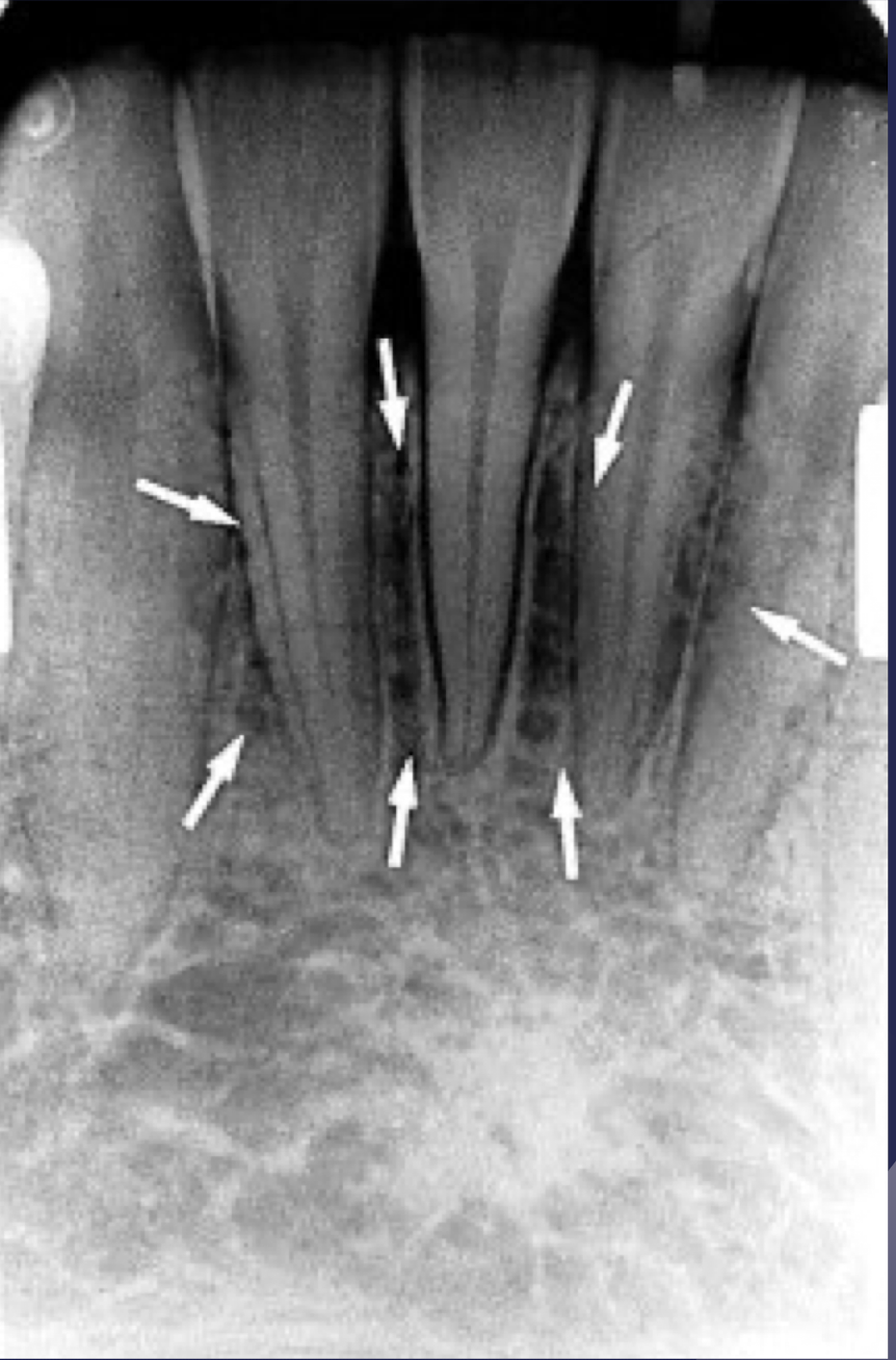
which landmark of the mandible?
Two radiopaque lines sweeping bilaterally forward and upward toward midline
Visualized in over-angulated views
mental triangle/ridge
mental triangle/ridge
mental triangle/ridge
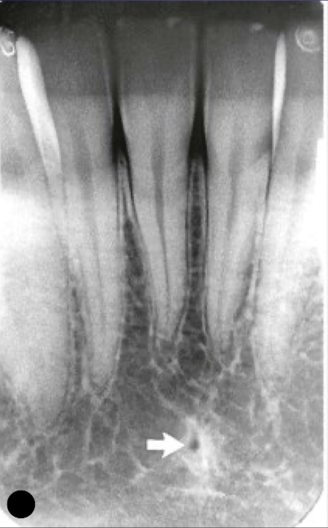
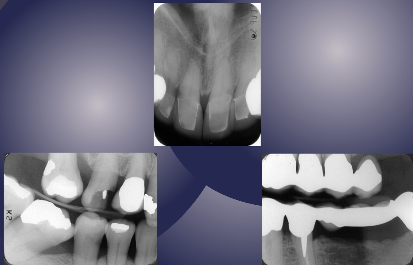
which landmark of the mandible?
Depression on labial aspect of anterior mandible, above mental ridge
Casts a radiolucent shadow over the roots of mandibular anterior teeth
Often mistaken for periapical/periodontal disease involving incisor
mental fossa
mental fossa
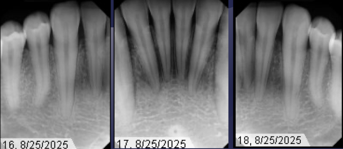
mental fossa
shows it’s more radiolucent bc it’s not as thick at mental fossa
T or F: the neurovascular bundles appear as radiolucent lines of varying widths
false *fairly uniform width
vascular/nutrient channels
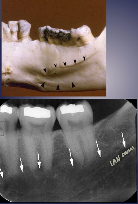
IAN canal
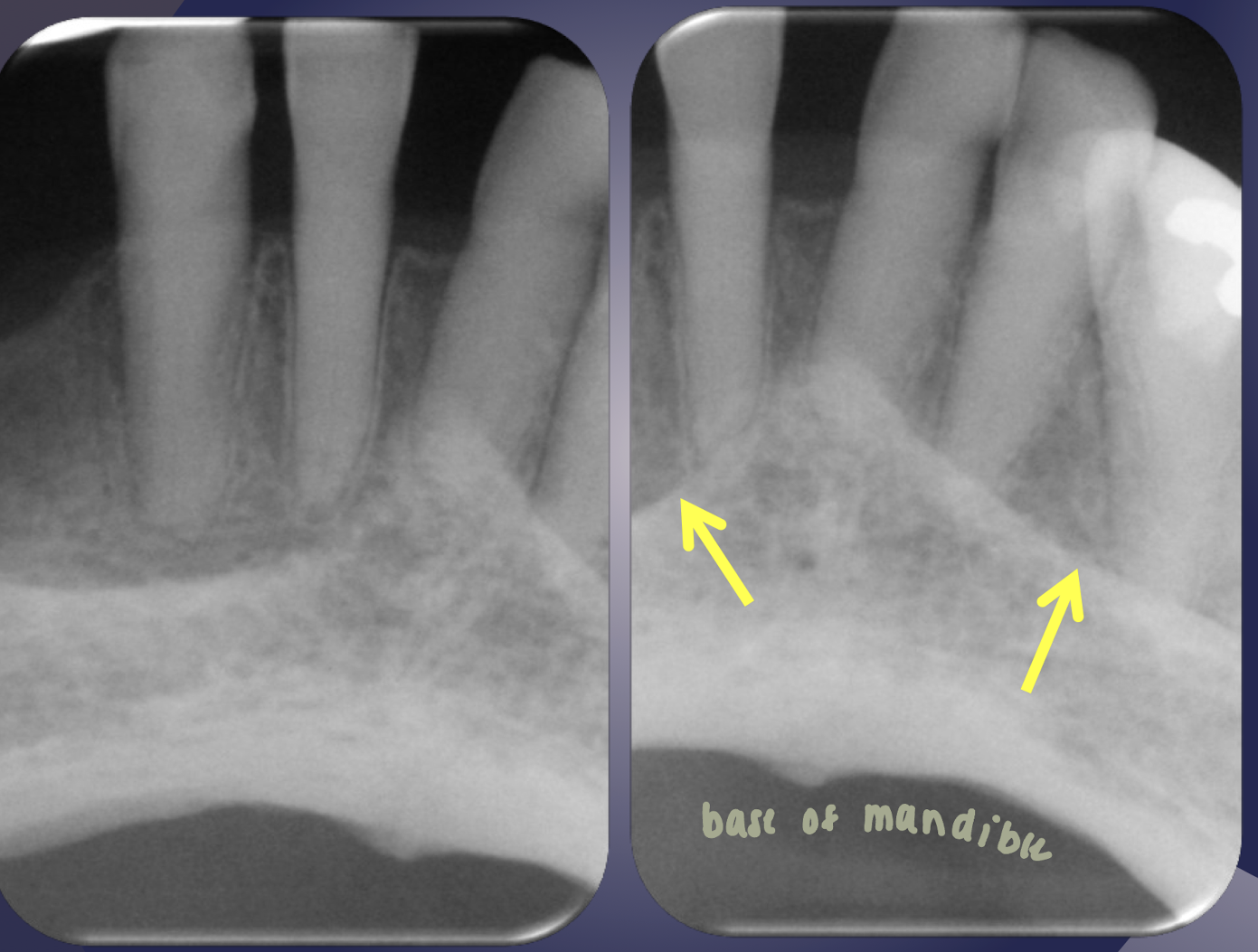
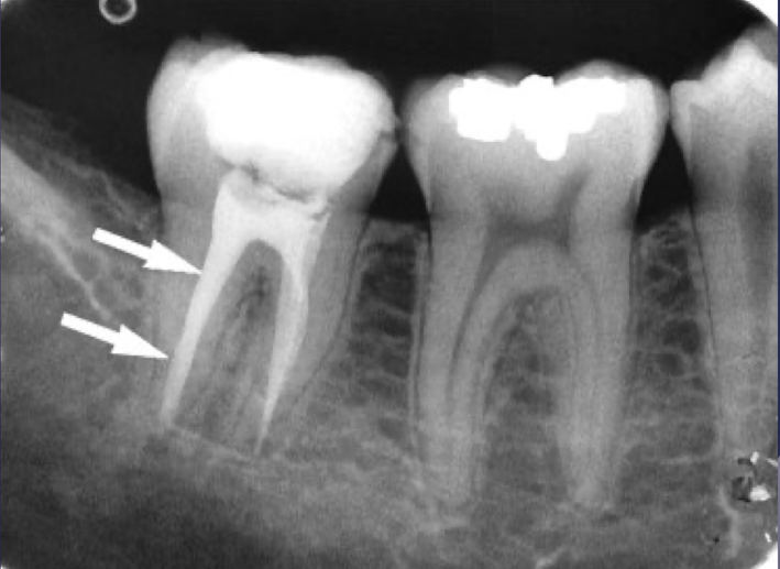
which structure of the mandible?
Radiolucent linear structure with thin radiopaque superior and inferior borders
Borders partially or not visualized sometimes
Anterior extension from mental foramen is rarely visible
IAN canal
IAN only inferior partially visualized
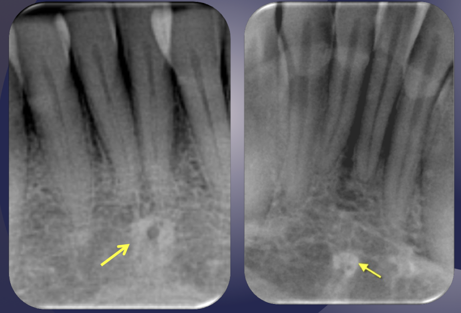
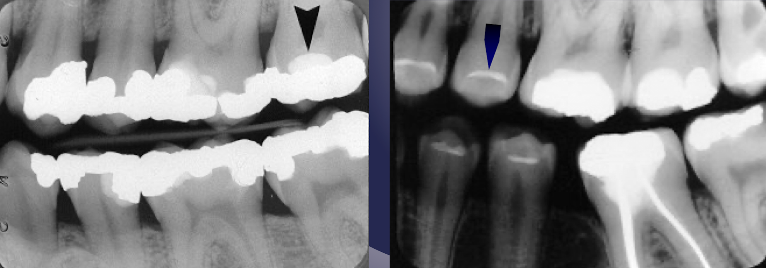
which landmark of the mandible?
Anterior limit of inferior alveolar nerve canal
Round/ovoid radiolucent structure
Apical to second premolar
May mimic periapical disease if projected over root apices
mental foramen
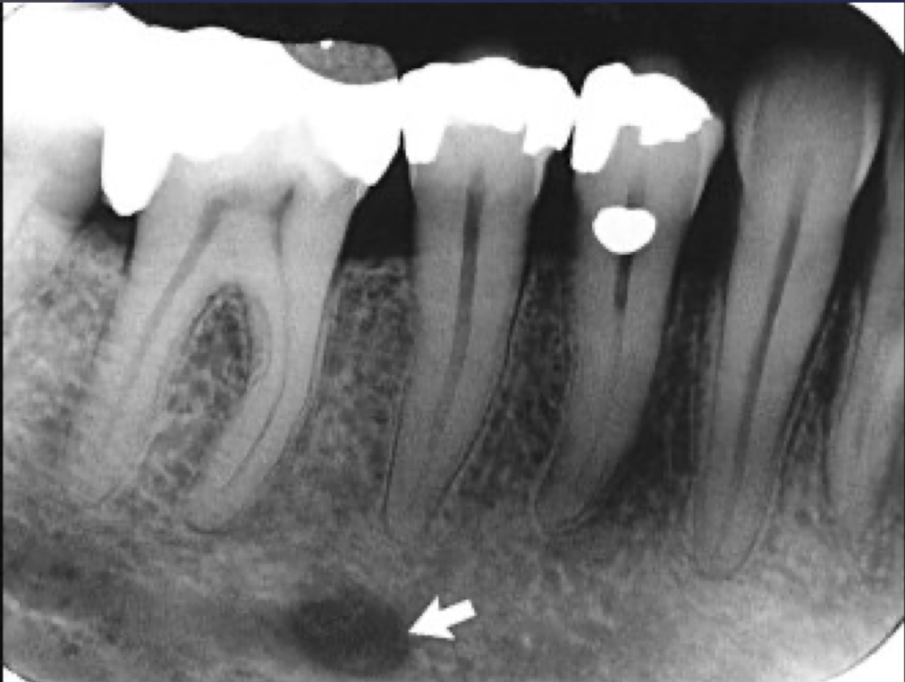
mental foramen
sagittal, coronal, axial of mental foramen
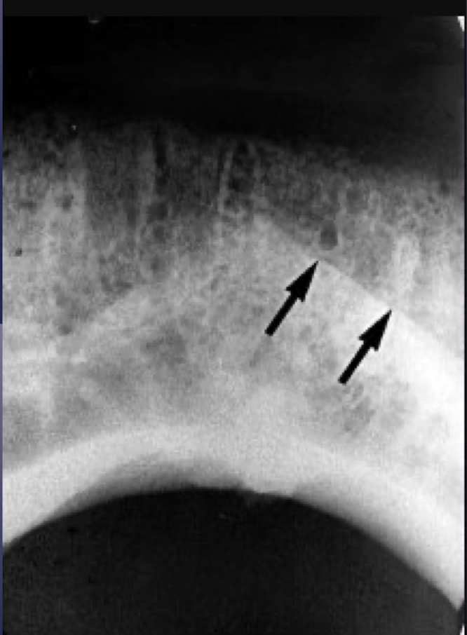
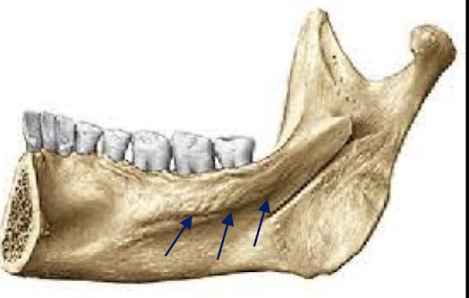
which landmark of the mandible?
Continuation of anterior border of ramus
Thick radiopaque line
Follows antero-inferior course from third molar area to first molar
external oblique ridge
external oblique ridge
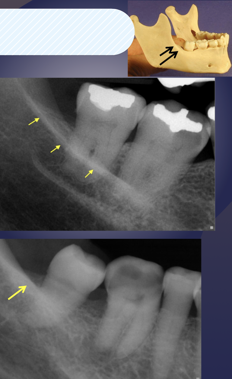
which landmark of the mandible?
Radiopaque line- sometimes defined but of variable width
Runs diagonally downward and forward at the level of apices of posterior teeth
internal oblique ridge/mylohyoid ridge
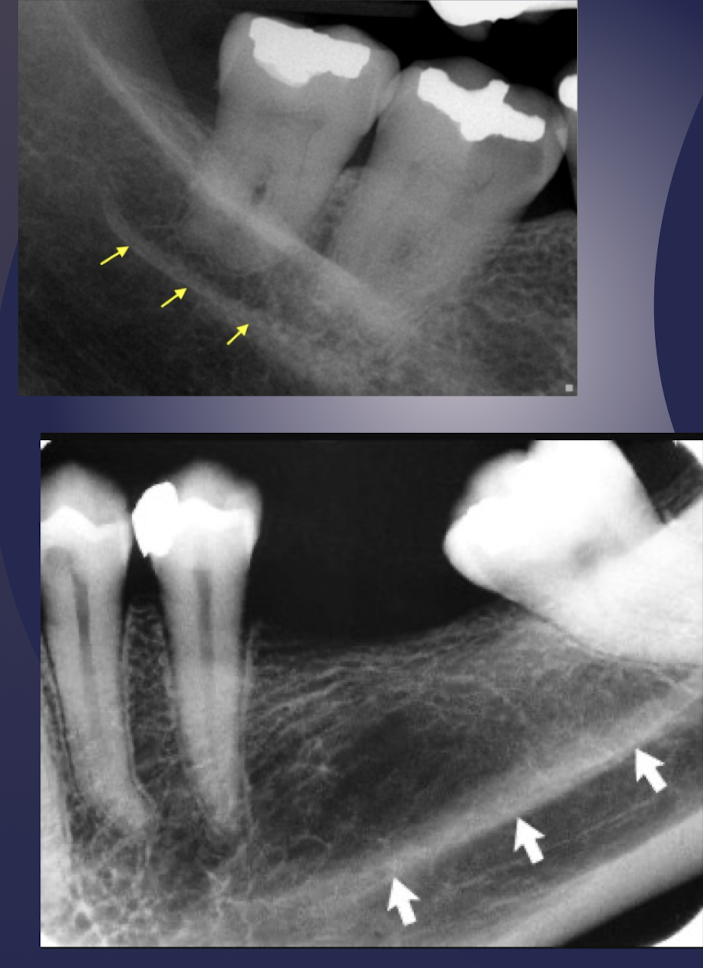
internal oblique ridge/mylohyoid ridge
internal oblique ridge/mylohyoid ridge
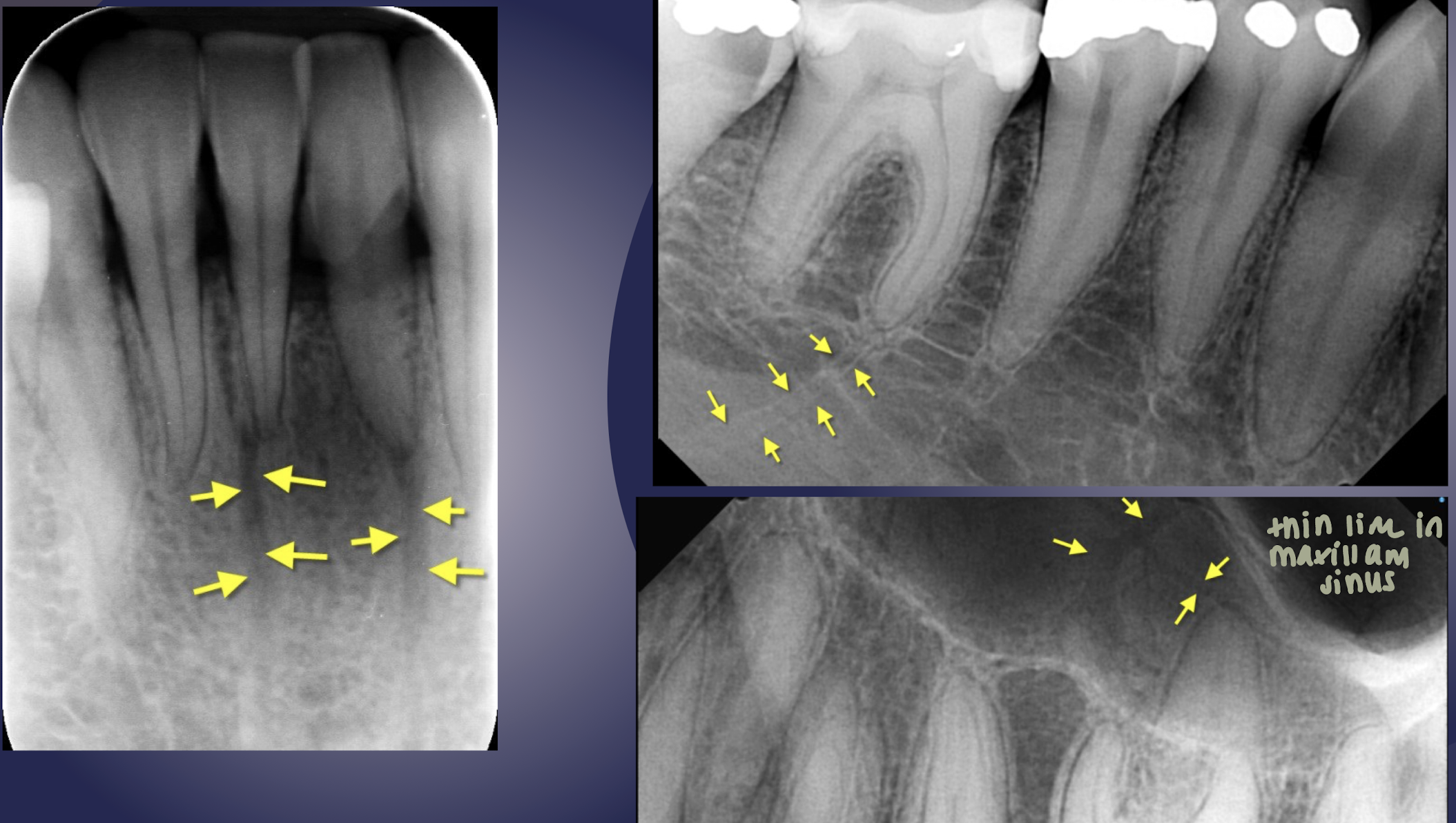
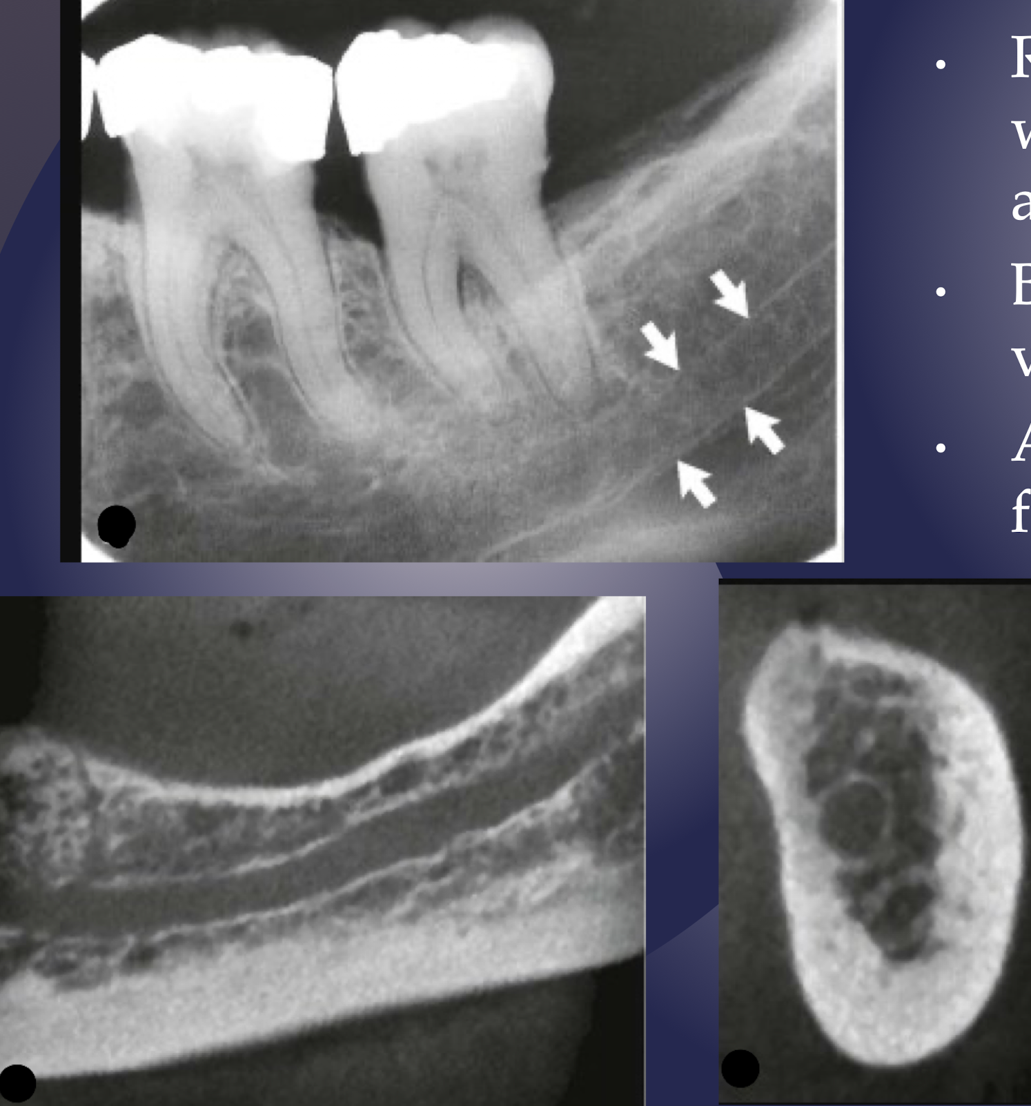
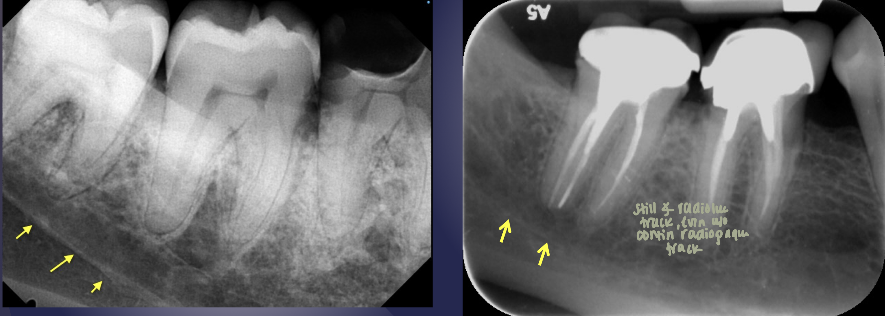
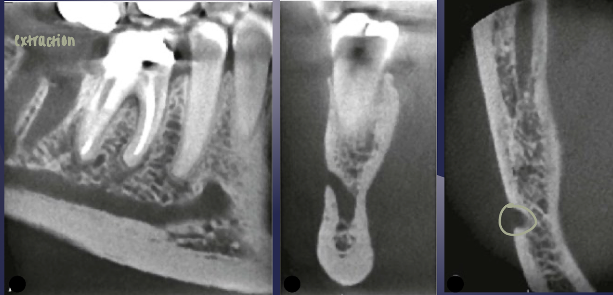
submandibular gland fossa
which landmark of the mandible?
Depression on lingual surface
Below mylohyoid ridge
Radiolucent area with sparse trabeculae
Mistaken for bony lesion
submandibular gland fossa
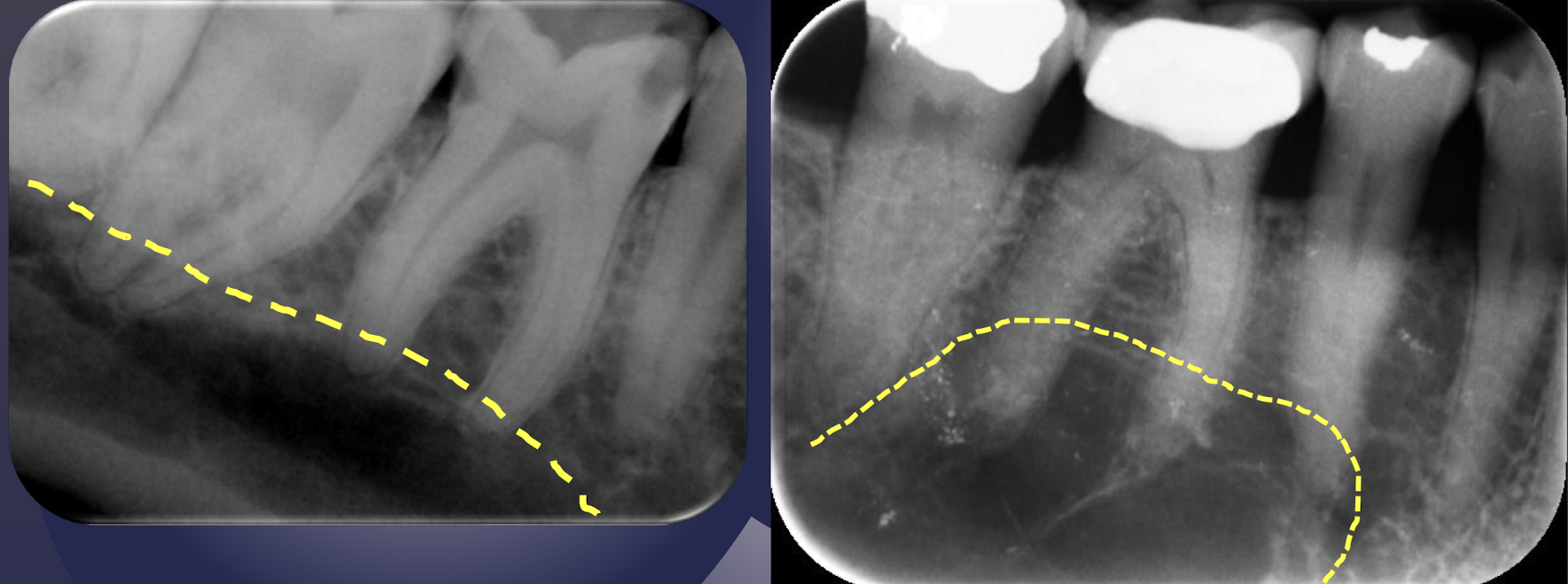
submandibular gland fossa
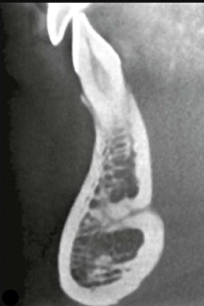
which landmark of the mandible?
Periapical radiograph of maxillary molars- mouth open position
Triangular radiopacity superimposed over third molar area
Mistaken for a root fragment
coronoid process
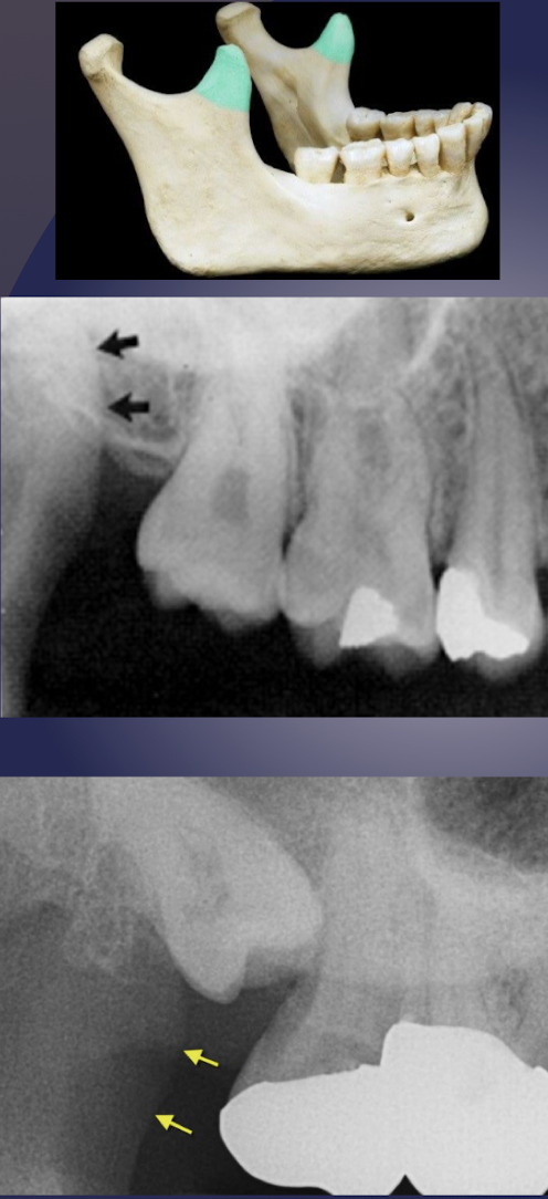
coronoid process of the mandible
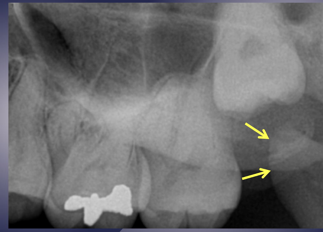
coronoid process of the mandible
inferior border of mandible
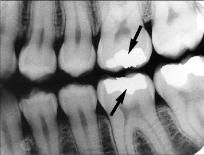
which restorative material?
amalgam
more radiopaque than enamel bc of metal content
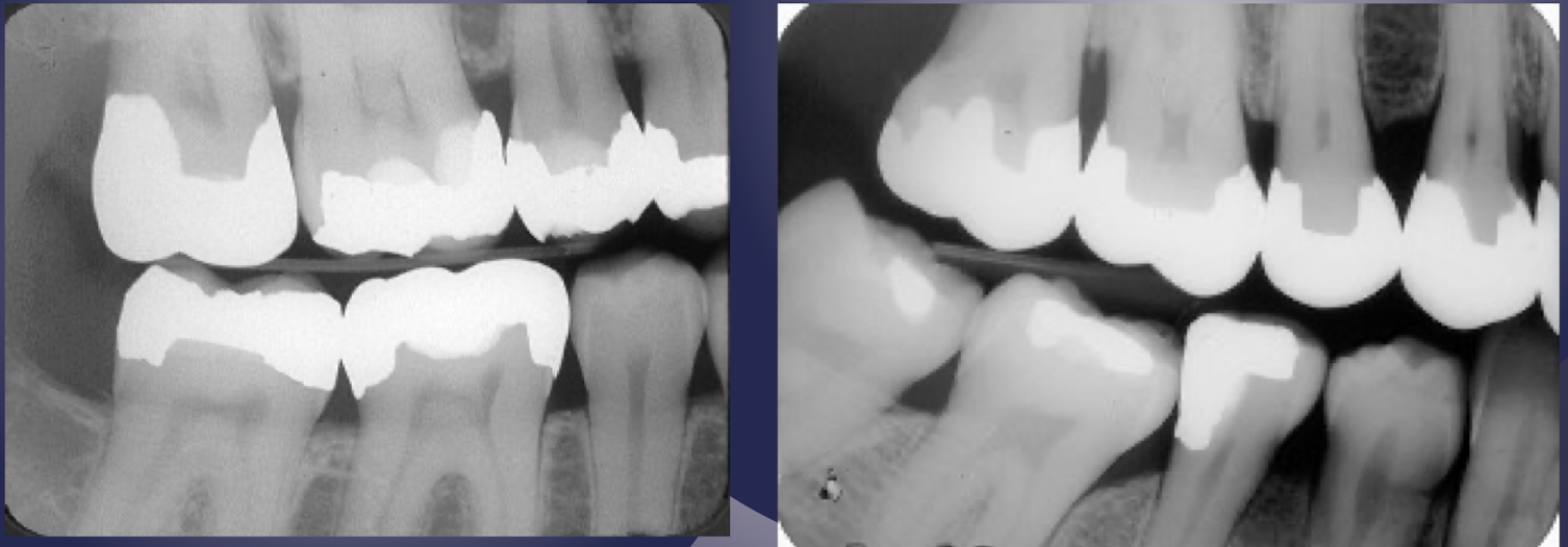
which restorative material?
metal inlays- crowns bc of coverage
more radiopaque than enamel bc of metal content
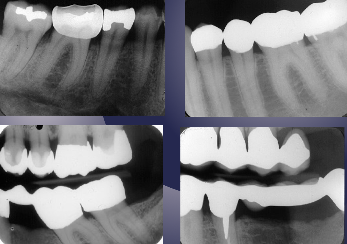
which restorative material?
stainless steel, gold crown and bridges
radiopacity like other metallic restorations
which restorative material?
porcelain crown and porcelain fused to metal crown, metallic cast post
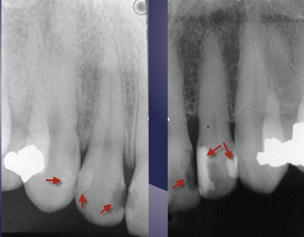
which restorative material?
Nonmetallic Restorative Materials (Composite-radiolucent and radiopaque types)
Note: the radiopacity of the radiopaque composite is less than that of amalgam seen in adjacent teeth
Radiolucent type of composite may mimic carious lesions- outline form and clinical exam helps distinguish the two
which restorative material?
cement liners and bases, under layer restorations so an issue would show as more radiolucent than the bone
which restorative material?
gutta percha (plastic)
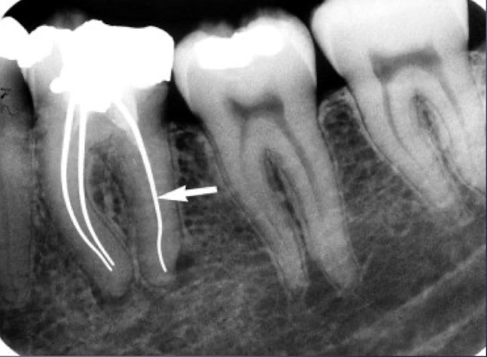
which restorative material?
silver point (metallic so more opaque than gutta percha)
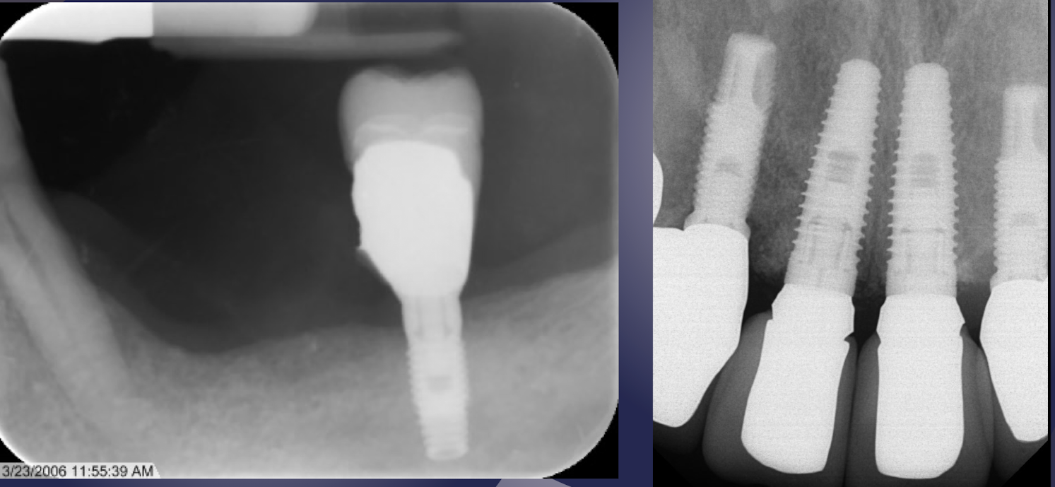
which restorative material?
implants