Neuropsychology Exam 2 Study Materials (LSU 4041)
1/66
There's no tags or description
Looks like no tags are added yet.
Name | Mastery | Learn | Test | Matching | Spaced |
|---|
No study sessions yet.
67 Terms
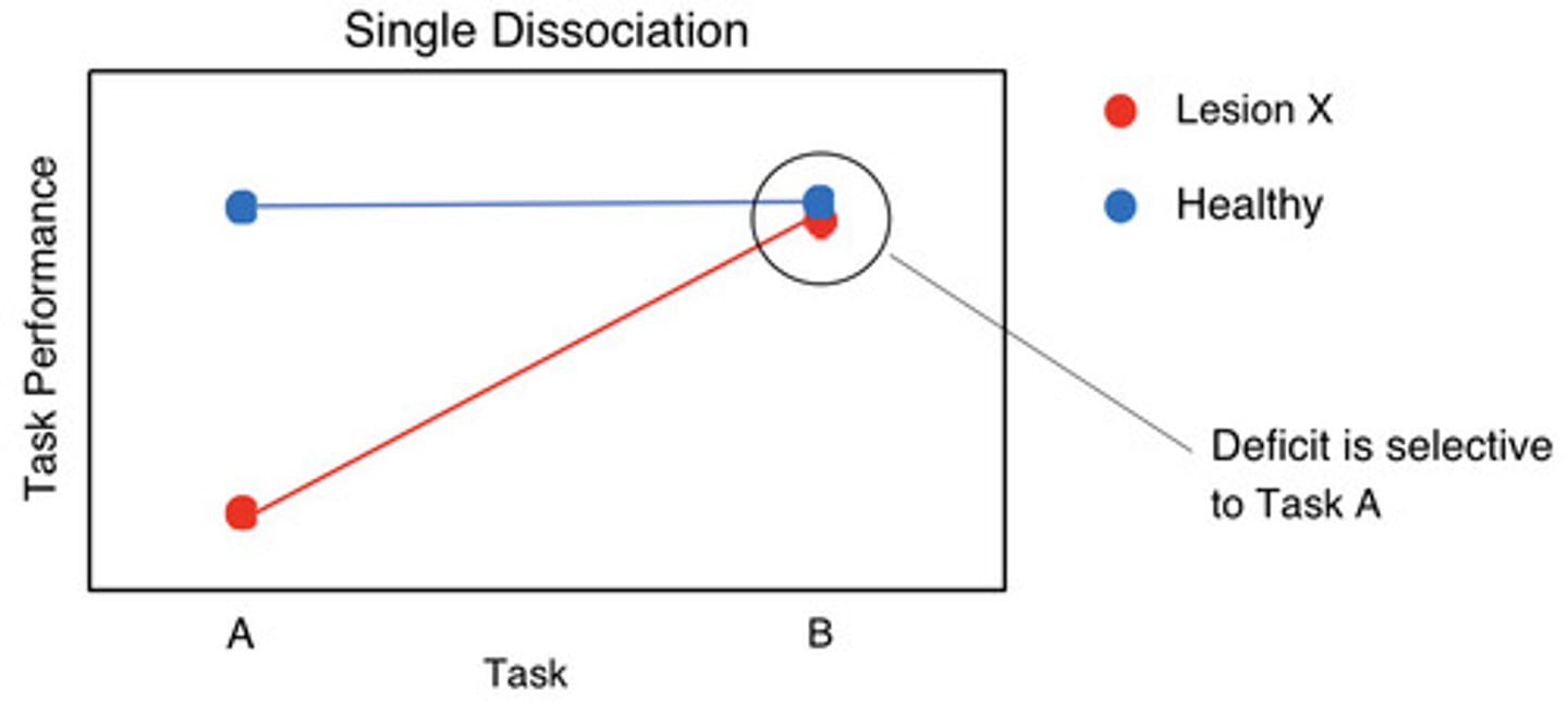
single dissociation
when a patient with damage to region "X" is impaired on a certain task (Task A), but unimpaired on another task (Task B)
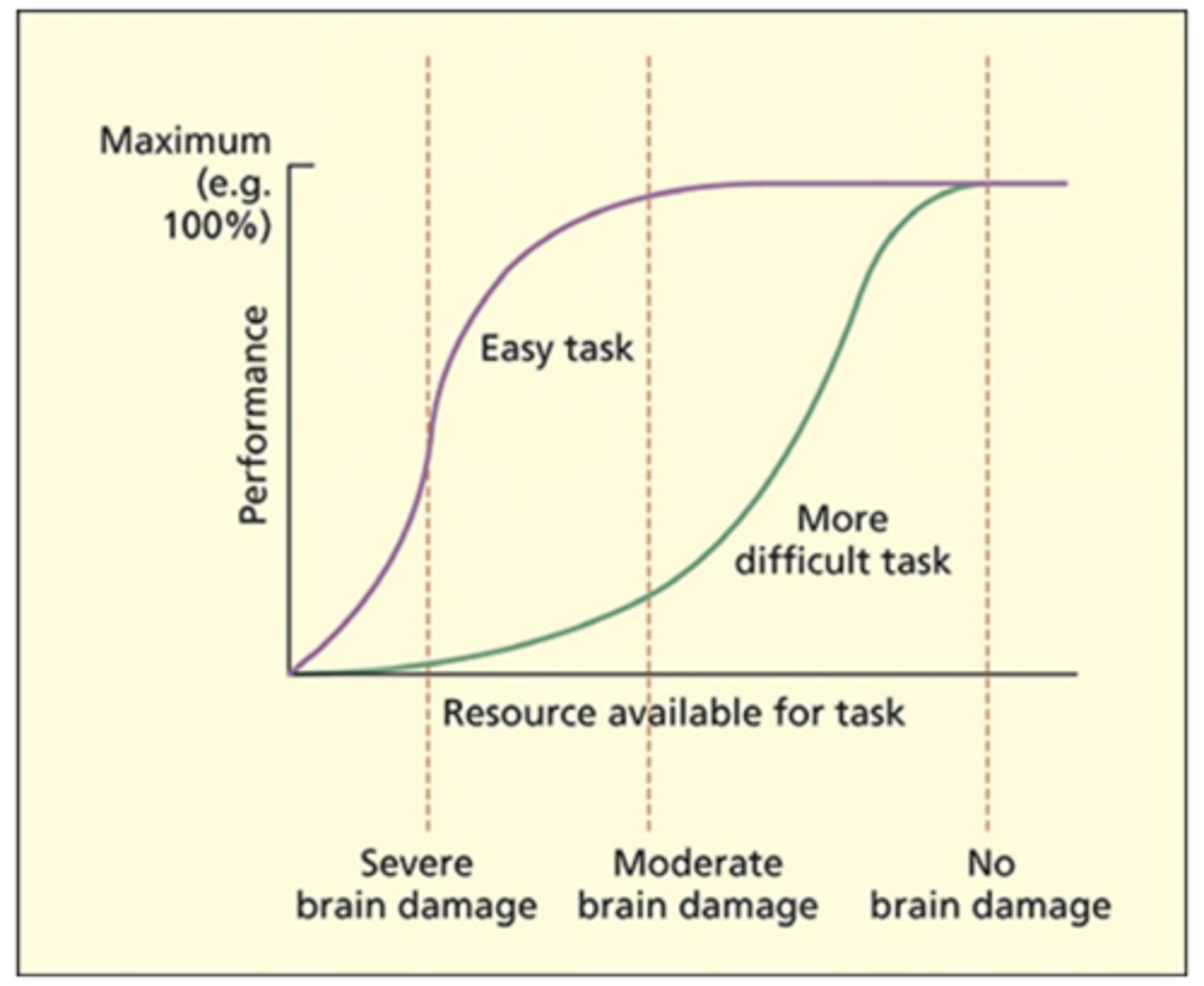
single dissociation challenges
- Task-resource artifact: If two tasks share the same neural/cognitive resource but one task uses it more → damage to this resource will affect the "harder" task more
- Task-demand artifact: A patient may perform worse on Task A versus Task B because they did not understand the instructions for Task A (or chose a bad strategy, etc.)
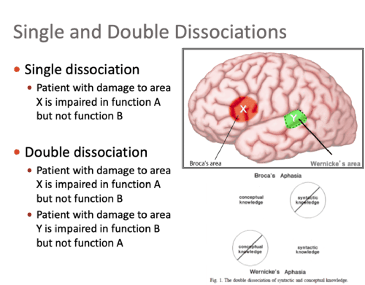
double dissociation
when there are two single dissociations with a complementary pattern:
➢Region "X" is important for Task A, but not Task B
➢Region "Y" is important for Task B, but not Task A
task-resource artifact & task-demand artifact
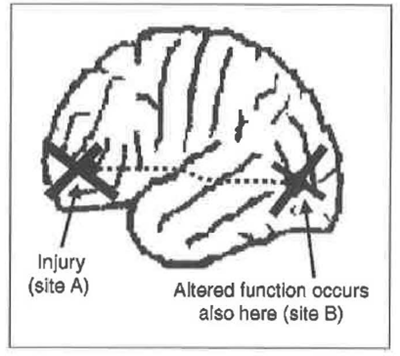
Diaschisis
- when a discrete, localized brain lesion disrupts functioning of spatially separate and structurally intact brain regions
classical neuropsychology
infer the function of brain regions by studying patients with lesions to that region and examining pattern of impaired and spared abilities
- favor group studies
cognitive neuropsychology
the pattern of spared and impaired abilities in and of themselves has been used to infer the building blocks of cognition—irrespective of where they are located in the brain
- favor single case study
Caramazza 3 assumptions
- The fractionation assumption. The first assumption is that damage to the brain can produce selective cognitive lesions
- The transparency assumption. The transparency assumption states that lesions affect one or more components within the preexisting cognitive system, but they do not result in a completely new cognitive system being created.
- The universality assumption. The universality assumption is that all cognitive systems are basically identical.
transparency assumption
- The transparency assumption refers to the comparability between premorbid and postmorbid cognitive systems, and not on where such systems are located.
- one needs to assume that brain damage removes one component of cognition, but does not create, from scratch, a rearranged or different cognitive system
vowel based lesion symptom mapping
for evaluating statistical relationships between damage to specific brain regions and resulting deficits.
transcranial magnetic stimulation (TMS)
- provides non-invasive stimulation of cortical neurons (action potentials) using brief electromagnetic fields
Advantages of TMS
- Non-invasive causal method
- No reorganization/plasticity (cf. transparency assumption)
- Stimulation target is focal and experimenter-controlled
- Multiple stimulation targets within the same participant
- On-line stimulation can determine timing of brain-behavior relationships & characterize ongoing neural processing
- Potential to improve behavioral performance
disadvantages of TMS
- Doesn't show changes in behavior/cognition are more apparent
- Subcortical lesions can't be studied
single pulse TMS
disruption of brain activity that occurs only during the brief period of stimulation
rTMS
the application of repeated pulses of magnetic energy to the brain; used to stimulate or suppress brain activity
(repetitive TMS)
practical use of TMS
- when to deliver the pulses, where to deliver the pulses and selection of appropriate control conditions
positioning TMS coil
- use skull land marks or structural and functional MRI
- MRI provides a better approach
sensation v perception
sensation- the effects of a stimulus on the sensory organs
perception - the elaboration and interpretation of that sensory stimulus by the brain
rods
Specialized for low level of light intensity, like those found at night
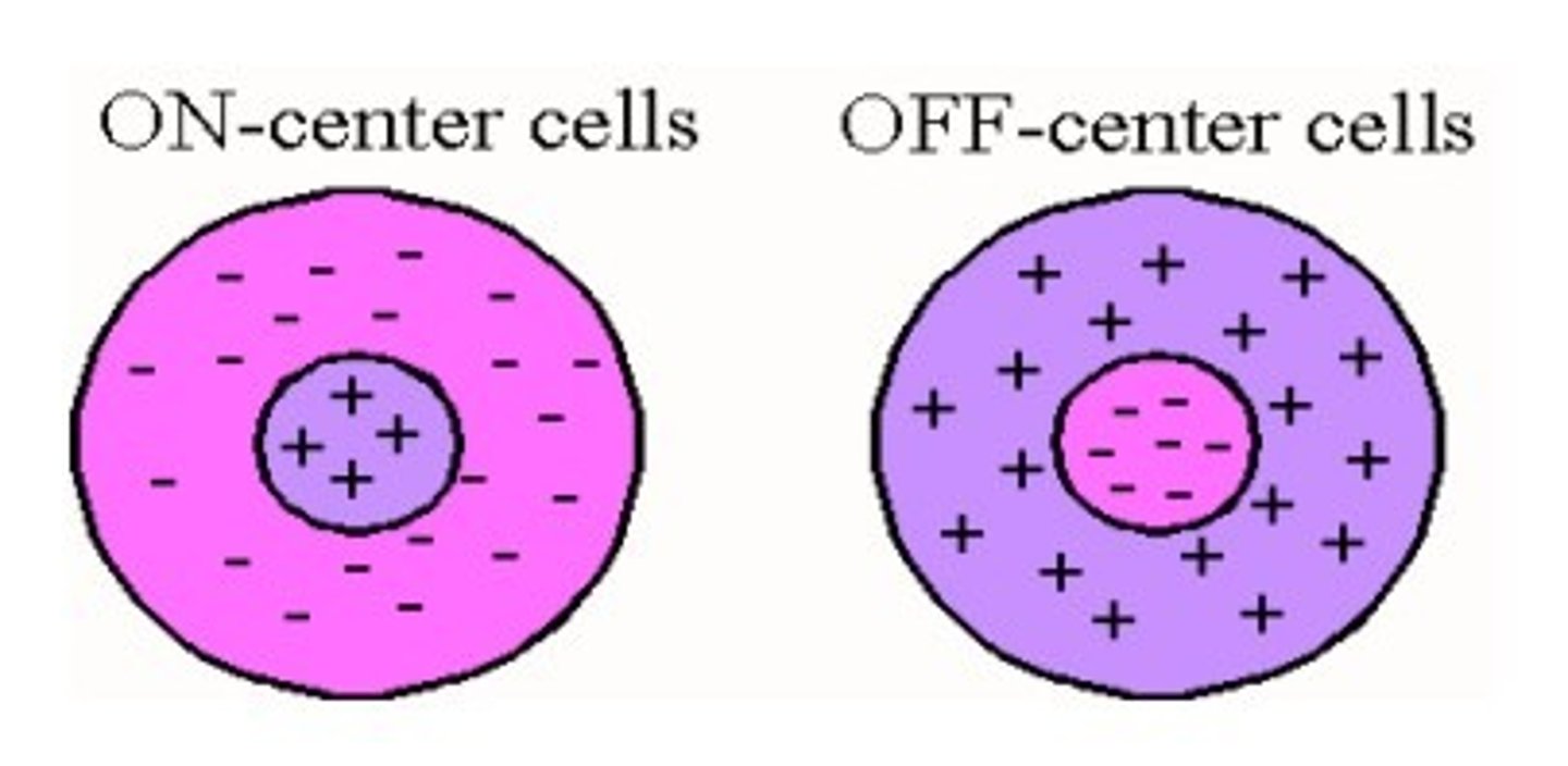
on-center/off-surround
referring to a concentric receptive field in which the center excites the cell of interest while the surround inhibits it

off-center/on-surround
referring to a concentric receptive field in which stimulation of the center inhibits the cell of interest while stimulation of the surround excites it
geniculostriate pathway
The neural pathway from the retina, via the lateral geniculate nucleus, to the primary visual cortex.
complex cells
- In vision, cells that respond to light in a particular orientation but do not respond to single points of light
hypercomplex cells
- In vision, cells that respond to particular orientations and particular lengths.
V1 renotopically organized
- spatial arrangement of light on the retina is retained in primary visual cortex
scotoma
- A small region of cortical blindness.
hemianopia
Cortical blindness restricted to one half of the visual field (associated with damage to the primary visual cortex in one hemisphere)
- Cortical blindness restricted to a quarter of the visual field
blindsight
- A symptom in which the patient reports not being able to consciously see stimuli in a particular region but can nevertheless perform visual discriminations (e.g., long, short) accurately
ventral "what"
Processes stimulus features and identities; memory
dorsal "where"
Processes motion and spatial attention; action
color constancy
- the color of a surface/object is perceived as constant even when illuminated by different lighting conditions
-V4 directly supports it
Achromatopsia
- A failure to perceive color (the world appears in grayscale), not to be confused with color blindness (deficient or absent types of cone cell).
hemiachromatopsia
- disorder of impaired color perception with relative preservation of form vision in one-half of the visual field.
Akinetopsia
damage causes failure to perceive visual motion
Perceptual grouping
The process by which the visual system combines separate regions of the retinal image that "go together" based on similar properties
- (simple/complex shapes; foreground/background)
5 principles by Gestalt psychologists
- The law of proximity states that visual elements are more likely to be grouped if they are closer together. For example, the dots in (a) in the figure tend to be perceived as three horizontal lines because they are closer together horizontally than vertically.
- The law of similarity states that elements will be grouped together if they share visual attributes (e.g., color, shape). For example, (b) tends to be perceived as vertical columns rather than rows, because elements in columns share both shape and color.
- The law of good continuation states that edges are grouped together to avoid changes or interruptions; thus, (c) is two crossing lines rather than > and <.
- The law of closure states that missing parts are "filled in"; thus (d) has circular properties in spite of the gap. This law, and the previous one, is important for recognizing objects that are partly occluded.
- The law of common fate states that elements that move together tend to be grouped together. A good example of this comes from studies of biological motion perception (e.g., Johansson, 1973). Light points attached to bodily joints are perceived as movement of a single human figure when viewed in the dark.
object constancy
- to be able to recognize an object across different viewpoints and different lighting conditions
object structural description
- description of an object from memory
- top down approach
Apperceptive agnosia
- deficit in object recognition due to impairment at the level of object perception
Associative agnosia
- deficit in object recognition due to impairment at the level of knowledge/semantic memory
Integrative agnosia
- deficit in object recognition due to impaired perceptual grouping of components into whole object
structural brain imaging
- Measures of the spatial configuration of different types of tissue in the brain (principally CT and MRI).
functional brain imaging
- Measures temporary changes in brain physiology associated with cognitive processing; the most common method is fMRI and is based on a hemodynamic measure.
CT (computed tomography) scan
- constructed according to the amount of X-ray absorption in different types of tissue. The amount of absorption is related to tissue density: bone absorbs the most (and so the skull appears white), cerebrospinal fluid absorbs the least (so the ventricles appear black) and the brain matter is intermediate (and appears gray). Given that CT uses X-rays, the person being scanned is exposed to a small amount of radiation. CT scans are typically used only in clinical settings, for example to diagnose tumors or to identify hemorrhaging or other gross brain anomalies. CT cannot distinguish between gray matter and white matter in the same way as MRI, and it cannot be adapted for functional imaging purposes.
PET (positron emission tomography) scan
a visual display of brain activity that detects where a radioactive form of glucose goes while the brain performs a given task
MRI (magnetic resonance imaging)
is a brain imaging technique that detects magnetic changes in the brain's blood flow patterns.
MRI advatages
• It does not use ionizing radiation and so is completely safe (people can be scanned many times).
• It provides a much better spatial resolution, which allows the folds of individual gyri to be discerned.
• It provides better discrimination between white matter and gray matter; this may enable early diagnosis of some pathologies, and can be used to explore how normal variation brain structure is linked to differences in cognitive ability.
• It can be adapted for use in detecting the changes in blood oxygenation associated with neural activity, and in this context is called functional MRI (fMRI)
How PET scan works
- A small amount of radioactive glucose (a sugar) is injected into a vein. The PET scanner takes a picture of where glucose is being used in the brain. PET scanning utilizes a radioisotope tracer that is an analog to glucose, called fluorodeoxyglucose (FDG).
BOLD singnal
- Blood oxygen-level-dependent contrast; the signal measured in fMRI that relates to the concentration of deoxyhemoglobin in the blood.
Hemodynamic response function (HRF)
- Changes in the BOLD signal over time.
three stages of HRF
- Initial dip. As neurons consume oxygen there is a small rise in the amount of deoxyhemoglobin, which results in a reduction of the BOLD signal
- Overcompensation. In response to the increased consumption of oxygen, the blood flow to the region increases. The increase in blood flow is greater than the increased consumption, which means that the BOLD signal increases significantly.
- Undershoot. Finally, the blood flow and oxygen consumption dip before returning to their original levels. This may reflect a relaxation of the venous system, causing a temporary increase in deoxyhemoglobin again.
process measured by fMRI
- uses a naturally occurring signal in the bloodstream to measure the change of bloodflow
voxel
- A volume-based unit (cf. pixels, which are 2D); in imaging research the brain is divided into many thousands of these.
cognitive subtraction
a type of experimental design in functional imaging in which activity in a control task is subtracted from activity in an experimental task
Baseline task
An activity that someone can complete easily
Pure insertion (pure deletion)
the assumption that adding a different component to a task does not change the operation of other components
parametric design v categorial design
an experimental design in which the amount of the independent variable is systematically varied across several levels
- the variable of interest is treated as a continuous dimension rather than a categorical distinction
brain region being active
- A region of "activity" refers to a local increase in metabolism in the experimental task compared with the baseline, but it does not necessarily mean that the region is essential for performing the task. Lesion studies might provide evidence concerning the necessity of a region for a task.
MVPA (Multi-voxel pattern analysis)
an fMRI analysis method in which distributed patterns of activity are linked to cognitive processes
evidence of reading minds
- For example, Haxby et al. (2001) gave participants pictures from eight different types of category, including cats, houses, faces and shoes. The neural activity from an individual trial was then compared to the previous known patterns of activity to determine the most probable category that was being viewed. This procedure could predict, given pairwise comparisons, what the person was seeing with 96 percent accuracy
-Other research has shown that activity in these regions can be used to accurately predict semantic categories when reading words (Mitchell et al., 2008) or when recalling previously seen images from memory (Polyn et al., 2005)
activation mapping analysis
- the most popular method for displaying the results of an fMRI experiment. They typically show colored voxels on an image of the brain, with color indicating whether neural responses at that location differ between experimental conditions.