1.2 and 1.3 (PSIO 202)
5.0(1)
5.0(1)
Card Sorting
1/36
Earn XP
Description and Tags
Heart Conduction, ECGs, Pacemaker and Cardiac APs
Study Analytics
Name | Mastery | Learn | Test | Matching | Spaced | Call with Kai |
|---|
No study sessions yet.
37 Terms
1
New cards
What is the structure of cardiac muscle?
branched, striated fibers and usually one centrally located nuclei
2
New cards
What does the branching pattern of cardiac muscle do?
forms a network that can facilitate the multi-directional transmission of electrical impulses in all directions
3
New cards
What connects cardiac muscles together?
intercalated disks
4
New cards
What are gap junctions and why are they important?
small channels that allow electrical impulses to pass quickly from one cell to the next
5
New cards
Where are gap junctions located?
in intercalated discs (lie between adjacent muscle fibers)
6
New cards
What are desmosomes and where are they located?
they hold adjacent cells together; located in intercalated discs
7
New cards
What do gap junctions do for the myocardium?
allow it to behave as a single unit (or functional system)
8
New cards
What is another name for the SA node?
pacemaker
9
New cards
What is the SA node and what does it do?
a mass of cells in the right atrial wall that spontaneously discharge action potentials at a rate of (greater than or equal to) 100-120 bpm
10
New cards
What do autonomic nerves do to the rate of discharge of APs from the SA node?
modify the rate so that HR is \~70 bpm at resting
11
New cards
What ion regulates sustained contraction?
Ca2+
12
New cards
What ion causes depolarization?
Na+
13
New cards
When do Na+ channels close?
at the peak; when the cell depolarizes
14
New cards
What causes the plateau of an action potential?
Ca2+ entering the cell
15
New cards
What is the last ion to enter the cell during an action potential?
K+
16
New cards
What does K+ do to an action potential?
causes a slight fall in the plateau and ultimately returns membrane to resting potential
17
New cards
What is the first step (of 3) of an AP?
rapid depolarization due to inflow of Na+ when Na+ channels open
18
New cards
What is the second step (of 3) of an AP?
plateau (or maintained depolarization) due to inflow of Ca2+ when slow Ca2+ channels open and some K+ channels open causing slight dip in plateau
19
New cards
What is the third step (of 3) of an AP?
repolarization due to Ca2+ channels closing and more K+ channels open causing K+ to leave the cell
20
New cards
What is the absolute refractory period?
the time when a cell will not respond regardless of the strength of stimulus
21
New cards
What is the relative refractory period?
the time when a cell will respond only if the stimulus is “supra-threshold”
22
New cards
When is the absolute refractory period in skeletal and heart muscle?
roughly the same time as the action potential (\~250 ms in heart muscle)
23
New cards
What type of cells are autorhythmic?
pacemaker cells
24
New cards
What does autorhythmic mean?
the cells can auto-initiate action potentials
25
New cards
What is a pacemaker potential?
unstable resting membrane potential
26
New cards
What do pacemaker cells use for the rising phase of the AP instead of sodium?
calcium
27
New cards
What is an ECG?
a composite record of action potentials of all active cells at points in time during a heartbeat
28
New cards
What happens during the P wave?
atrial depolarization
29
New cards
What happens during the P to Q interval?
signal conduction from SA node to AV node; atrial systole begins
30
New cards
What happens during the QRS complex?
atrial repolarization and diastole; repolarization concealed by QRS wave
31
New cards
What happens during the T wave?
ventricular repolarization
32
New cards
What happens during the PR interval?
signal conduction through the AV node, before activating ventricles
33
New cards
What happens during the QT interval?
duration of ventricular depolarization; shorter during exercise
34
New cards
What happens during the ST segment?
ventricular systole and ejection of blood; corresponds to plateau of cardiomyocyte action potential
35
New cards
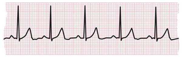
What is the reading of this ECG?
normal sinus rhythm
36
New cards
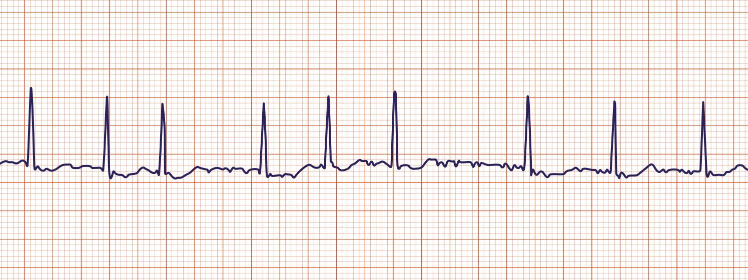
What is the reading of this ECG?
atrial fibrillation (multiple P waves)
37
New cards
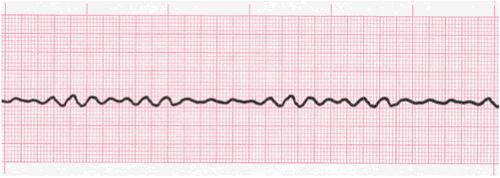
What is the reading of this ECG?
ventricular fibrillation (heart attack)