Vitreous-choroid-retina
1/13
There's no tags or description
Looks like no tags are added yet.
Name | Mastery | Learn | Test | Matching | Spaced | Call with Kai |
|---|
No analytics yet
Send a link to your students to track their progress
14 Terms
What wavelength of visible light is associated with the development of macular dengeration?
Blue-violet light between 415 nm and 455 nm
Death of RPE cells
Blue-turquoise (465 to 495) doesn’t affect ocular health and is actually important for the pupillary reflex and wake/sleep cycle
Fluorescent lamps, LED lights, and sunlight
Blood vessels and their resistance and blood flow rate
Retinal blood vessels:
small
high resistance
low flow rate (due to small lumen)
Choroidal vessels:
larger
lower resistance
higher flow rate
Ophthalmic/carotid arteries:
Larger than retinal and choroidal vessels
Even higher flow rates
How do these conditions present on FA?
Pigment epithelial detachment
Central serous retinopathy
Cystoid macular edema
Choroidal neovascular membrane
Pigment epithelial detachment:
well defined pooling of dye underneath the RPE that doesn’t expand in size overtime but gets brighter instead
Central serous retinopathy:
“smoke-stack” presentation due to leakage of fluorescein dye through the retinal pigment epithelium
Causes a hyperfluorescent spot in the early stages
During the later venous stage, dye will continue to pass into the subretinal space and ascend vertically to the upper limit of the detachment and then extending laterally
Cystoid macular edema:
“Flower petal” pattern of hyperfluorescence
Accumulation of fluorescein dye inside the microcystic spaces that developed within the retina
Classic Choroidal neovascular membrane:
Well defined membrane that fills with fluorescein in the early phase
Dye eventually leaks after 1-2 minutes in the sub retinal space surrounding the neovacular membrane
Rank how the posterior hyaloid face of the vitreous attaches to the following from strongest to weakest:
A. Retinal Blood Vessels
B. Macula
C. Optic Nerve Head
D. Vitreous Base
E. ILM
Vitreous base
Optic Nerve head
Macula (fairly weak attachment)
Retinal Vasculature (weaker)
ILM (weakest)
(VOM-RI)
Which vitamin when taken in excess amounts can interfere with the uptake of Vitamin A?
Vitamin A is used to manage retinitis pigmentosa
Vitamin E
Drusen typically deposits between which layers of the retina?
RPE and Bruch’s Membrane
The RPE has an important role in phagocytosis of shed outer segments of the photoreceptors
If it fails to rid the debris, it will accumulate and show up as drusen → impacts vision and leads to macular degeneration
The rhodopsin molecule is found at what location in a rod photoreceptor?
The membrane of the otuer segment
Rhodopsin is embedded in the discs of the outer segment of the rod photoreceptor and absorbs a photon of light, causing and electrical change in the membrane of. therod
Need several rods to summate in order to signal the presence of a stimulus
Rods then release glutamate post-synaptically to bipolar and horizontal cells
What is the Area of Martegiani
Signifies the funnel-shaped dilation surrounding the optic disc
Represents the posterior termination of Cloquet’s Canal (aka hyaloid canal)
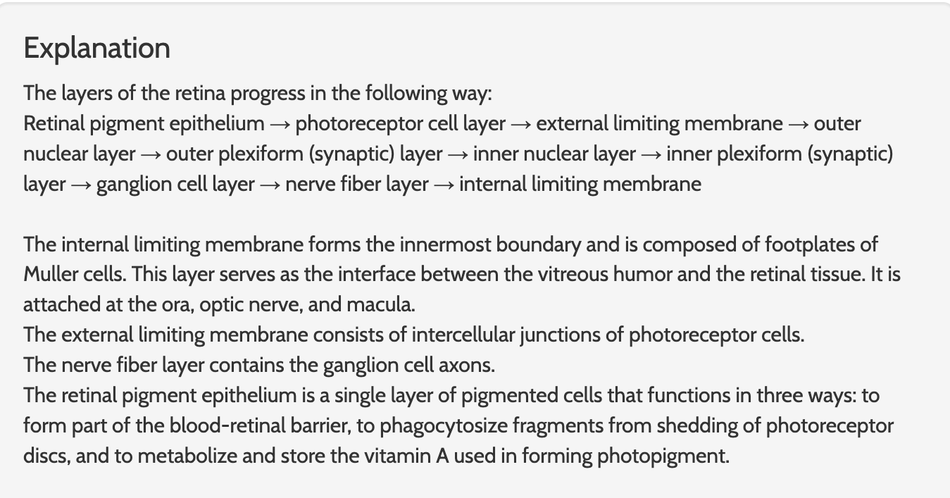
List the layers of the retina starting from the retinal pigment epithelium
Plexiforms layers are closer to the vitreous