3. Pacemaking - generation and regulation of cardiac myocyte contractions
1/9
There's no tags or description
Looks like no tags are added yet.
Name | Mastery | Learn | Test | Matching | Spaced | Call with Kai |
|---|
No analytics yet
Send a link to your students to track their progress
10 Terms
What are the similarities between cardiac myocytes and skeletal myocytes?
similar myofibrils that contract when intracellular Ca2+ is elevated
intracellular Ca2+ is elevated when a cardiac myocyte fires an action potential
What are the differences between cardiac and skeletal myocytes?
cardiac myocytes NOT directly driven by NS innervation. they are initiated by the electrical activity of other cardiac myocytes
if heart is taken out briefly, it can beat on its own
does not mean the heart cannot respond to the NS because it can → has receptors for NTs
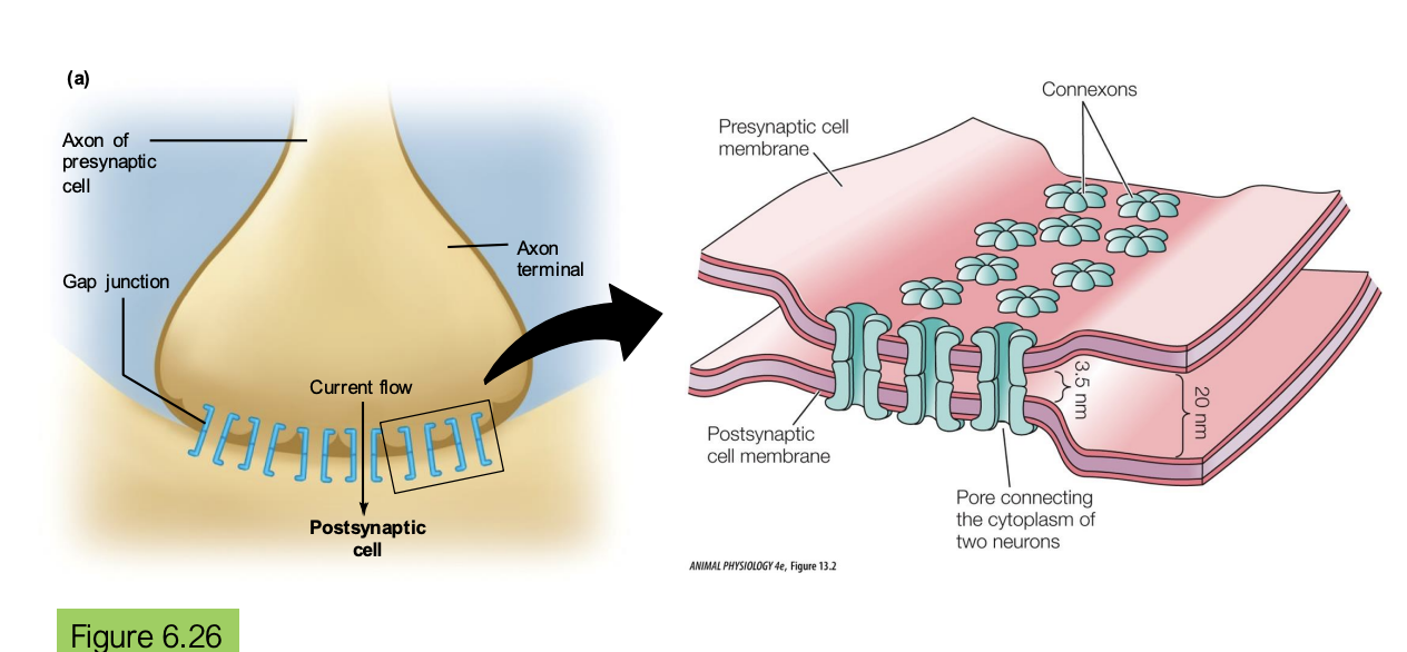
cardiac myocytes are electrically coupled to each other via gap junctions
cardiac action potentials are prolonged, so cardiac twitches do not summate → think of one heartbeat as one twitch
What does the gap junctions allow the heart to do?
Allows ions to flow across electrical synapses through gap junctions so that if one cell is depolarized, the others depolarize → synchronous depolarization
What are the two functionally specialized kinds of cardiac myocytes?
contractile cardiac myocytes → typical muscle cells that contract and generate tension. is responsible for the pumping action of the heart
pacemaker cells of the conducting system: controls the timing and rhythm of heart contractions
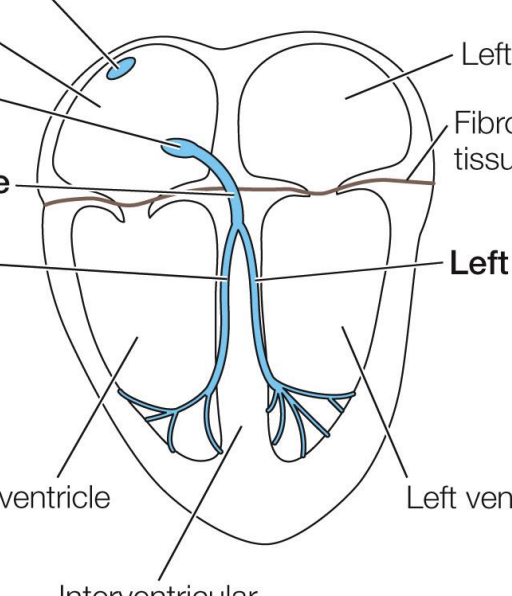
what is the stuff highlighted in blue? are they neurons or muscle cells? what system is this part of?
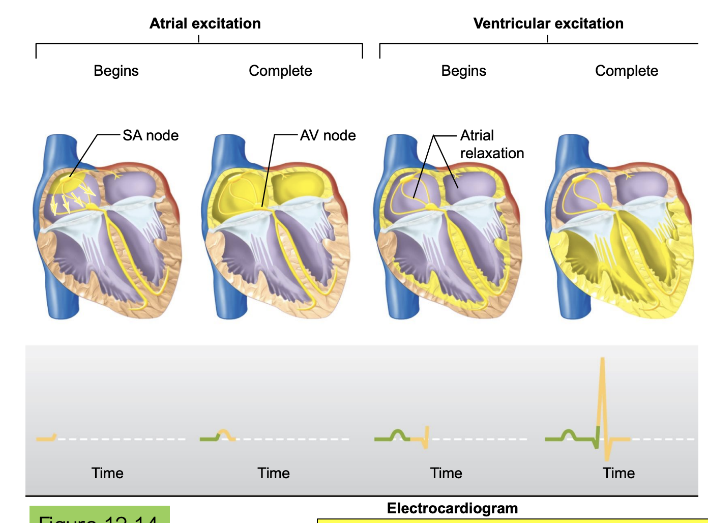
the SA node cells (circular blue dot) are in the right atrium and they are the most important pacemakers that set the heart rate in healthy adults
the SA node is what depolarizes the AV node and the right/left bundles to the right/left ventricles
they are NOT NEURONS. they are MUSCLE CELLS
this is part of the conducting system

what do gap junctions further link? so what does the action potential of each look like?
they link cells of the conducting system to each other and to surrounding contractile cardiac muscle fibers
the action potential of the SA node is similar to that of a neuron: it depolarizes then depolarizes right after
since gap junctions are linked, the depolarization of the SA nodes trigger the depolarization of the contractile myocytes
the action potential of a contractile myocyte depolarizes and stays depolarized for a long time, and then repolarizes
druing the depolarization, it is contracting and pulling
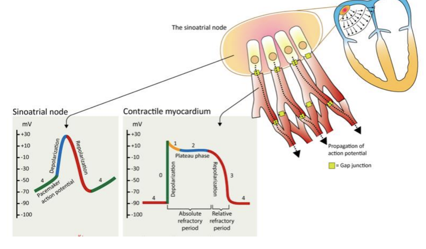
although the action potentials of the pacemaker cells are similar to that of a neuron, what is the biggest difference? what is this called? what is each action potential?
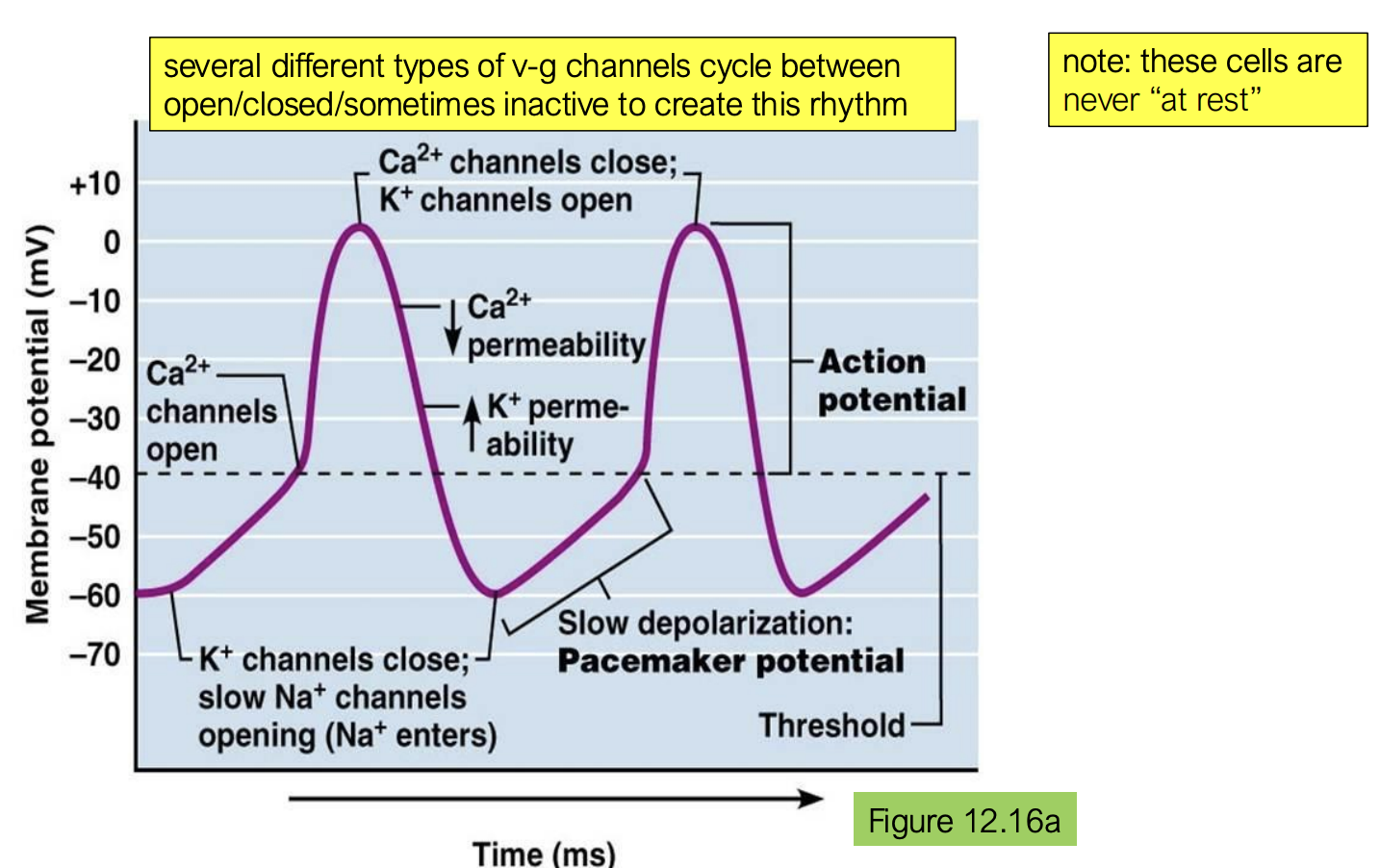
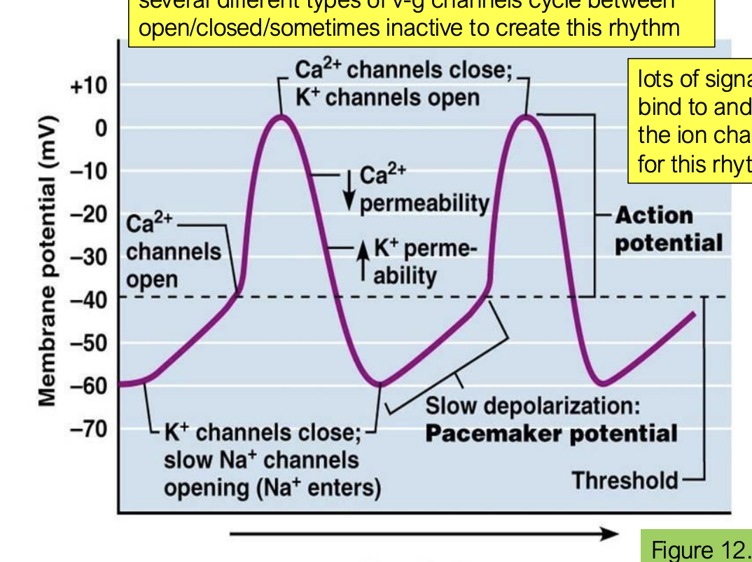
pacemaker cells are never at rest! they have no resting membrane potential and are constantly cycling
they slowly get depolarized, then they fire, and the hyperpolarized, and then slowly depolarize again and continually forever
this is called endogenous bursters that always fire regardless of NS activity
each action potential is a heartbeat
what can bind and what can they do? how would the action potentials be different?
a lot of signaling compounds can bind and alter the activity of the ion channels responsible for this rhythm
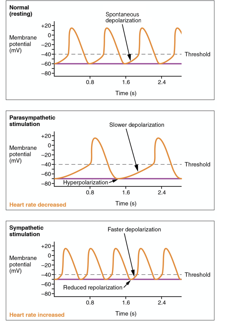
acetylcholine (parasympathetic) would bind to slow the bursting frequency of SA node cells → slow down heart beat
action potential would have slower depolarization
norephinephrine or epinephrine would bind to speed up bursting frequency of SA node cells → quicken heart beat
action potential would have faster depolarization and reduced repolarization
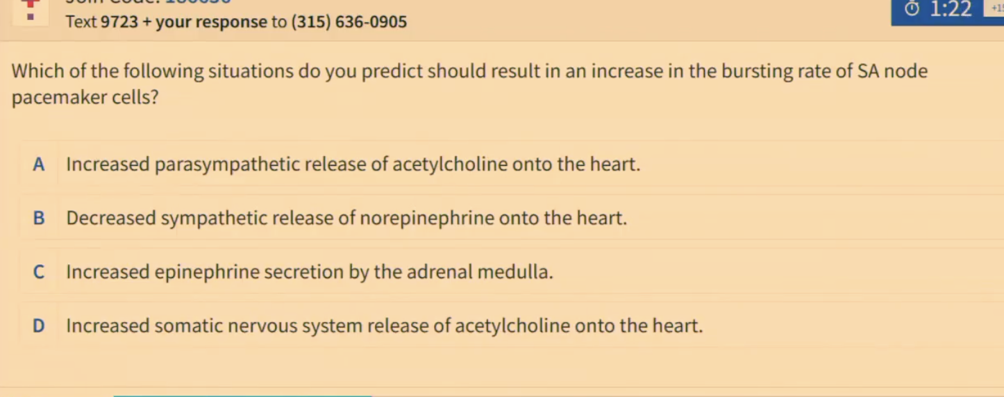
TH: which of the following situations do you predict should result in the bursting rate of SA node pacemaker cells?
increased epinephrine secretion by the adrenal medulla
When the heart is relaxed, what happens?
SA pacemaker cells fire
depolarization spreads because cardiac muscle cells are coupled with gap junctions
depolarization cannot propagate directly from atria to ventricles because fibrous tissue separates the two
so in essence, SA node fires, right and left atria contract at the same time
then, the wave of depolarization can propagate down the bundle of His into the ventricles that occurs slightly later
the bundle of His depolarizes the ventricles, initiating ejection of blood from the heart