Chapter 8: The Skeletal System
1/81
Earn XP
Description and Tags
Merged flashcards from Chapter 8, McGraw Hill Anatomy and Physiology Ninth Edition, by Kenneth S. Saladin.
Name | Mastery | Learn | Test | Matching | Spaced | Call with Kai |
|---|
No analytics yet
Send a link to your students to track their progress
82 Terms
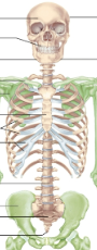
Axial skeleton
The central supporting axis of the body; includes the skull, vertebrae, sternum, ribs, sacrum and hyoid
Appendicular skeleton
The appendages of the body; includes the pectoral girdle, upper extremities (arms), pelvic girdle, and lower extremities (legs)
Number of bones
270 at birth, but reduces to 206 by adulthood and may vary depending on new growths
Articulated skeleton
Held together by wire and rods, shows spatial relationships between bones
Disarticulated bones
A skeleton taken apart to study surface features and markings
Sutures
Immovable joints that hold the 22 skull bones
Cavities of skull
Cranial cavity (brain case)
Orbits (eye sockets)
Nasal cavity
Oral (buccal) cavity
Middle and inner ear cavities
Paranasal sinuses
Foramina
Holes that allow passage for nerves and blood vessels
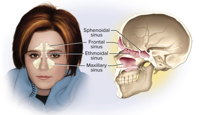
Paranasal sinuses
Air-filled holes lined by a mucous membrane; adds voice resonance and lightens skull
Cranium (braincase)
Consists of the calvaria (skullcap) and cranial case and has membranes separating brain from bones
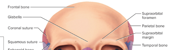
Frontal bone
Forms the forehead and roof of the cranium
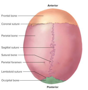
Parietal bones
Forms most of the cranial roof and part of its lateral walls
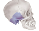
Temporal bones
Forms the lateral walls and floor of the cranial cavity
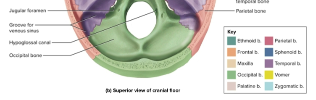
Occipital bone
Forms the rear and base of the skull
Sphenoid bone
Located at the anterior base of the skull, made of the body and greater and lesser wings
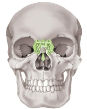
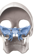
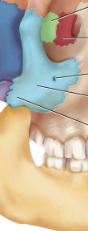
Ethmoid bone
Located between the eyes; contributes to the medial wall of the orbit, walls of nasal cavity, and nasal septum
Facial bones
Gives shape to and supports the face
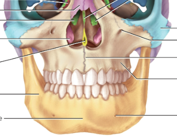
Maxillae
The largest facial bones; forms the upper jaw
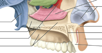
Palatine bones
L-shaped bones dividing the oral and nasal cavities
Zygomatic bones
Forms part of the angles of the cheekbones and lateral orbital wall

Lacrimal bones
Forms part of the medial wall of each orbit; smallest bones of the skull

Nasal bones
Forms the bridge of the nose and supports cartilages that give shape; often fractured
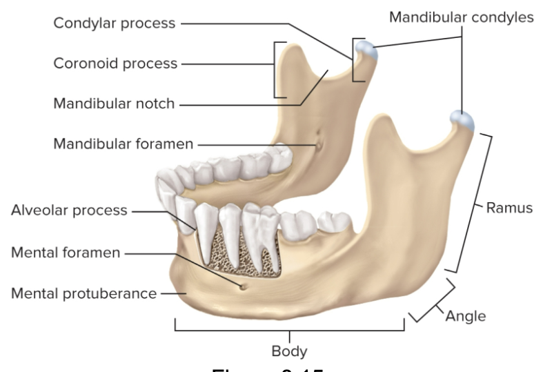
Mandible
The strongest bone of the skull and the only one to noticeably move; supports the lower teeth
Auditory ossicles
Three in each middle-ear cavity for hearing
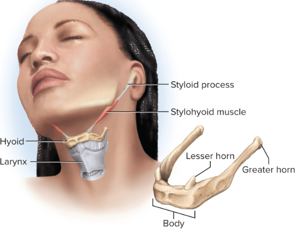
Hyoid bone
Slender U-shaped bone between the chin and larynx, not articulated and has horns
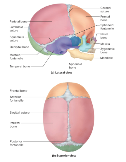
Fontanelles
Spaces between the unfused cranial bones childhood for growth
Spine functions
Supporting and protecting the skull, trunk, and spinal cord by absorbing stresses
Vertebral regions
7 cervical vertebrae
12 thoracic vertebrae
5 lumbar vertebrae
5 sacral vertebrae
4 vertebrae in the coccyx
Primary curvature
The first C-shaped convex curve at birth; persists as the thoracic and pelvic spine
Secondary curvatures
Develop with crawling and walking in childhood; creates the cervical and lumbar areas concave curves
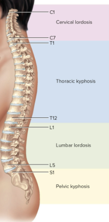
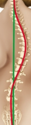
Final curvatures
Spine turns into an S shape: cervical lordosis, thoracic kyphosis, lumbar lordosis, and pelvic kyphosis
Lordoses curve in, kypohses curve out
Abnormal curvatures
Can result from disease, paralysis, posture, or congenital defects like scoliosis (sideways)
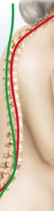
Hyperkyphosis
An exaggerated thoracic curvature usually from osteoporosis, the spine curve goes overly out
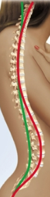
Hyperlordosis
An exaggerated lumbar curvature usually caused by pregnancy or obesity, the spine curve goes overly in
Spinous process
Projection upward from one vertebra to meet another articular process above
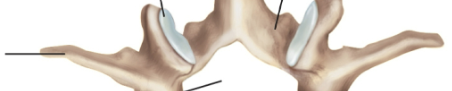
Transverse process
Lateral extension from a vertebtra
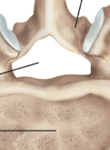
Intevertebral foramen
Opening between pedicles of two adjoining vertebrae
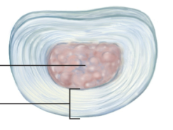
Intervertebral discs
Pads between the vertebrae that bind them together to support the weight of the body
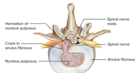
Herniated disc
The slipping or rupturing of a disc; can put painful pressure on the spinal nerve or cord
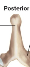
Cervical vertebrae
Notated as C1 to C7; C2 to 6 have forked spinous process while C1 and C2 are the atlas and axis
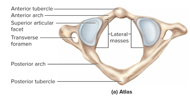
Atlas
The first cervical vertebrae (C1), supports the head and allows nodding “yes” (pitch) with anterior and posterior arches
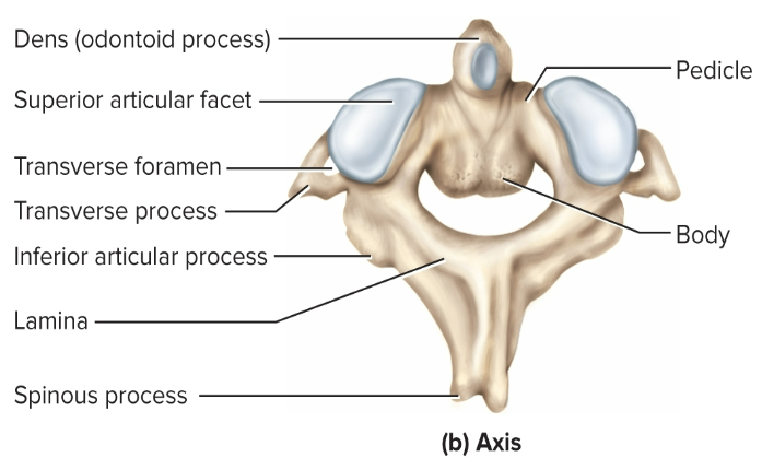
Axis
The second cervical vertebrae (C2), allows nodding “no” (yaw)
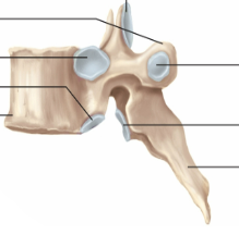
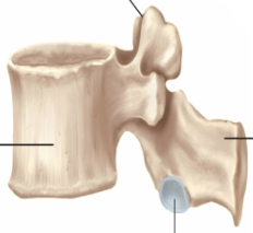
Thoracic vertebrae
Notated as T1 to T12; has downward angled spinous processes that correspond to the 12 pairs of ribs they are attached to
Lumbar vertebrae
Notated as L1 to L5; have thick, stout bodies and blunt, squarish spinous processes
Sacrum
Bony plate forming the posterior wall of the pelvic girdle, notated as S1 to S5 that begin fusing around age 16
Anterior surface is smooth and concave while posterior surface is very rough
Coccyx
Consists of four smaller vertebrae notated as Co1 to Co4, fuses into single, triangular bone by age 20 to 30
Can be fractured during childbirth or hard fall and provides pelvic muscular attachment
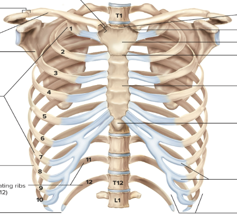
Thoracic cage
Consists of the thoracic vertebrae, sternum, and ribs to enclose the lungs and heart
Sternum
The bony plate anterior to the heart, divided into the manubrium (superior portion), body (long mdidle portion), and xiphoid (inferior point)
Ribs
12 pairs with ends attached to vertebral column and sternum; costal cartilages attach ribs to sternum
True ribs
Ribs 1 to 7, each directly connected to the sternum
False ribs
Ribs 8 to 12, lacking independent connections to the sternum
Floating ribs
Ribs 11 and 12 (also false), no connection at all to sternum or cartilages
Pectoral girdle
Supports the arm, consists of the clavicle (collarbone) and scapula (shoulder blade)
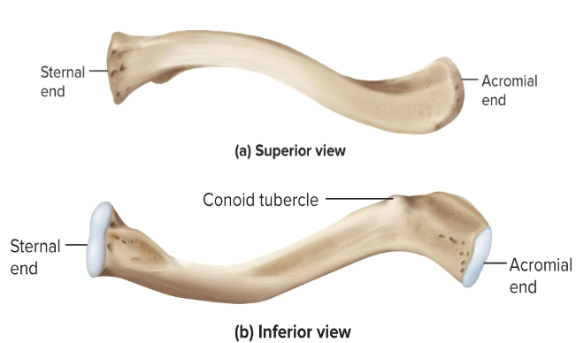
Clavicle
An S-shaped, somewhat flattened bone that is frequently fractured
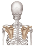
Scapula
A shovel-like, triangular plate posteriorly overlying ribs 2 through 7
Upper limb
Contains 30 bones in three regions: the brachium (humerus), antebrachium (radius and ulna), and hand (5 metacarpals, 14 phalanges)
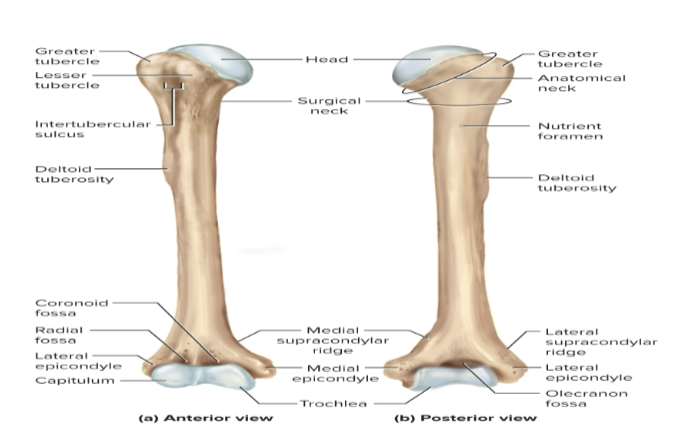
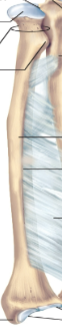
Humerus
The arm proper, articulates with the scapula, clavicle, ulna, and radius
Radius
Bone that is part of the antebrachium; has a disc-shaped head for rotation
Ulna
Bone that is part of the antebrachium; has a hook to the humerus
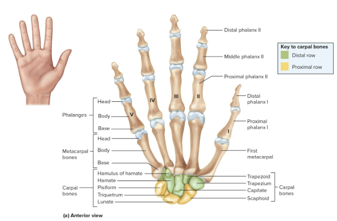
Carpals
Eight bones that form the wrist to allow for flexion, extension, abduction, and adduction in two rows
Metacarpals
Bones of the palm
Phalanges
Bones of the fingers
Pelvic girdle
A ring composed of three bones: two hip (coxal) bones and one sacrum (also part of the vertebral column)
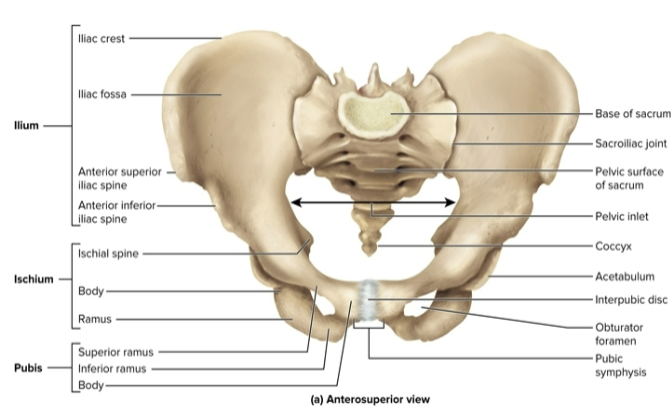
Pelvis
The pelvic girdle, ligaments, and muscles that line the pelvic cavity and its floor
Sacroiliac joint
Joins the coxal bone to the vertebral column
Pubic symphysis
The interpubic disc of fibrocartilage joining the pubic bones anteriorly
Iliac crest
The superior crest of the hip
Acetabulum
The socket of the hip
Ilium
The largest bone in the hip, extends from the iliac crest to the acetabulum
Ischium
The inferioposterior portion of the hip; heavy body with a prominent spine
Pubis
The pubic bone, most anterior portion of the hip bone
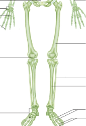
Lower limb
Divided into three regions with 30 bones: the thigh (femoral region), leg proper (crural region), and foot (tarsals, metatarsals, and toes)
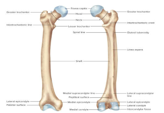
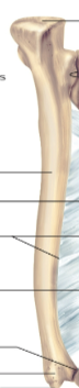
Femur
The longest and strongest bone of the body; the head articulates with the acetabulum of the pelvis
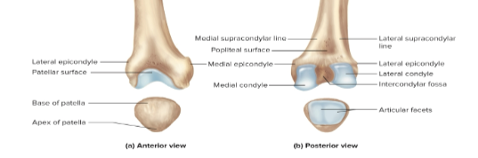
Patella
The triangular sesamoid bone embedded in the tendon of the knee; cartilaginous at birth and ossifies from ages 3 to 6
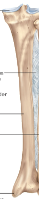
Tibia
The thick, medial, weight-bearing leg bone in the leg proper (crural region)
Fibula
The slender, lateral strut that helps stabilize the ankle but does not bear any body weight
Calcaneus
The largest tarsal bone forming the heel
Talus
The most superior tarsal bone sitting upon the calcareous and forms a joint with the tibia and fibula
Metatarsals
Similar to the metacarpals in the hand
Phalanges
Similar to the digits in the hand
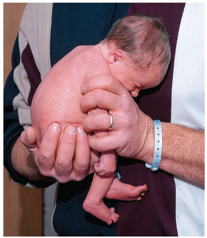
Embryonic limb rotation
The rotation of the upper and lower limbs in opposite direction in week 7 of embryonic development; explains posterior and anterior flexion differences
Bipedal adaptation
Bipedalism is advantageous — strong and springy foot arches to counter stress, posture requires less effort, and muscles are supported well