Chapter 19 THE CARDIOVASCULAR SYSTEM: THE HEART
1/43
There's no tags or description
Looks like no tags are added yet.
Name | Mastery | Learn | Test | Matching | Spaced | Call with Kai |
|---|
No analytics yet
Send a link to your students to track their progress
44 Terms
Serous Membranes
Thin membranes that reduce friction between organs; surround the heart and other thoracic organs.

Pericardial Sac (Pericardium)
A double-walled membrane that encloses the heart; protects and anchors it.

Pericardial Space
The fluid-filled space between the layers of the pericardial sac; contains serous fluid to reduce friction during heartbeats.

Goal: to get the blood RIGHT to the lungs so it can become oxygenated.
Shelby and I Very Carefully
Run Away
To Vegas
Rarely Visiting
Peaceful Villages
Party Absurdly Loud
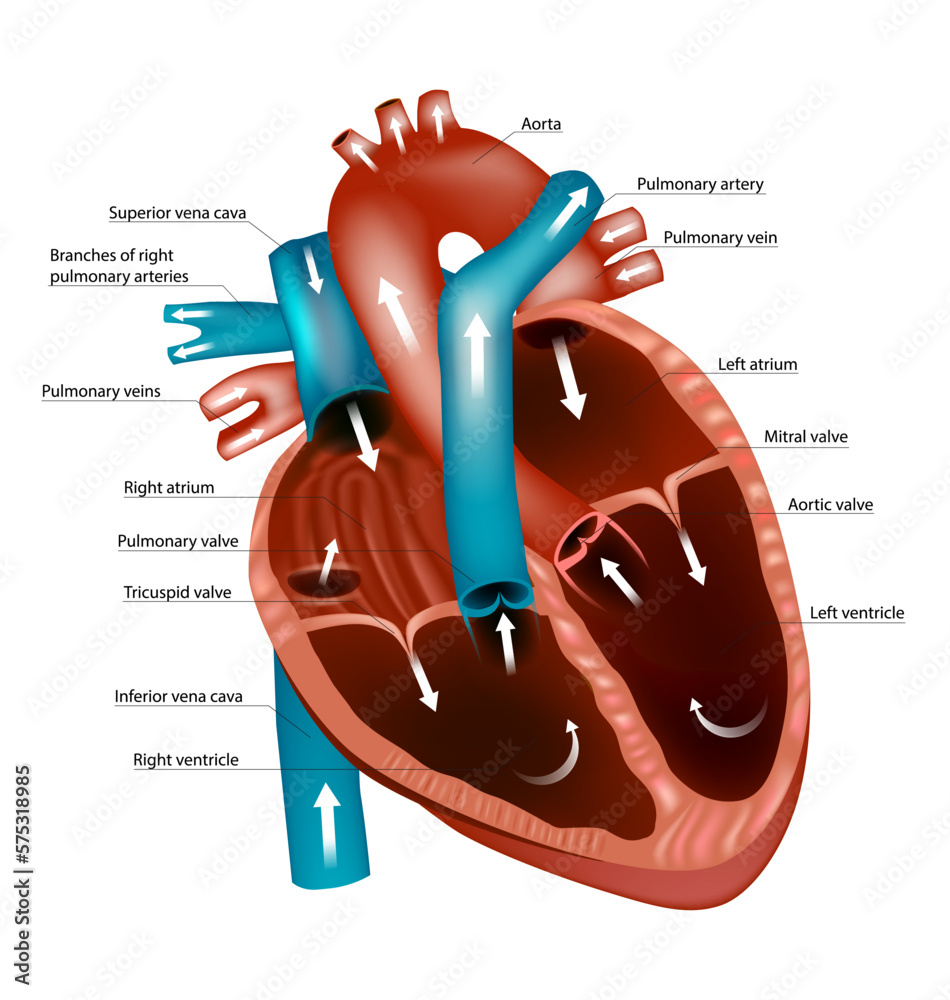
1. The un-oxygenated blood (this is blood that has been “used up” by your body and needs to be resupplied with oxygen) enters the heart through the SUPERIOR AND INFERIOR VENA CAVA.
2. Blood enters into the RIGHT ATRIUM
3. Then it is squeezed through the TRICUSPID VALVE
4. Blood then enters into the RIGHT VENTRICLE
5. Then it is squeezed into the PULMONIC VALVE
6. Blood is then shot up through the PULMONARY ARTERY and then enters into the LUNGS for some oxygen

Goal: to get the richly, oxygenated blood that LEFT the lungs to the body to feed the brain, tissue, muscles, organs etc.)
Please Visit
Laila And
Bianca’s Very
Lame Volleyball
At Vegas
Annually
7. Blood enters from the lungs through the PULMONARY VEIN
8. Blood then enters into the LEFT ATRIUM
9. Down through the BICUSPID VALVE (also called mitral valve)
10. Then blood is squeezed into the LEFT VENTRICLE
11. Up through the AORTIC VALVE
12. Lastly up through the AORTA, where it pumped throughout the body
Fibrous Pericardium
The outermost tough layer of the pericardium; anchors the heart to surrounding structures and prevents overfilling.

Parietal Pericardium
The inner layer of the pericardial sac; lines the fibrous pericardium.
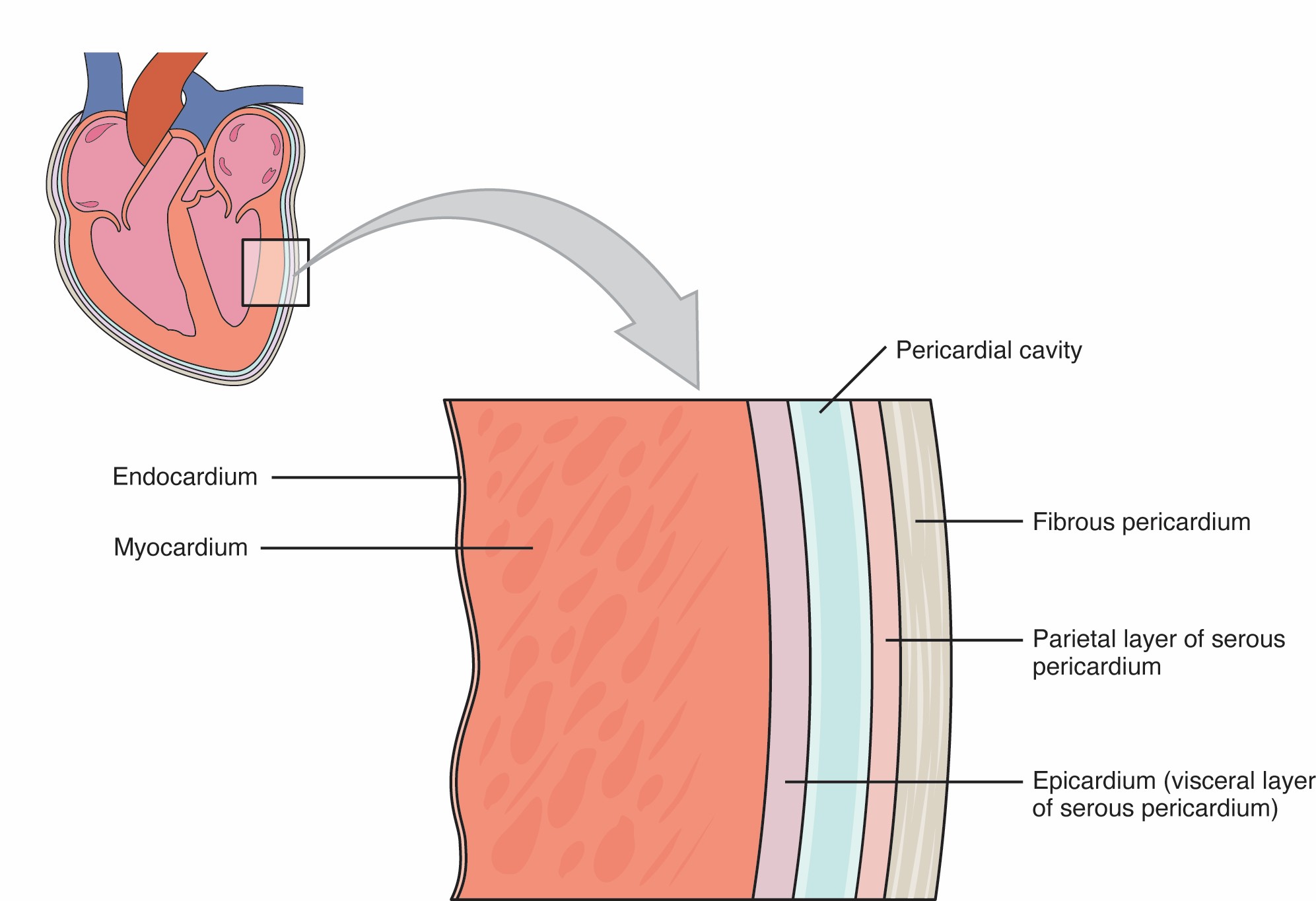
Visceral Pericardium (Epicardium)
The outermost layer of the heart wall; also part of the pericardial membrane. It’s continuous with the parietal layer but directly touches the heart.

Pericardial Cavity
The space between the parietal and visceral pericardium; filled with serous fluid to reduce friction as the heart beats.

Epicardium
Also called the visceral pericardium; outer layer of the heart wall.

Myocardium
The thick, muscular middle layer of the heart wall; made of cardiac muscle responsible for contractions.

Endocardium
The smooth inner lining of the heart chambers and valves; helps reduce friction and prevent clotting.

Cardiac Muscle Arrangement
Cardiac muscle fibers are arranged in a spiral or swirling pattern, allowing the heart to contract efficiently and eject blood effectively.
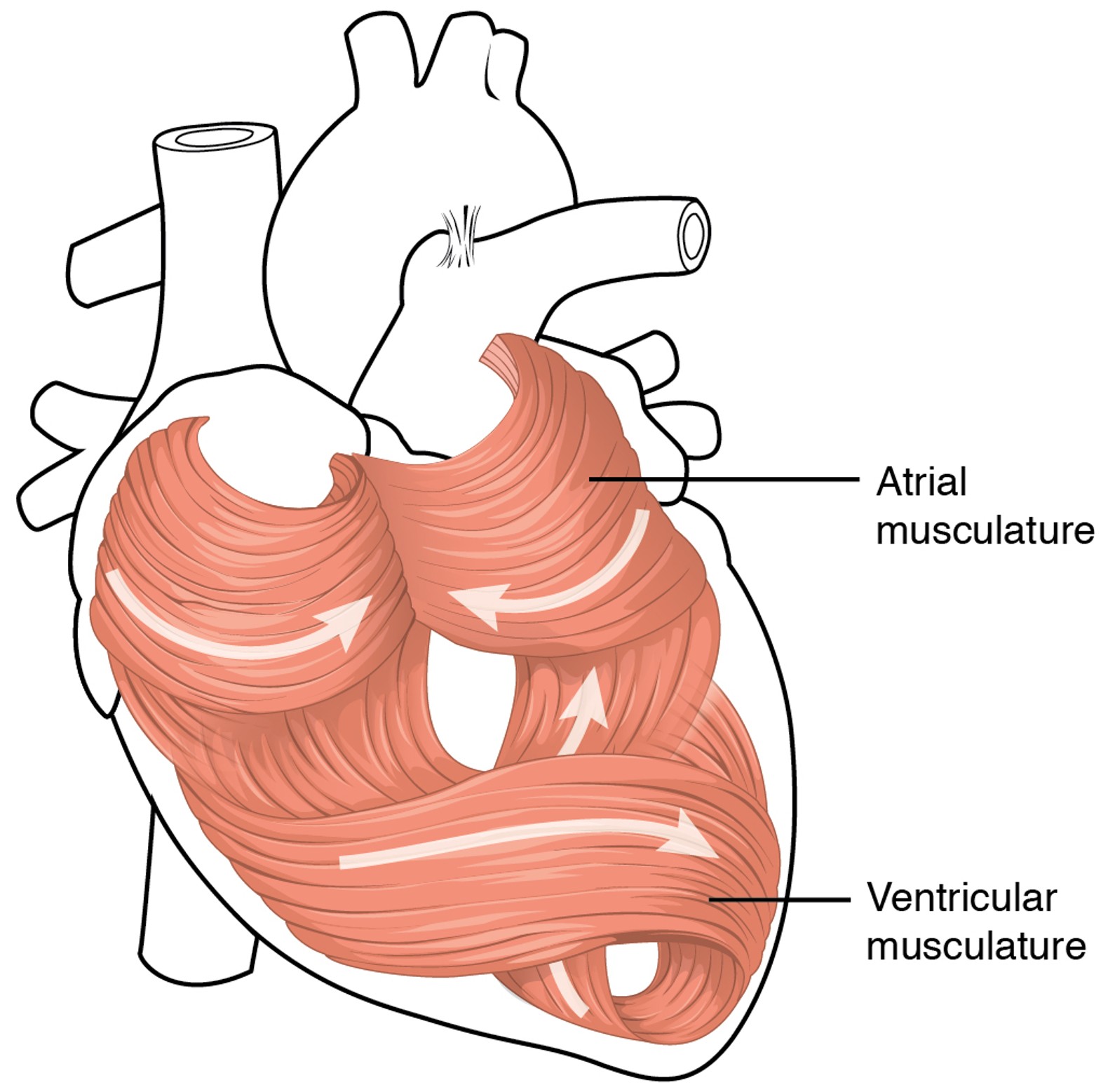
Atrial Musculature
Muscle fibers in the atria are circularly arranged, helping to squeeze blood downward into the ventricles

Ventricular Musculature
Arranged in spiral and figure-eight patterns, enabling a wringing motion that forces blood upward into the arteries.

Left Ventricle Wall Thickness
This is much thicker than the right because it must pump blood with greater force to overcome the higher resistance in the systemic circuit.
Ventricular Lumen (Cavity)
Changes between relaxed and contracted states and is generally smaller in the left ventricle during contraction due to its stronger force.
Patent Foramen Ovale
A hole in the interatrial septum that results from the failure of the foramen ovale to close after birth. Can allow blood to mix between the atria.
Atherosclerosis
A condition where fatty plaques build up inside arteries, narrowing them and reducing blood flow.
Plaque Buildup
Composed of cholesterol, fat, calcium, and cellular waste; contributes to arterial narrowing and stiffness.
HDL (High-Density Lipoprotein)
"Good" cholesterol; helps remove excess cholesterol from arteries and transport it to the liver for disposal.
LDL (Low-Density Lipoprotein)
"Bad" cholesterol; can deposit cholesterol in artery walls, contributing to plaque formation and atherosclerosis.
Ischemia
A reduction in blood flow to tissues, leading to lack of oxygen (hypoxia).
Hypoxia
A condition where tissues are deprived of oxygen, often due to poor blood flow.
Myocardial Infarction (Heart Attack)
Occurs when blood flow to part of the heart is blocked, causing cardiac muscle cells to die.
Angioplasty with Stent
A procedure where a balloon-tipped catheter is used to open narrowed arteries, often followed by placement of a stent (small mesh tube) to keep the artery open.
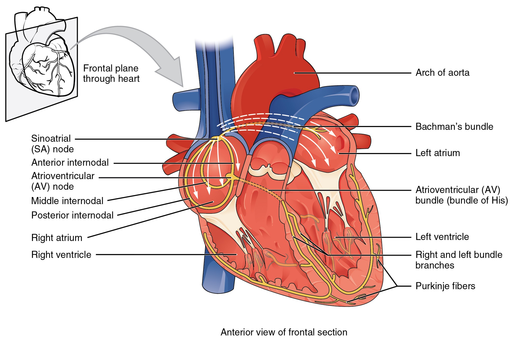
Cardiac Cycle
A complete heartbeat, lasting about 0.8 seconds, including:
SA node fires → atrial systole (atria contract)
Ventricular diastole (ventricles fill)
AV node fires → ventricular systole (ventricles contract)
Atrial diastole (atria refill)
Reset → both chambers relax and prepare for the next cycle
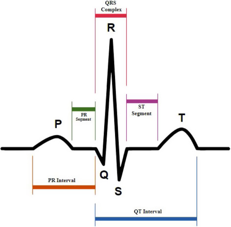
PQRST Complex (on ECG)
P wave: Atrial depolarization (atria contract)
QRS complex: Ventricular depolarization (ventricles contract)
T wave: Ventricular repolarization (ventricles relax)
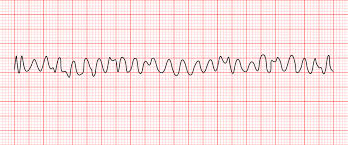
Ventricular Fibrillation (V-Fib)
A life-threatening condition where the ventricles quiver instead of contracting due to disorganized electrical activity—results in no effective blood flow.

Angina Pectoris
Chest pain or discomfort due to reduced blood flow to the heart muscle; often a warning sign of coronary artery disease.
Pallor
Unusual paleness of the skin, often caused by reduced blood flow or oxygenation, and may be a symptom of heart problems or shock.

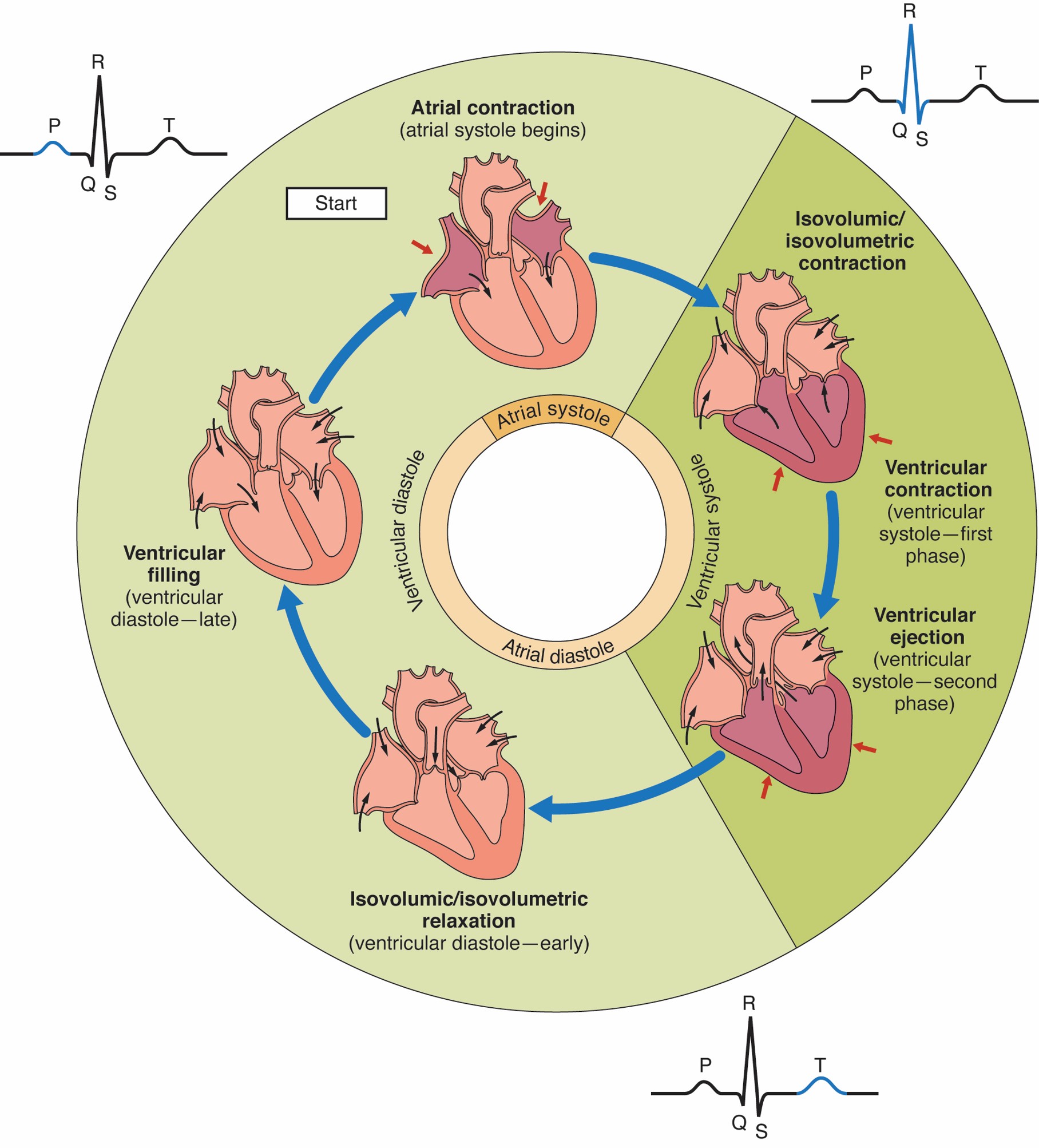
Atrial Contraction (Atrial Systole Begins)
Blood is pushed from the atria into the ventricles.
🩺 Correlates with the P wave on an ECG.

Isovolumic Contraction (Ventricular Systole – First Phase)
Ventricles begin to contract, but all valves are closed, so no blood leaves yet.
🩺 Occurs just after the QRS complex begins

Ventricular Ejection (Ventricular Systole – Second Phase)
Pressure builds, semilunar valves open, and blood is ejected into the aorta and pulmonary artery.

Isovolumic Relaxation (Ventricular Diastole – Early)
Ventricles relax, all valves close, and pressure drops.
🩺 Follows the T wave on the ECG.

Ventricular Filling (Ventricular Diastole – Late)
AV valves open, and blood flows passively from atria into ventricles.
Atria are in diastole and filling.
Prolapse
When a body structure slips or moves out of its normal position.
Mitral Valve Prolapse
A condition where the mitral valve bulges (goes backward) into the left atrium during ventricular contraction; may cause leakage or a heart murmur
Stenosis
The narrowing of a tubular structure in the body, such as a blood vessel, valve, airway, or spinal canal, which can restrict flow.
Valve Replacement
A surgical procedure to replace a damaged heart valve with a mechanical or biological valve.
Cardiomegaly
An enlarged heart, often due to chronic high blood pressure or heart disease; may weaken the heart’s pumping ability.
Congestive Heart Failure (CHF)
A condition where the heart can’t pump blood effectively, leading to fluid buildup in the lungs, legs, or other tissues.
Atherosclerosis
A disease where plaque builds up in artery walls, narrowing them and reducing blood flow; can lead to heart attacks and strokes
Fetal Circulation
The circulatory system of a fetus, which bypasses the lungs using special structures like the foramen ovale and ductus arteriosus, since oxygen is received through the placenta, not the lungs.