module 3 - cardiac muscle
1/6
There's no tags or description
Looks like no tags are added yet.
Name | Mastery | Learn | Test | Matching | Spaced | Call with Kai |
|---|
No analytics yet
Send a link to your students to track their progress
7 Terms
cardiac muscles are also called _____ cells
myocardial; the specialized muscle cells of the heart
typically striated bc contractile fibres organized into sarcomeres (striated = banded pattern = A/I bands)
cardiac muscle compared to skeletal muscle
cardiac muscle cells are much smaller, single nucleus, 1/3 of cell is occupied by mitochondria (tf generates ATP thru oxidative metabolism)
T tubules are much larger and branched, and SR is much smaller
adjacent cells are joined via intercalculated discs w desmosomes (anchor)
around 1% cardiac muscle cells are not involved contraction, what is their PURPOSE
electrical excitation of the heart - aka the electrical conducting system of the heart
initiates heartbeat, and allow electrical excitation to spread rapidly thru the heart
connected to other cardiac cells via gap jxns
CALLED: PACEMAKER CELLS
steps of contraction for cardiac muscle
similar to skeletal contraction except:
Ca enters thru Ca channels on CM and the SR
Ca enters thru external Ca channels, then Ca induced Ca release (releases stored Ca from SR, which supplies 90% needed for contraction)
cardiac cells have Na Ca antiport (passive) in addition to Ca-ATPase, removes Ca from cytosol and pumps into the EC space (calcium ions (Ca2+) out of a cell in exchange for sodium ions (Na+) moving into the cell,)
exhibit graded (not all or none) contraction - force generated is proportional to number of ACTIVE CROSSBRIDGES which is proportional to cytosolic Ca (depends on the amount of Ca bound to troponin)
factors influencing cardiac muscle contraction force
CHANGES IN [CA]
regulated by epinephrine (adrenal medulla) and norepinephrine (from sympa post gang neurons) - bind to beta 1 adrenergic receptors, activating cAMP second mssnger (GPROTEIN) signalling pathway causing
phosphorylation of voltage gated Ca channels - increases probability of channel to open and increase [Ca] in cytosol (lowers threshold for opening + open more often)
phosphorylation of phospholamban - causes increase SR Ca-ATPase activity - increase SR Ca - to make next ‘beat’ stronger
SARCOMERE LENGTH
tension generated proportional to length of muscle fibre
due to degree of overlap of actin and myosin (optimal amt of overlap)
stretching a myocardial muscle cell can allow more Ca to enter thru CM Ca channels - more forceful next contraction (stretching = ex. filling heart w blood)
steps of cardiac muscle contraction
Most of the calcium in heart muscle cells is stored in the sarcoplasmic reticulum (SR).
When the heart contracts, calcium is released from the SR
an excitable tissue that can generate APs
FAST THEN SLOW K; SLOW Ca
RMP - at -90mV
DEPOLARIZATION - (all channels are triggered) AP opens voltage gated Na channels, cause increase in membrane permeability to Na, at peak = close (20mV)
INITIAL REPOLARIZATION - open fast K channels
PLATEAU - initial depolarization triggers voltage gated Ca channels to slowly open, cause increase in Ca permeability (in) and fast K channels close
RAPID REPOLARIZATION - Ca channels close, slow voltage gated K channels open triggered by initial depolarization, and RMP is restored
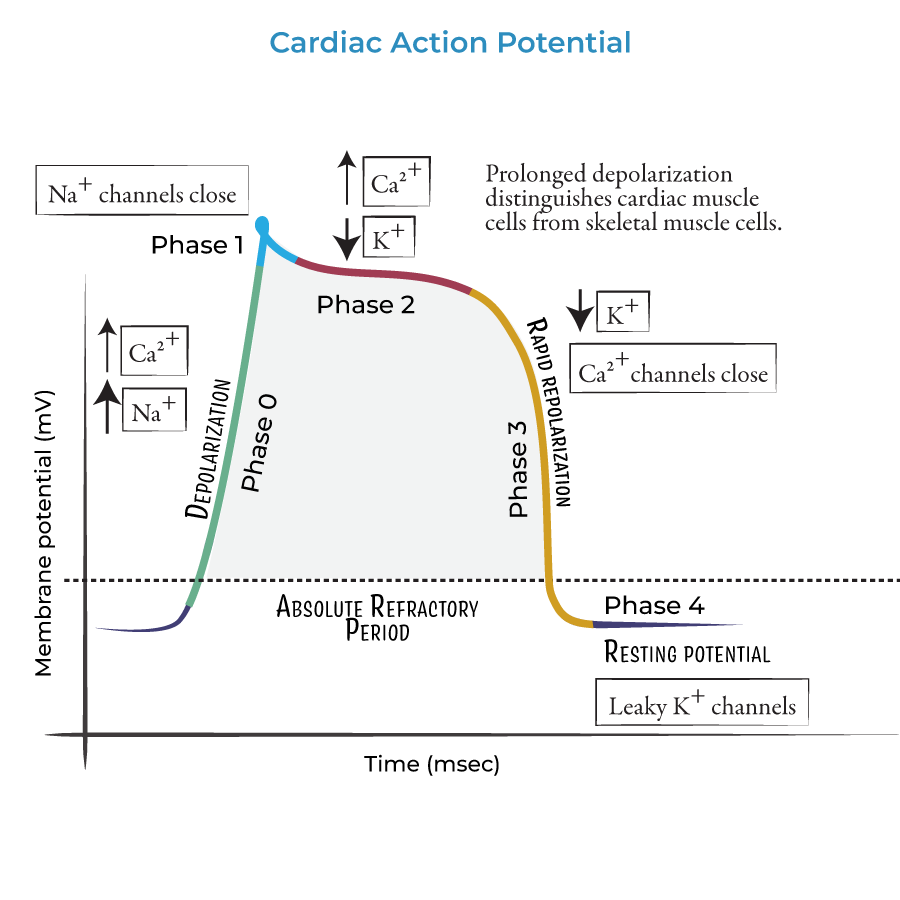
sustained depolarization is due to
slow opening of the voltage gated Ca channels
causing - longer AP (200msec) which is important - gives time heart time to relax btw contractions and prevent tetanus (prolonger contraction meaning not rapid contract-relax)
cardiac muscle cells dont undergo summation/tetanus - bc of longer refractory period, meaning cell finishes contracting before next AP