2.1.1 cell structure and microscopy
1/36
There's no tags or description
Looks like no tags are added yet.
Name | Mastery | Learn | Test | Matching | Spaced |
|---|
No study sessions yet.
37 Terms
Light microscopes- how to prepare a dry mount and what it's used for
-used for hair, pollen, dust, insect parts, muscle tissue, plants
-solid specimens are cut thinly with a sharp blade (sectioning)
-hold with tweezers
-covered with a cover slip
Light microscope- how to prepare a smear slide
-used for blood samples to view cells
-edge of slide is used to smear sample on another slide
-cover slip place
light microscopes
-poor resolution due to long wavelength of light (200nm)
-living samples can be examined
-a colour image is obtained
-medium is air
-magnification up to x2,000
Transmission electron microscopes
-high magnification (x 1 000 000) and resolution (0.2nm)
-electrons pass through specimen to create an image
-2D image created
-thin specimens are placed in a vacuum
-beam of electrons are focused by the electromagnet
-some parts of specimen absorb electrons, making them appear darker
Scanning Electron Microscope
-high resolution (2nm) and magnification x500 000
-electrons reflect off surface of specimen to create an image
-electrons are scattered in different ways depending on contours
-producing a 3D image
-specimen doesn't need to be thin
-source is electron beam
-medium is vacuum
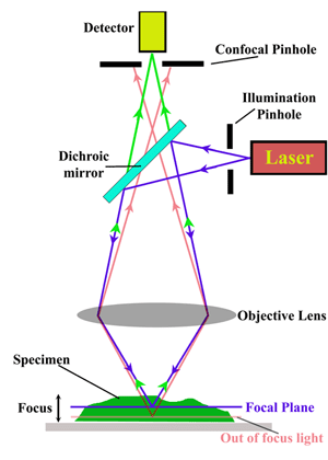
Laser Scanning Confocal microscope
-high resolution
-3D images
-uses laser beam
-a type of fluorescent microscope
-image created using high light intensity to illuminate specimen stained with a fluorescent dye
-light is filtered through a pinhole aperture
-enables scientists to view sections of tiny structures that would be hard to physically section off, like embryos
-as light is emitted from specimen it causes fluorescence
What is resolution?
-the ability to distinguish 2 objects from one another
-(determined by wavelength if light or wavelength of beam of electrons)
-determined by quality of equipment used, medium used (better in vacuum, no particles), source (light, laser beam)
What is magnification and the equation?
-How many times larger an image is under microscope compared to the actual size of the object
-image size= actual size x magnification
-convert units so they're the same, mm to micrometers, x1000
Light microscopes-How to prepare a wet mount
-when specimens are added to water or a stain
-coverslip is lowered/tilted with a mounted needle at an angle to prevent air bubbles froming
-used for viewing aquatic organisms
Light microscopes-How to prepare a squash slide
-they are wet mounts which you then push down on the coverslip to squash sample
-to ensure you have a thin layer to enable light to pass through
-used when creating a root tip squash sample to view chromosomes in mitosis
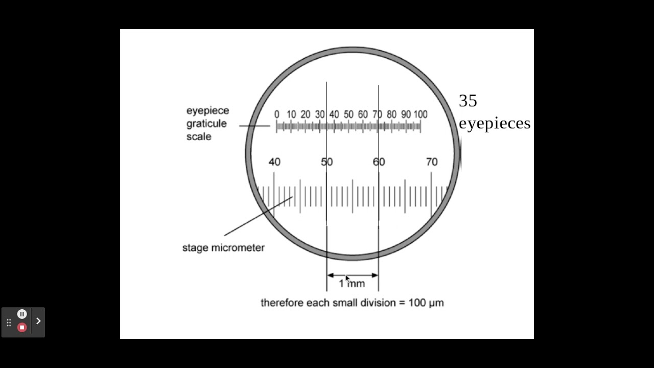
How do you calibrate the eyepiece graticule?
1- line up stage micrometer and eyepiece graticule
2- count how many divisions on epg fit into one division of stage micrometer
3- each small division on micrometer is 100 micrometers / 0.1mm
-use this to calculate what one division on epg is at that current magnification
One eyepice division= number of micrometers/number of eyepiece divisions
What is differential staining?
-a technique involving many chemical stains being used to stain different parts of a cell in different colours and distinguish between different organelles
What are crystal violet and methylene blue?
-stains used in light microscopy
-they're positively charged, so are attracted to and stain negatively charged materials in a cell
What are congo red and nigrosin?
-stains that are negatively charged
-can't enter cells as cytosol repels them
-this creates a stained background
-making unstained cells stand out
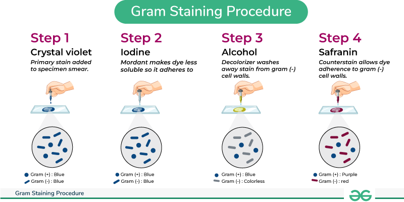
What is gram staining and what is it used for?
-a use of differential staining to prescribe an appropriate antibiotic
-used to identify the type of bacteria
-2 different stains are used, crystal violet and safranin
-crystal violet is added, then iodine to fix the stain, then alcohol to wash any unfixed stain
-gram-positive bacteria appear blue/purple as stain is retained due to thicker peptidoglycan cell wall absorbing the dye
-gram-negative bacteria can't absorb crystal violet as cell wall is thin so they can't retain it
-safranin is used as a counterstain, turning them red
-gram positive are susceptible to penicillin, which inhibits cells wall formation
What are the rules of a scientific drawing?
-draw with a sharp pencil
-title to indicate what the specimen is
-state magnification or add a scale
-annotate cell components, cells and sections of tissue visible
-don't sketch, use solid lines that overlap
-don't colour/shade
-aim is to show size, location and proportion
Describe Electron microscopes
-a beam of electrons has a very short wavelength = high resolution, small organelles can be visualised
-image is created using an electromagnet to focus the beam of negative electrons
-vacuum is used as air would absorb beam of electrons so living samples can't be used
-image is black and white so stain has to be added
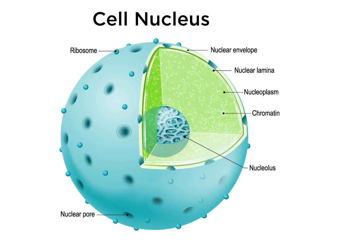
What is the struture and function of the nucleus?
-nuclear envelope, double membrane structure
-nuclear pores
-nucleoplasm,
-chromosomes, protein-bound, linear DNA
-nucleolus, site of rRNA and ribosome production
-site of DNA replication and transcription (making mRNA)
-contains genetic information for each cell in forms of DNA, whcih directs synthesis of proteins
-controls metabolic activities of cell
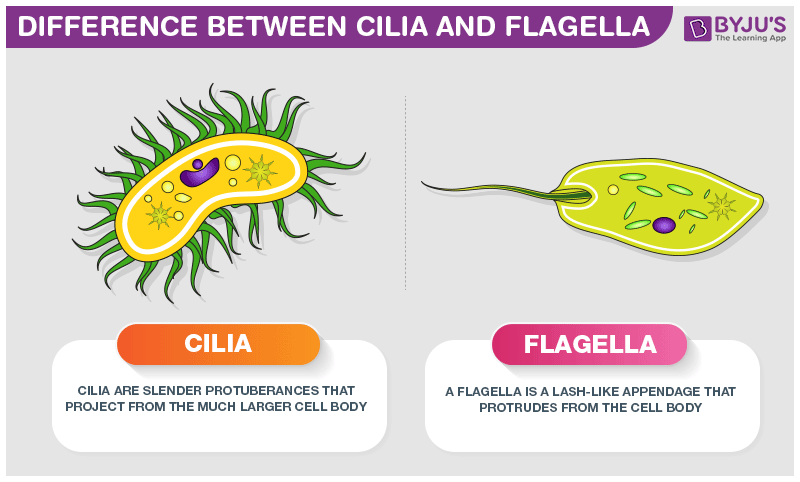
What is the function and structure of flagella
-found in some eukaryotic cells
-whip-like tail structure
-for mobility and sometimes a sensory organelle for chemical stimuli

What is the structure and function of cilia?
-found on some eukaryotic cells
-hair-lke projections out of cells
-mobile cilia help move substances in a sweeping motion (e.g. in trachea to move mucus)
-stationary cilia are more important in sensory organs like the nose
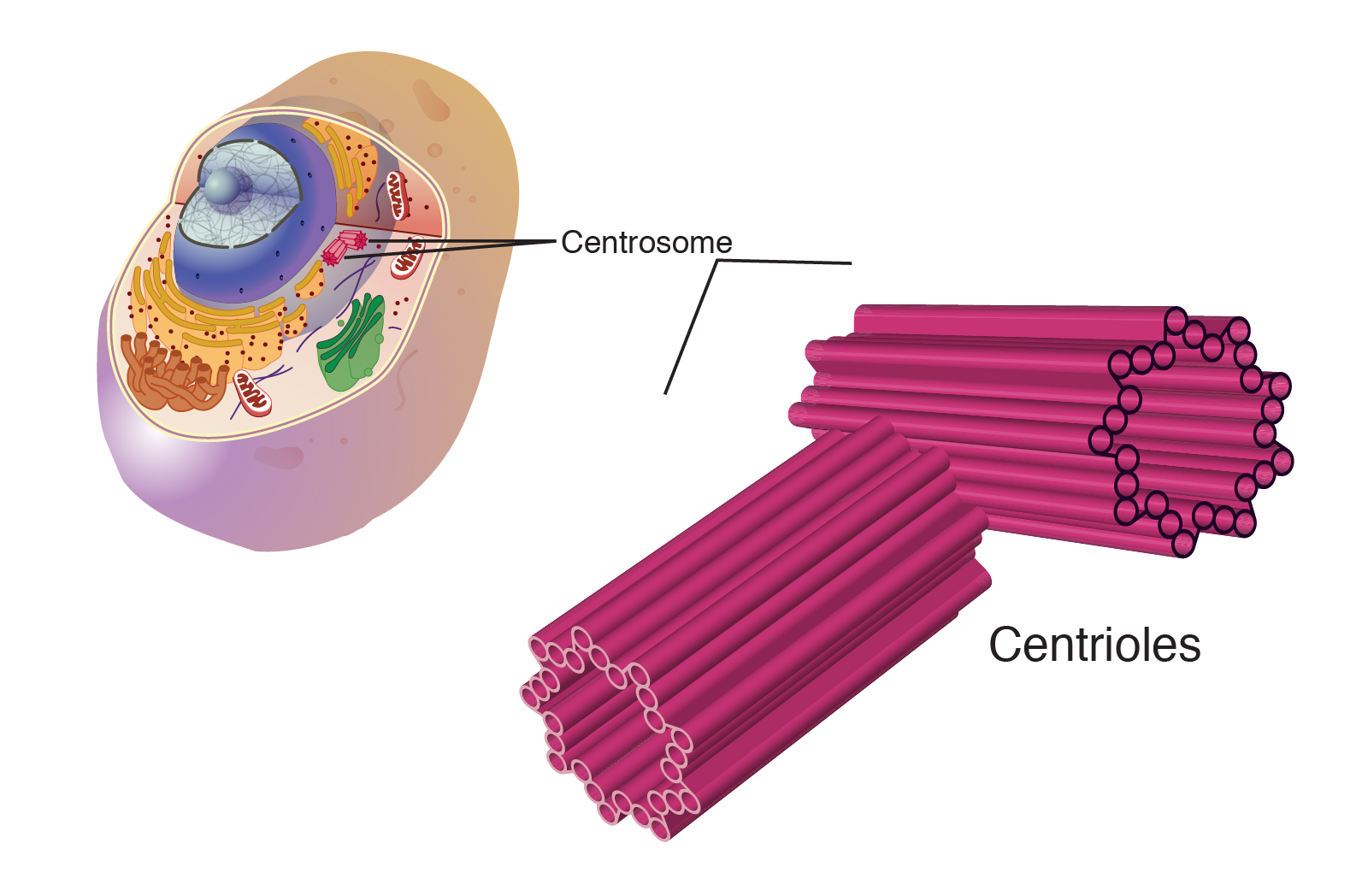
What is the struture and function of centrioles?
-made of microtubules
-occur in pairs to form a centrosome
-involved in production of spindle fibre to separate chromosomes in cell division
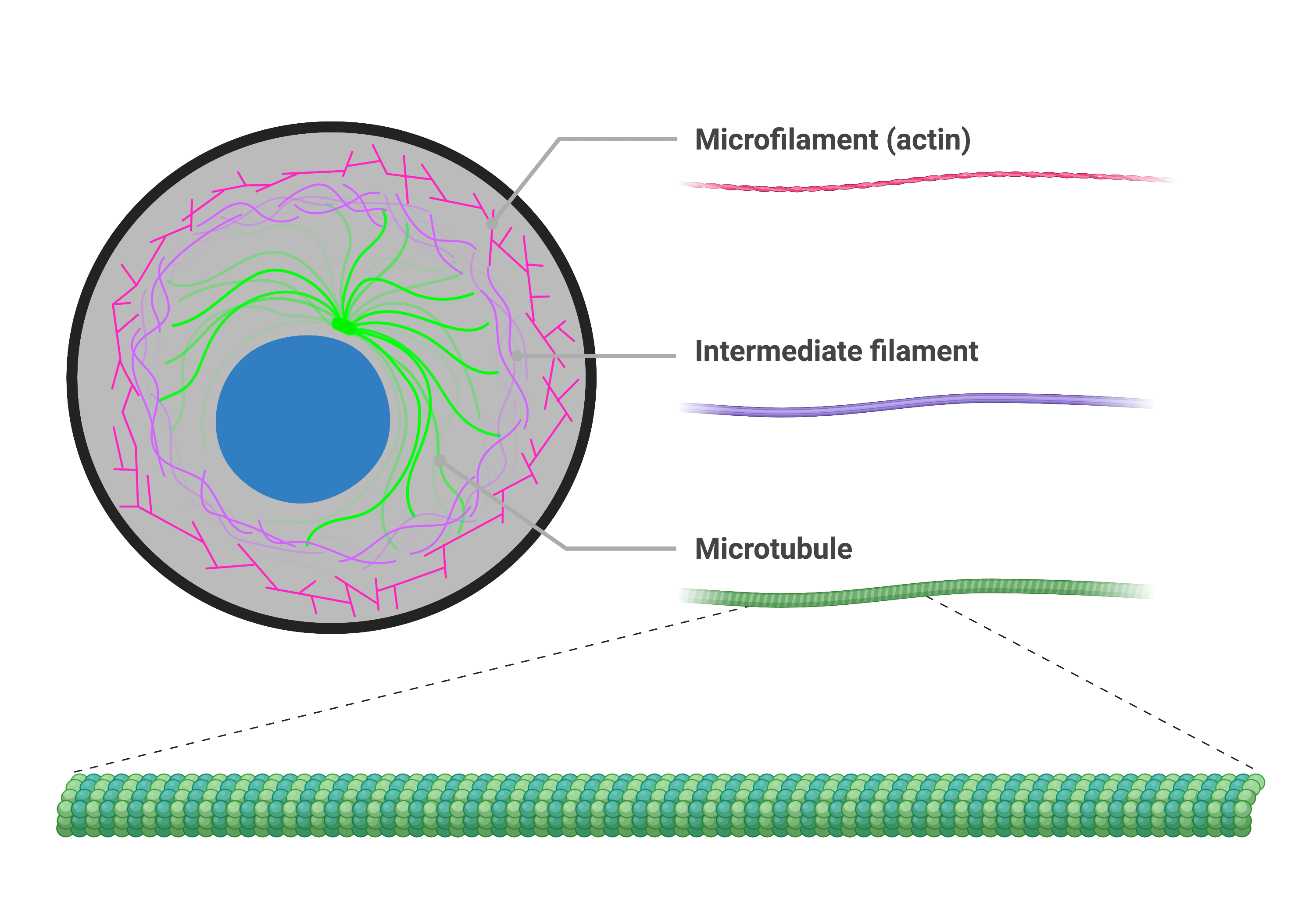
What is the struture and function of the cytoskeleton?
-a network of fibres for mechanical strength, cell movement, transport within cell
-consists of microfilaments, microtubules and intermediate fibres
-many organelles are bound to hold them in place
-movement of vesicles
-changing cell shape
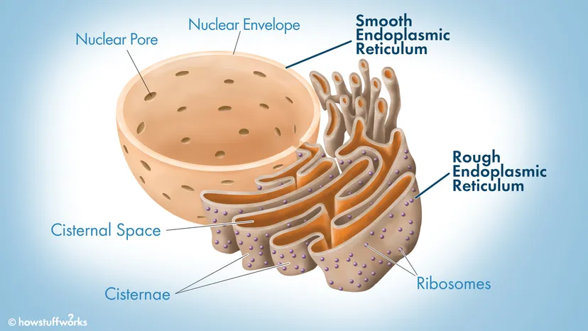
What is the structure and function of the endoplasmic reticulum?
-Rough and smooth ER both have folded membranes calles cisternae made from phospholipid bilayer for compartmentalisation
-rough ER cisternae hold ribosomes in place
-function of rough ER is protein synthesis
-function of smooth ER is synthesis and storage of lipids and carbohydrates
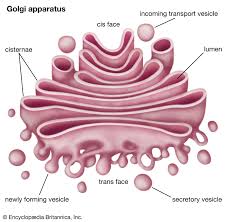
What is the structure and function of golgi apparatus and vesicles?
-folded membranes called cisternae
-secretary vesicles pinch off from cisternae
-adds carbohydrate to proteins to make glycoproteins
-produce secretory enzymes
-secrete carbohydrates
-transport, modify and store proteins/lipids
-form lysosomes
-finished products are transported to cell surface in golgi vesicles where they fuse with the cell membrane and contents are released
What is the structure and function of lysosomes?
-bags/vesicles of digestive enzymes
-break down pathogens ingestes by phaocytic cells
-break down dead cells (autolysis) and worn out organelles for reuse of material
-exocytosis -release enzymes to outside of cell to destroy material
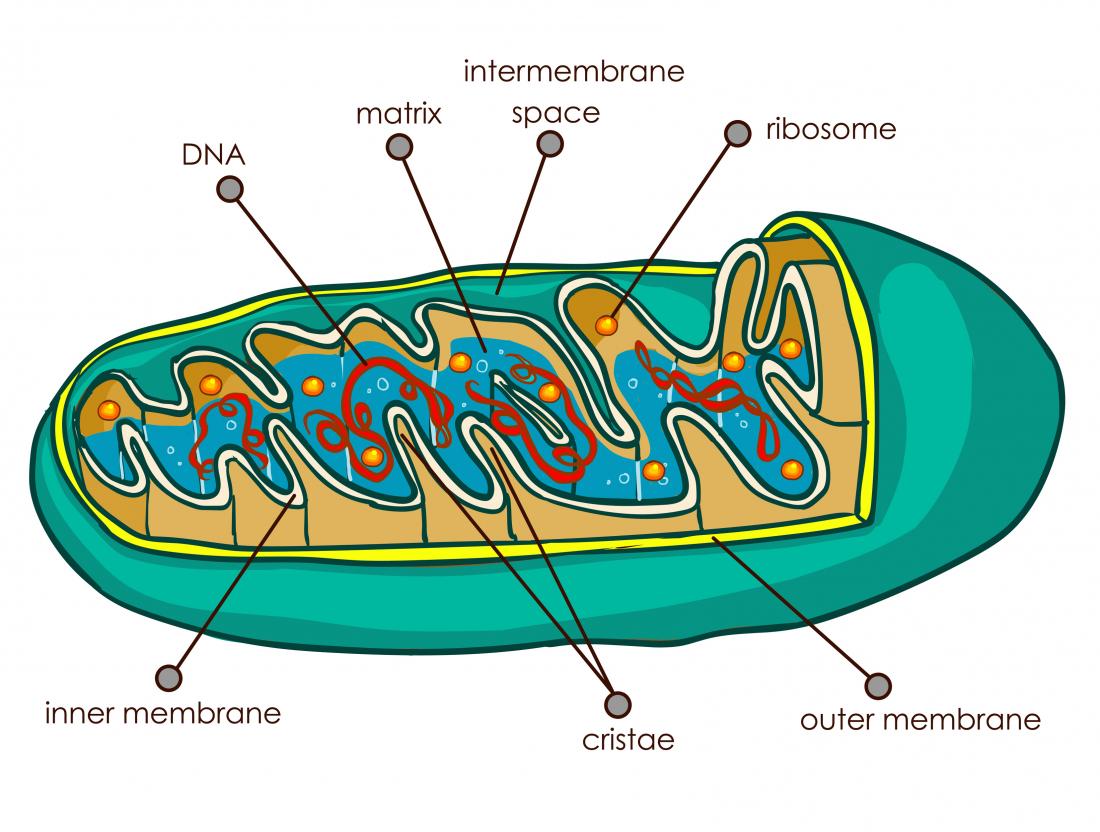
What is the structure and function of mitochondria
-double membrane bound
-inner membrane called cristae
-fluid centre called the mitochondrial matrix
-contain loop of mitochondra DNA
-site of aerobic respiration and ATP production
-DNA to code for enzymes needed in respiration
What is the structure and function of ribosomes?
-small, made up of proteins and rRNA
-80s, large ribosomes found in eukaryotic cells (25nm)
-70S, smaller found in projaryotic cells, mitochondra and chloroplasts
-site of protein synthesis
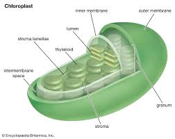
What is the structure and function of chloroplasts?
-surrounded by a double membrane
-contains thylakoids (folded membranes embedded with a pigment)
-stroma contains enzymes for photosynthesis
-site of photosynthesis
What is the structure and function of the cell wall?
-in plants and fungi cells
(plants) made of microfibrils of the cellulose polymer
(fungi) made of chitin, a nitrogen-containing polysaccharide
(bacteria) made of peptidoglycan
-provides structural strength for cell
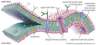
What is the structure and function of plasma (cell surface) membrane?
-phospholipid bilayer- molecules embedded within and attached on outside (proteins, carbohydrates, cholesterol)
-controls entrance and exit of molecules
Which organelles are involved in production and secretion of proteins?
-Instructions fro protein syntehsis in genes is transcribed/copid into mRNA
-mRNA leaves nucleus via nuclear pores and travels to ribisomes on RER
-polypeptide chains are synthesised on rough endoplasmic reticulum
-these chains are packages into transport vesicles to be sent to golgi apparatus via cytoskeleton
-in golgi apparatus, the proteins are modifies and packaged into the vesicles
-secretory vesicles carry the proteins to cell surface membrane, where it fuses and releases the protein by exocytosis
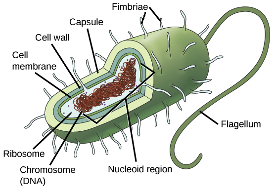
What are the key differences between prokaryotic cells and eukaryotic cells?
-prokaryotic cells are much smaller (2micrometres vs 10-100)
-no membrane-bound organelles
-smaller ribosomes, 70S
-DNA isn't contained within a nucleus and is circular rather than linear (not plasmids)
-a cell wall made of polysaccharide, vs no cell wall in animals, chitin in fungi, cellulose in plants
-they may also contains plasmids and flagella
-don't contains mitochondria, chloroplasts, endoplasmic reticulum or golgi apparatus
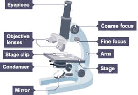
light microscope structure
Why is staining cells necesaary?
-increases contrast as different components within cells take up stain to different degrees
-so can identify organelles
What is the acid-fast technique?
-used to differntiate species of mycrobacterium from other bacteria
-a lipid solvent is used to carry carbol fuchsin dye into cells
-cells are washed with dilute acid-alcohol solution
-mycrobacterium arent's affected by it and retain carbolfuchsin stain (red)
-other bacteria lose stain
-other bacteria are counterstained with methylene blue
What are artefacts and how are they created?
-visible structural details caused by processing the specimen
-not a feature of the specimen, e.g. air bubble
compartmentalisation
-It's created by the use of internal membranes to rpovide distinct environments
-it allows each area to have different conditions for different enzymes