Orbits, Mandible, and TMJ
1/80
There's no tags or description
Looks like no tags are added yet.
Name | Mastery | Learn | Test | Matching | Spaced | Call with Kai |
|---|
No analytics yet
Send a link to your students to track their progress
81 Terms
Orbits are what shape?
Cone-shaped
What angle are the orbits at?
37 degree medial (in) and 30 degree cephalic (up)
What bones make up the orbits?
7 bones - frontal, sphenoid, ethmoid, maxilla, zygoma, lacrimal, and palatine
What Cranial Bones are apart of the Orbit?
Frontal, Sphenoid, and Ethmoid
What facial bones are apart of the Orbit?
Maxilla, zygoma, lacrimal, and palatine
What cranial nerves go through the openings of the orbits?
CN II through Optic Foramen
CN III-VI through Superior Orbital Fissure
CN V through Inferior Orbital Fissure

What is A?
Orbital plate of Frontal bone

What is B?
Sphenoid Bone
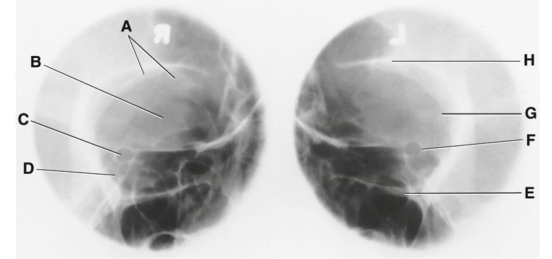
What is C?
optic foramen and canal

What is D?
Superior Orbital Fissure

What is E?
Infraorbital Margin (IOM)

What is F?
Sphenoid strut

What is G?
Lateral Orbital Margin

What is H?
Supraorbital Margin (SOM)
Parieto-Orbital Oblique Projection: Optic Foramina is also called
Rhese Method
Rhese Method Technique
75-80 @ 10-16
Rhese Method Positioning
Rotate head 37 degrees toward affected side
Tripod - chin, cheek, and nose touch the table
AML perpendicular to the IR

What is Red?
Frontal Sinus

What is Orange?
Optic Foramen and canal

What is Yellow?
Inferior Orbital Rim

What is Green?
Maxillary Sinus
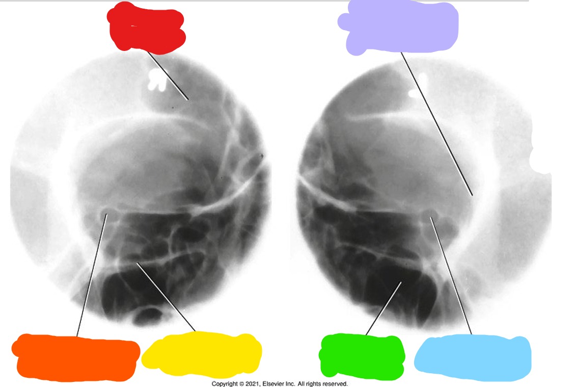
What is Blue?
Optic foramen and canal

What is Purple?
Lateral Orbital Margin

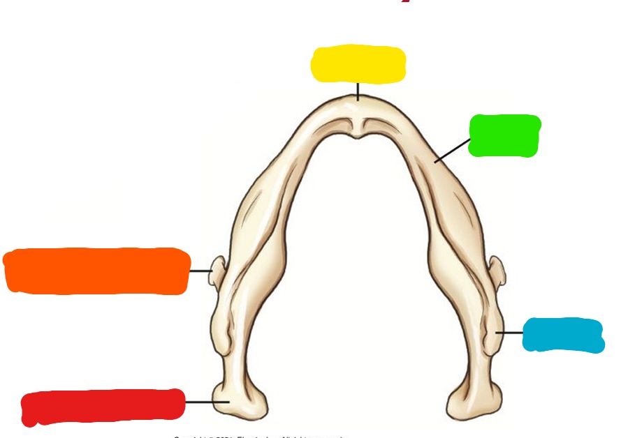
What is Red?
Mental point

What is Orange?
symphysis (symphysis menti)

What is Yellow?
Mentum (mental protuberance)

What is Light Green?
alveolar process

What is Dark Green?
Mental foramen

What is Light Blue?
Body

What is Dark Blue?
Angle or gonion
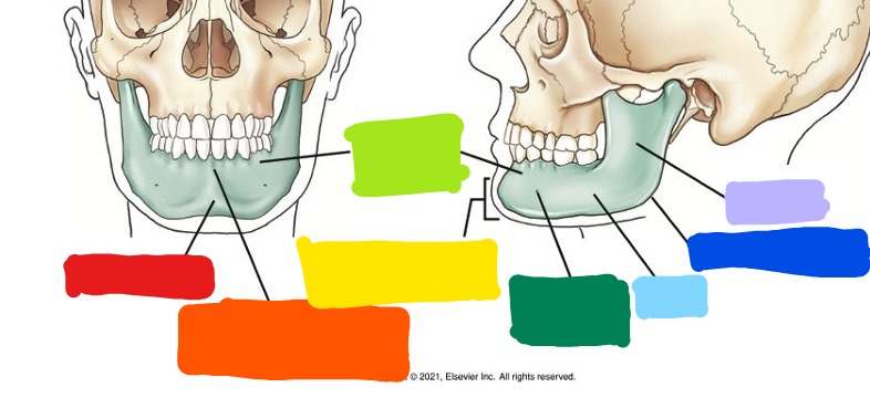
What is Purple?
Ramus

What is Red?
Coronoid process

What is Orange?
mandibular notch

What is Yell9ow?
Condyloid process
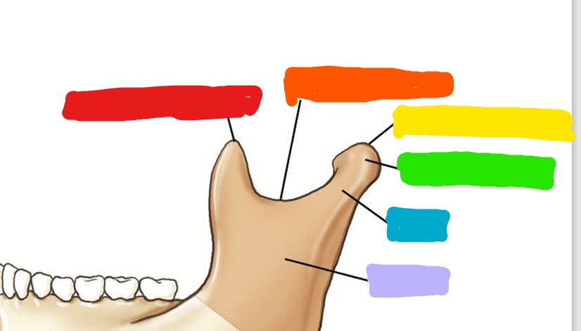
What is Green?
Condyle or head

What is Blue?
Neck

What is Purple?
Ramus

What is Red?
condyle or head

What is Orange?
Coronoid process
What is Yellow?
Mentum

What is Green?
Body

What is Blue?
Ramus

What is Red?
Temporal bone

What is Orange?
Capsule
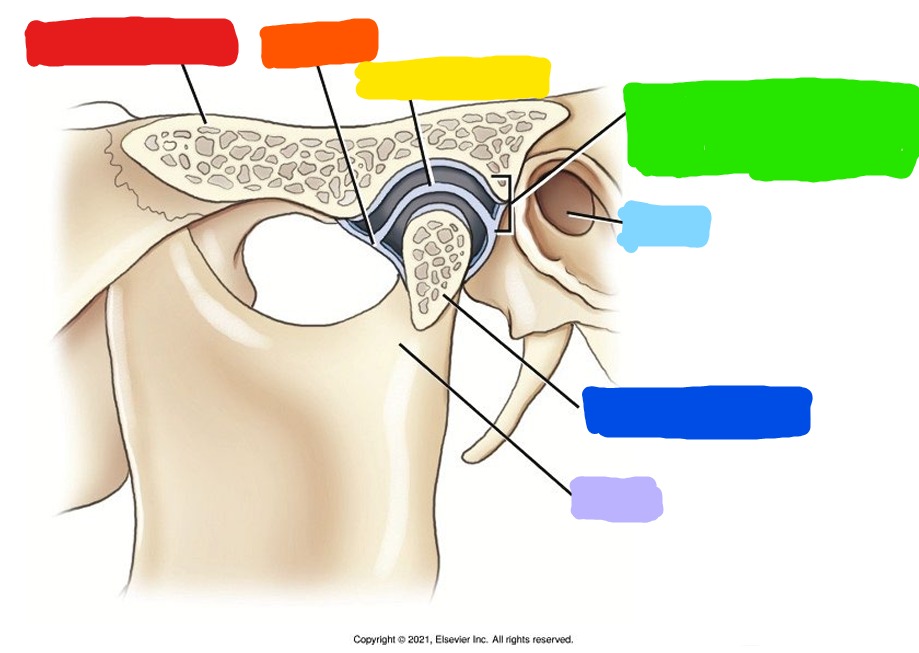
What is Yellow?
articular disk

What is Green?
temporomandibular fossa

What is Light Blue?
EAM

What is Dark Blue?
Condyle or head

What is Purple?
Neck
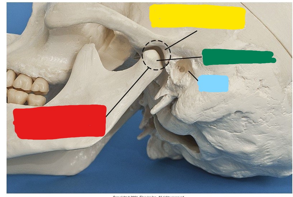
What is Red?
Temporomandibular joint (TMJ)

What is Yellow?
Temporomandibular fossa

What is Green?
Condyle or head

What is Blue?
EAM
To image the mandibular Ramus - what lateral?
True lateral
To have a general mandibular survey - what lateral
10-15 degree rotation
To image the mandibular body - what lateral?
30 degree rotation
To image the mandibular mentum - what lateral?
45 degree
Lateral Mandible Technique
75-80 @ 10-16
Lateral Mandible CR
side of interest against IR
25 degree cephalic angle or angle head 25 degree towards the IR
Exit mandible of interest
PA or PA Axial Mandible Technique
75-80 @ 10-16
PA Mandible CR
horizontal, center to junction of lips
PA or PA Axial Mandible Part Positioning
forehead on IR
OML perpendicular to IR
MSP perpendicular to IR
PA Axial Mandible CR
exit at acanthion, angled 20-25 degree cephalic
Anatomy visualized in PA Mandible
mandibular rami and lateral portion of the body
Anatomy visualized in PA Axial Mandible
TMJ region and heads of condyles visible through mastoid processes, condyloid processes well visualized
AP Axial (Towne) Mandible Technique
75-80 @ 10-16
AP Axial (Towne) Mandible CR
OML used - 35 degree caudal
IOML used - 42 degree caudal
Centered 1 in superior to glabella
AP Axial (Towne) Mandible Anatomy Visualized
condyloid process of mandible and TM fossae
Panorex is also called
Orthopantomogram
Panorex positioning
IOML parallel to floor
bite block between patient teeth
tongue to roof of mouth
What is the only moveable joint in the skull
TMJ
Classify TMJ
Synovial, Diarthrodial, Hinge-type and gliding type
What does the condyle do as you open your mouth
condyle moves forward
AP Axial (Towne) TMJ Technique
75-80 @ 10-16
AP Axial (Towne) TMJ CR
OML used - 35 degree caudal
IOML used - 42 degree caudal
Center 3 inches superior to nasion
Axiolateral TMJ - Schuller Method Technique
75-80 @ 10-16
Axiolateral TMJ - Schuller Method CR
center to projected TMJ
25-30 degree caudal angle
Axiolateral TMJ - Schuller Method Positioning
Erect or semi prone - head in true lateral
IPL perpendicular to IR
IOML perpendicular to front edge of IR
Closed and open mouth
Axiolateral Oblique TMJ - Modified Law Method Technique
75-80 @ 10-16
Axiolateral Oblique TMJ - Modified Law Method CR
15 degree caudal
centered 1.5 in superior to upside EAM
Axiolateral Oblique TMJ - Modified Law Method Part Positioning
head in lateral position, side of interest closest to IR
Rotate face 15 degree towards the IR
IPL perpendicular to IR
MSP parallel to IR
IOML perpendicular to front edge of IR