Looks like no one added any tags here yet for you.
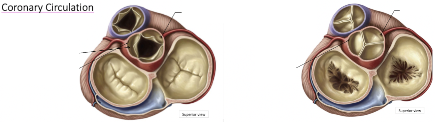
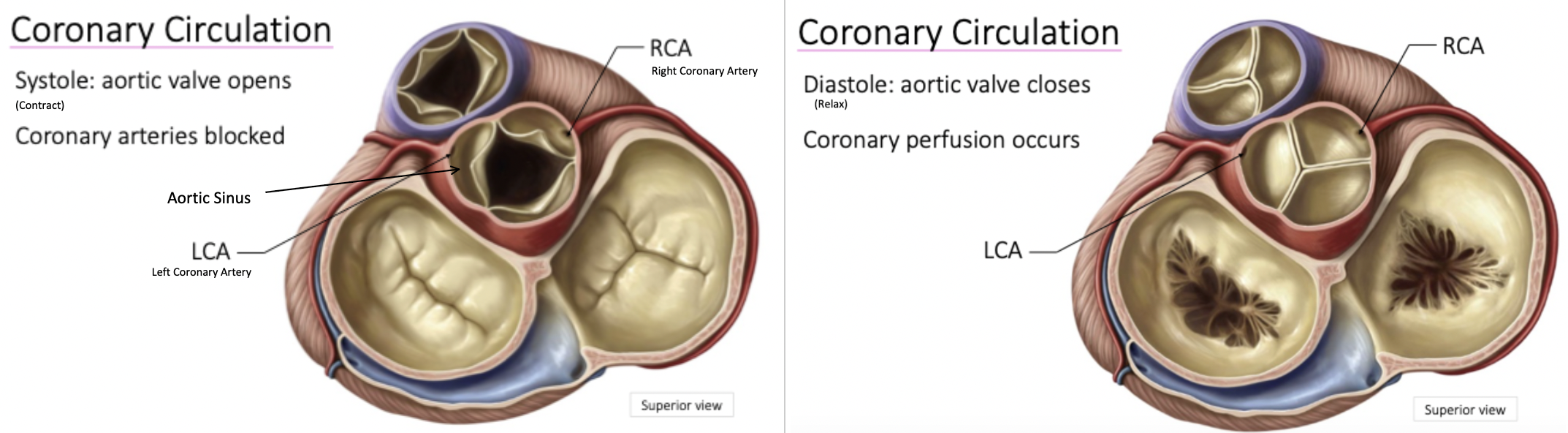
Aortic Valve activity: systole vs diastole
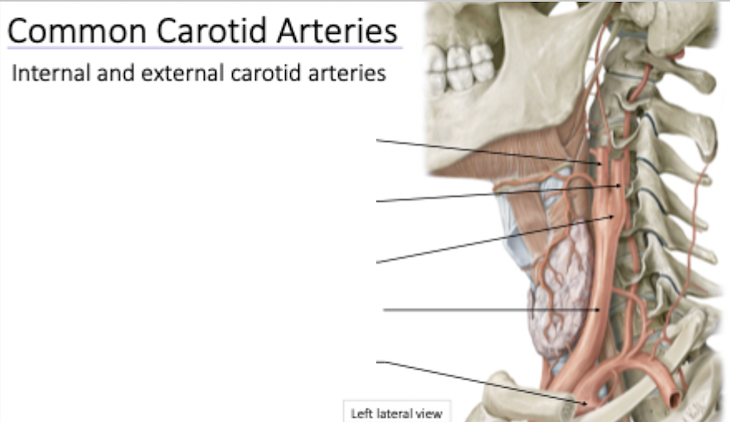
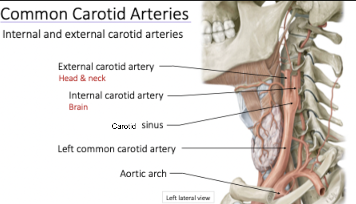
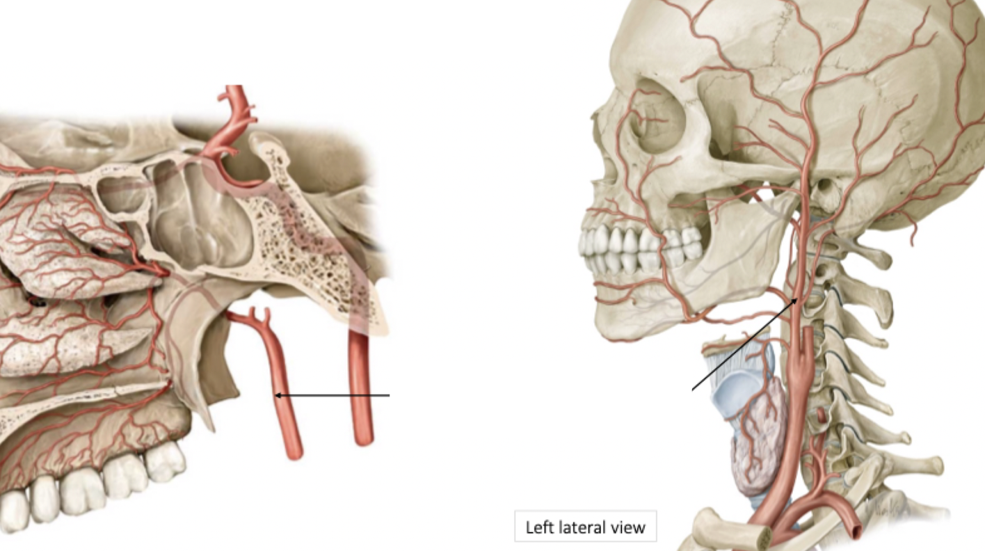
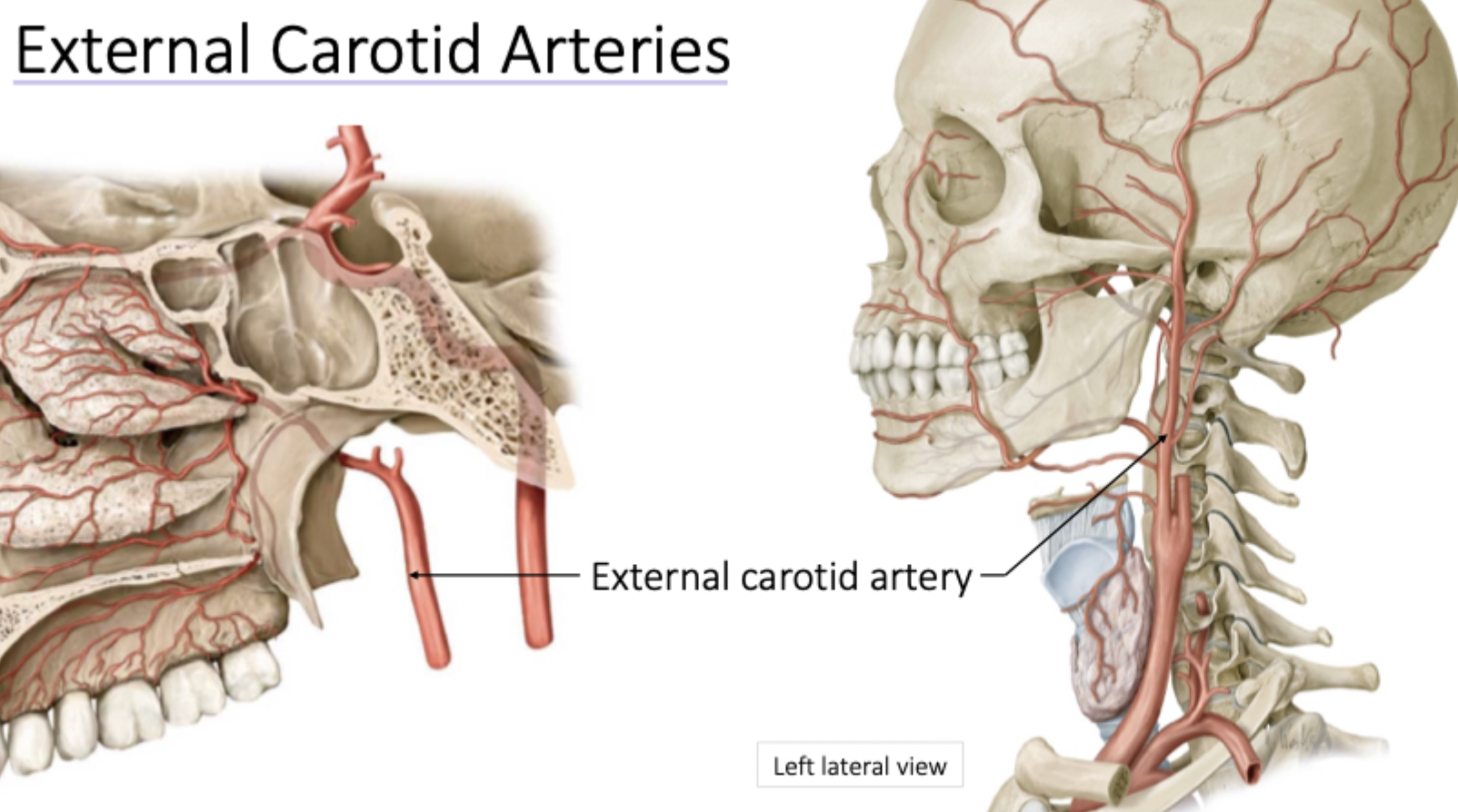
Common carotid bifurcates at carotid sinus
Travels upward and gives off several branches to head and neck
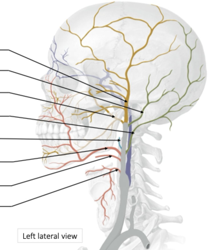
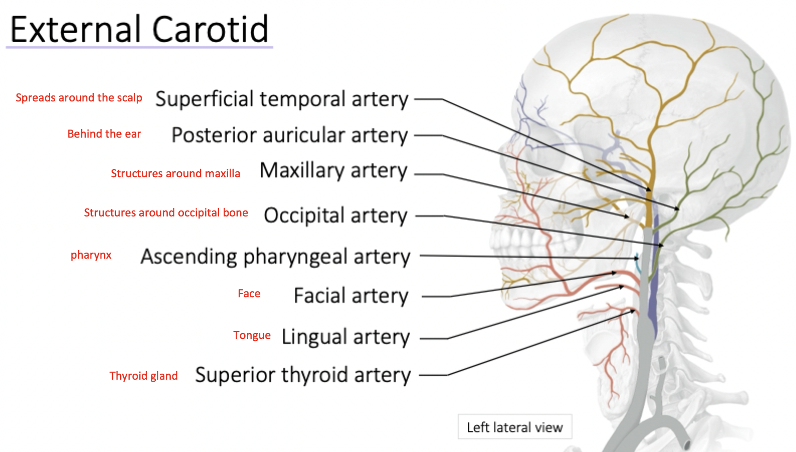
External Carotid Artery Branches
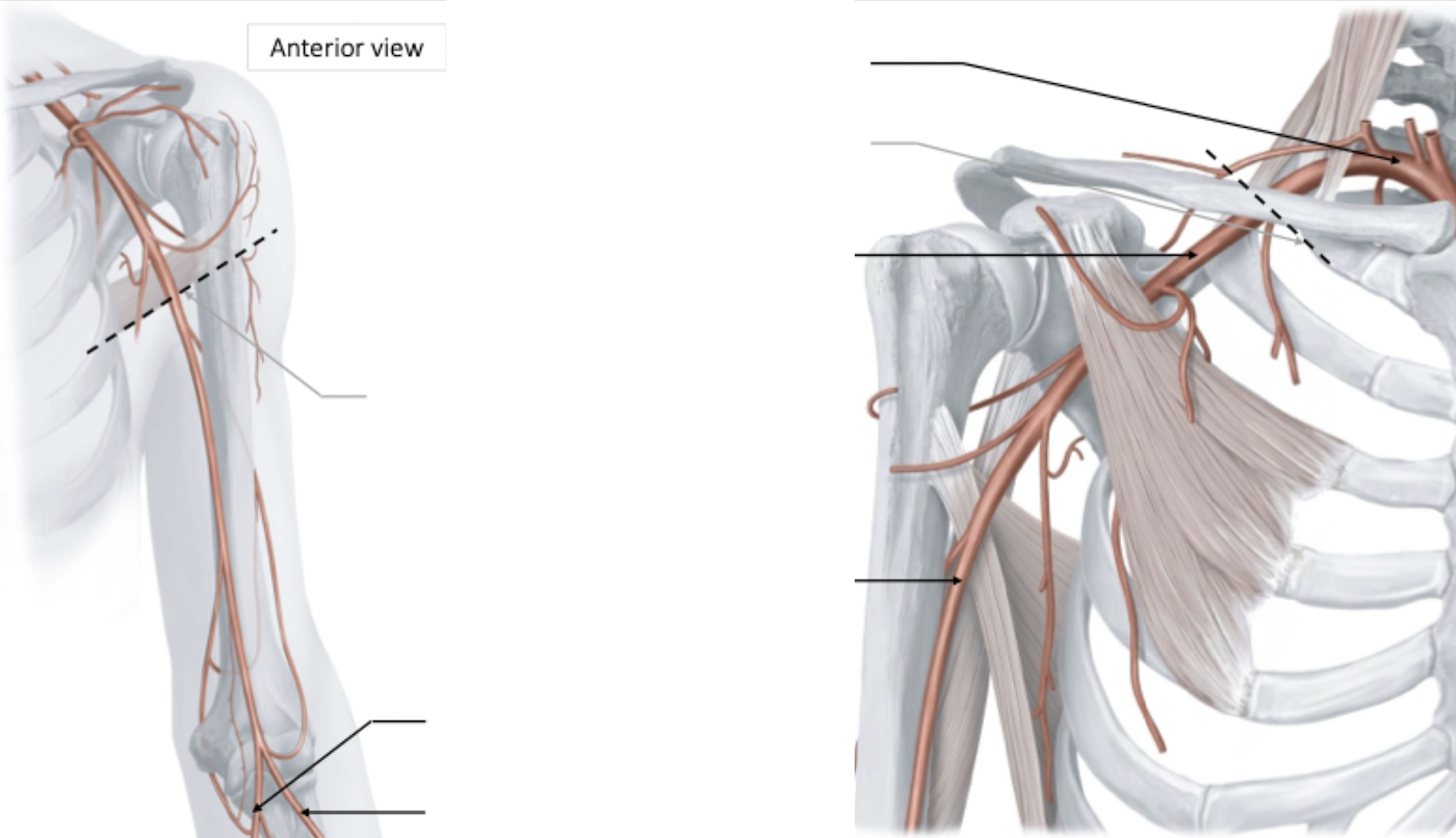
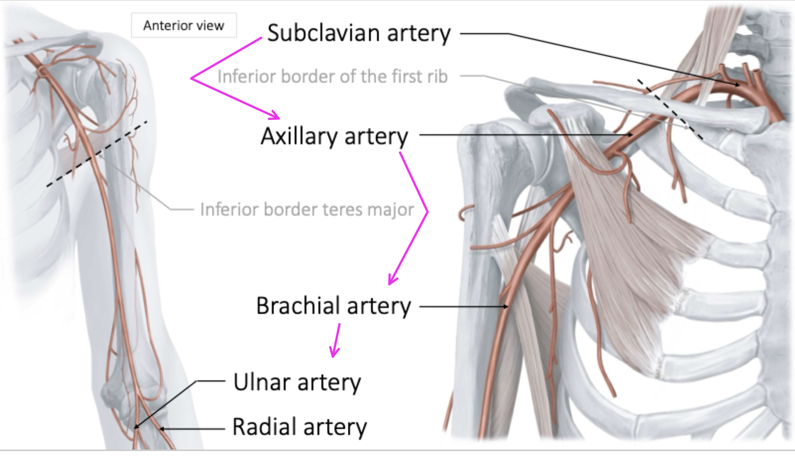
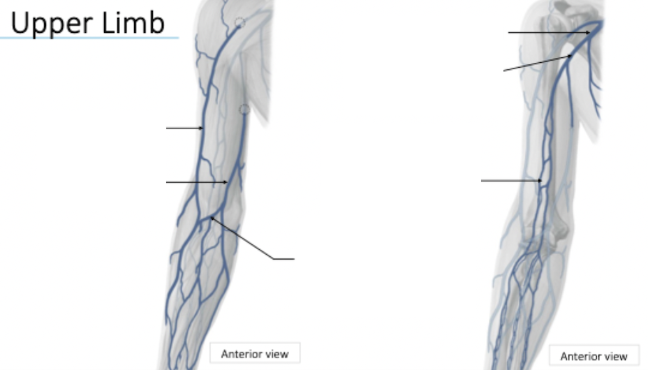
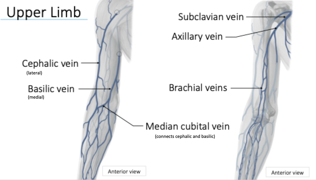
Upper limb blood supply
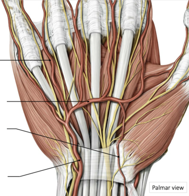
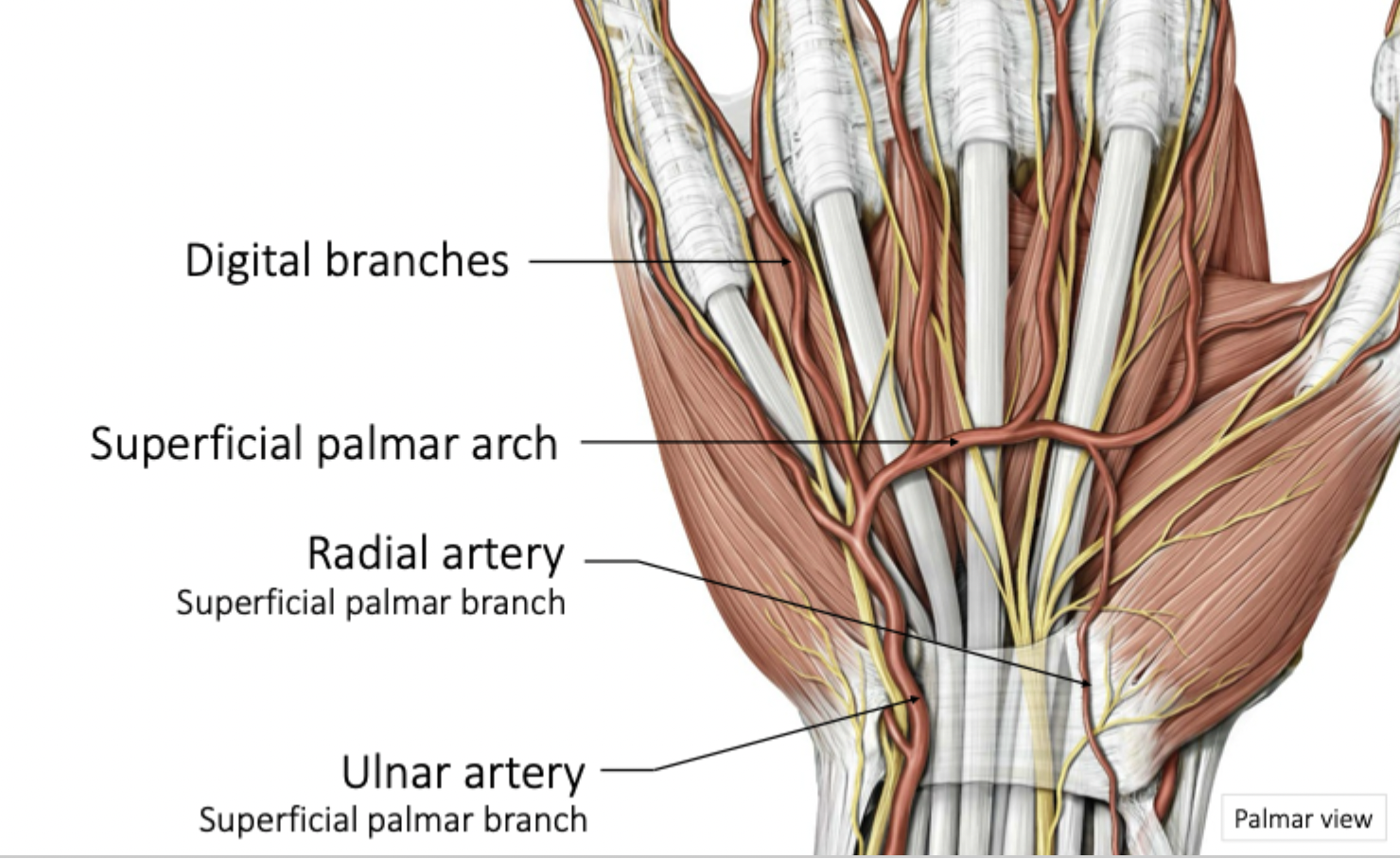
Hand blood supply
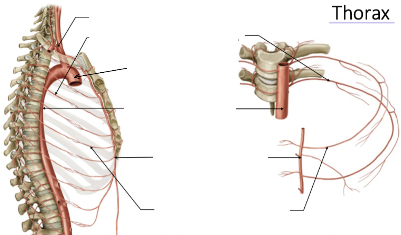
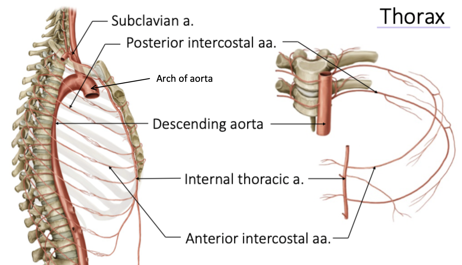
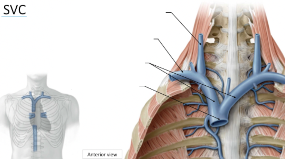
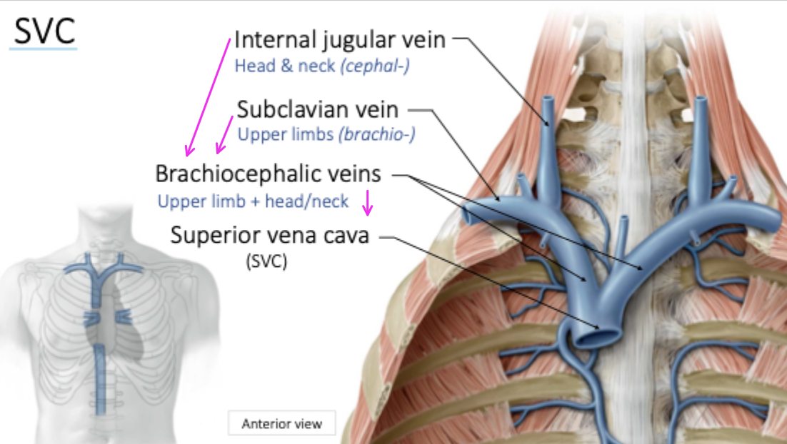
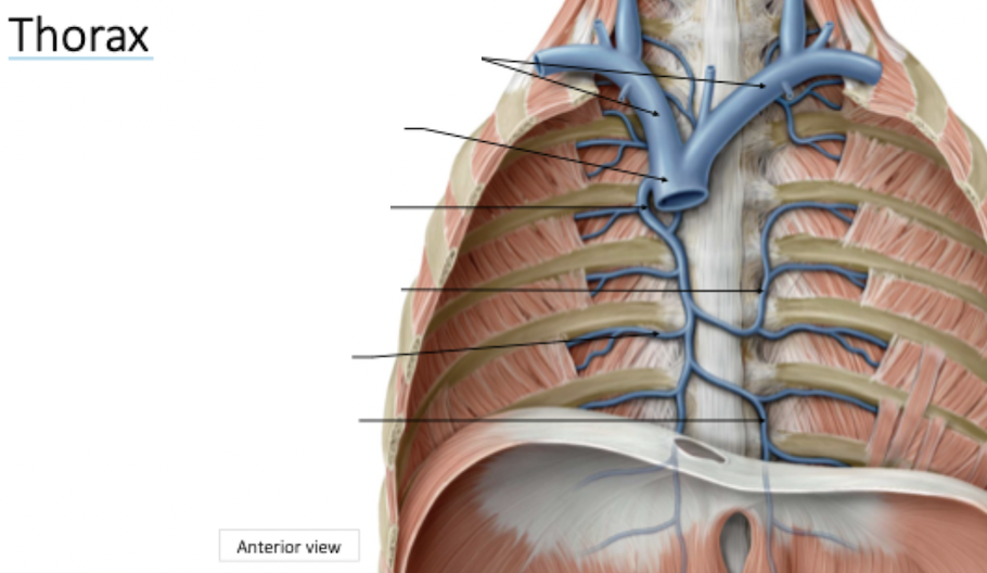
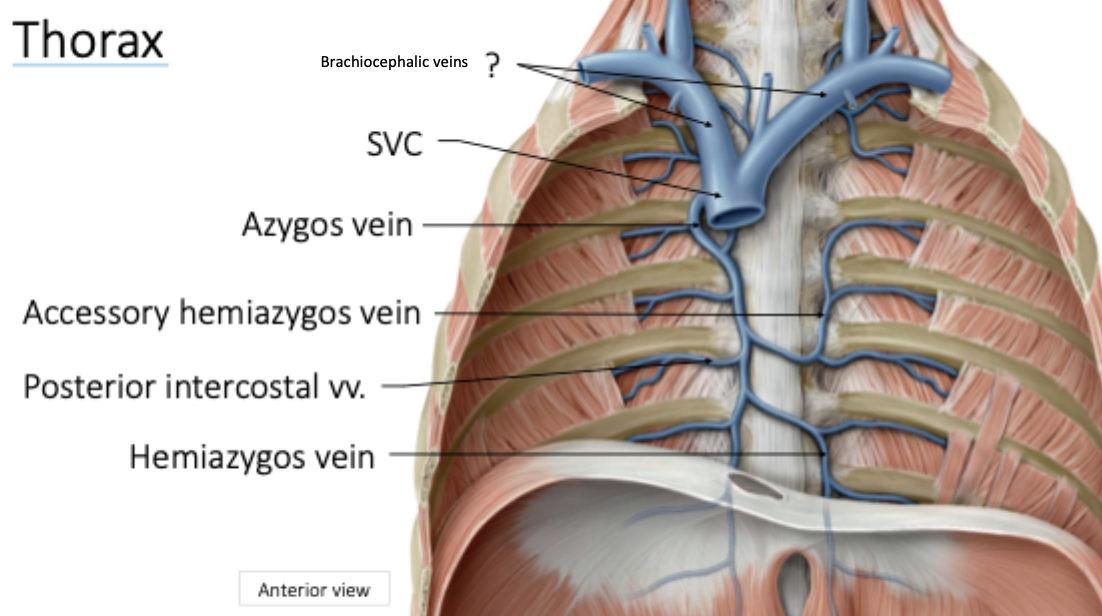
Thorax blood supply
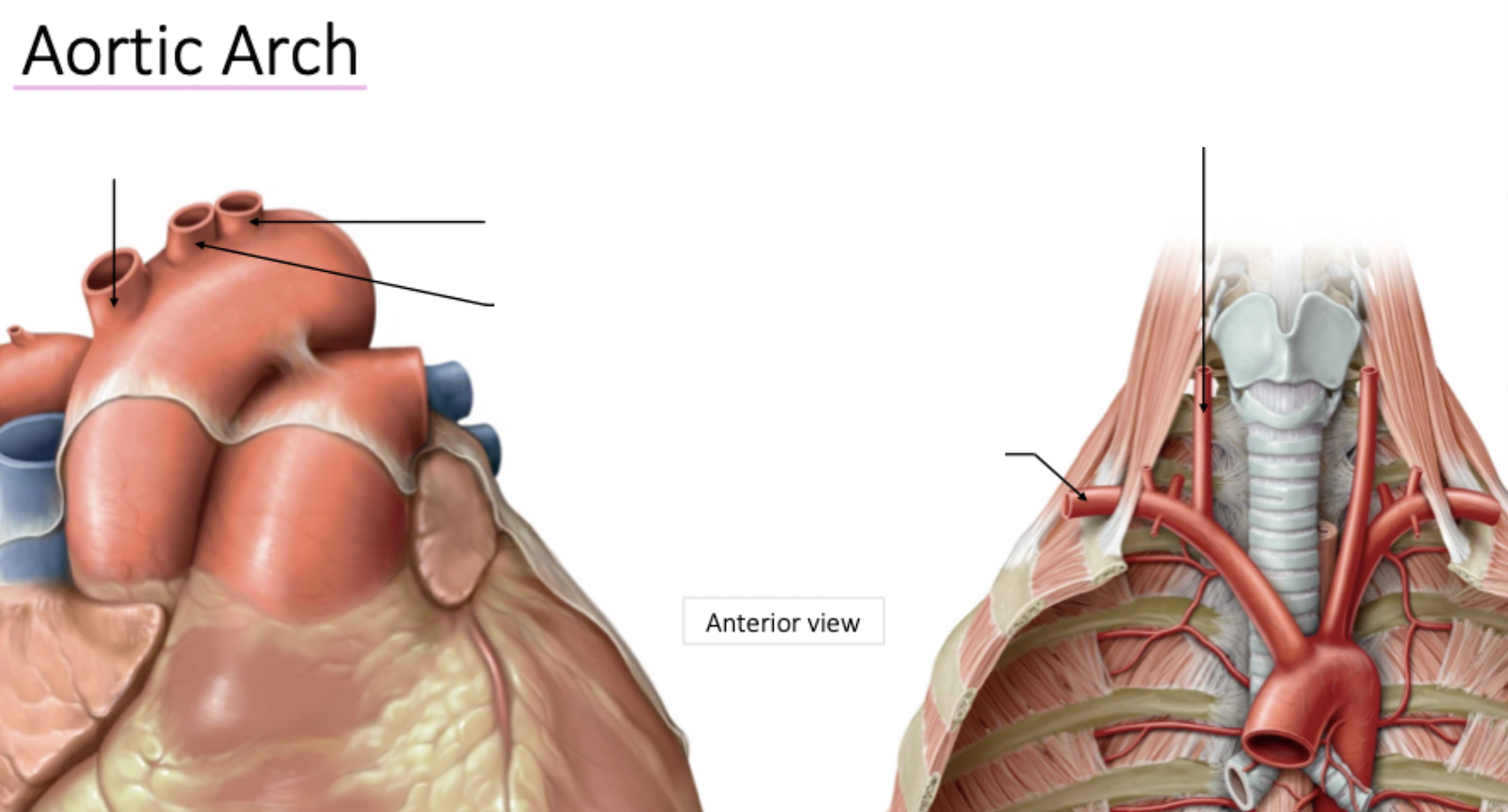
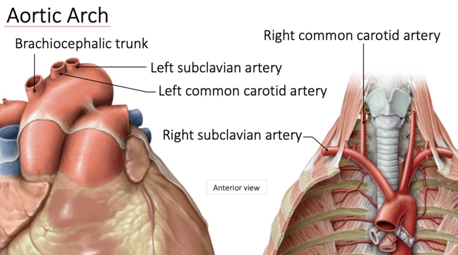
Arch of Aorta → Subclavian → Internal Thoracic → Anterior Intercostal
Arch of Aorta → Descending Aorta → Posterior Intercostal
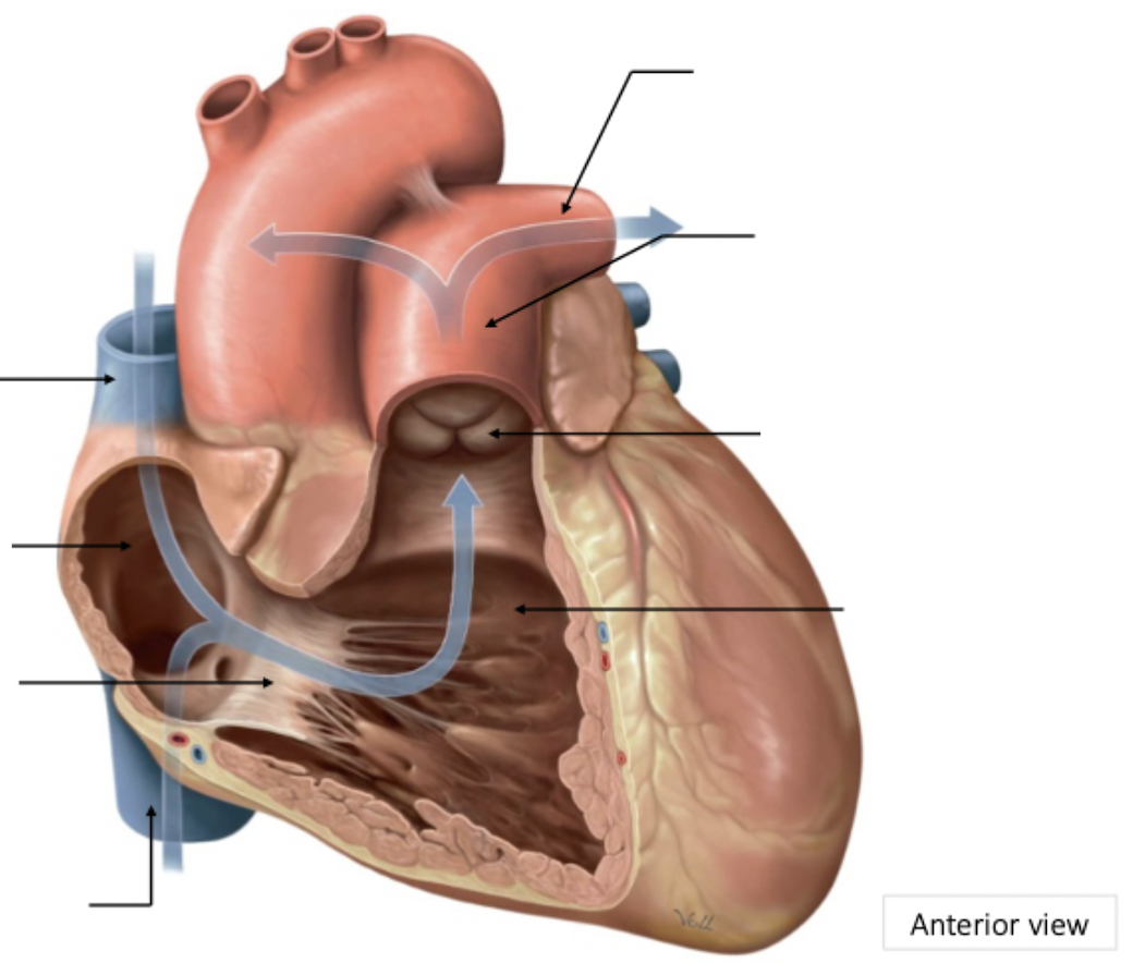
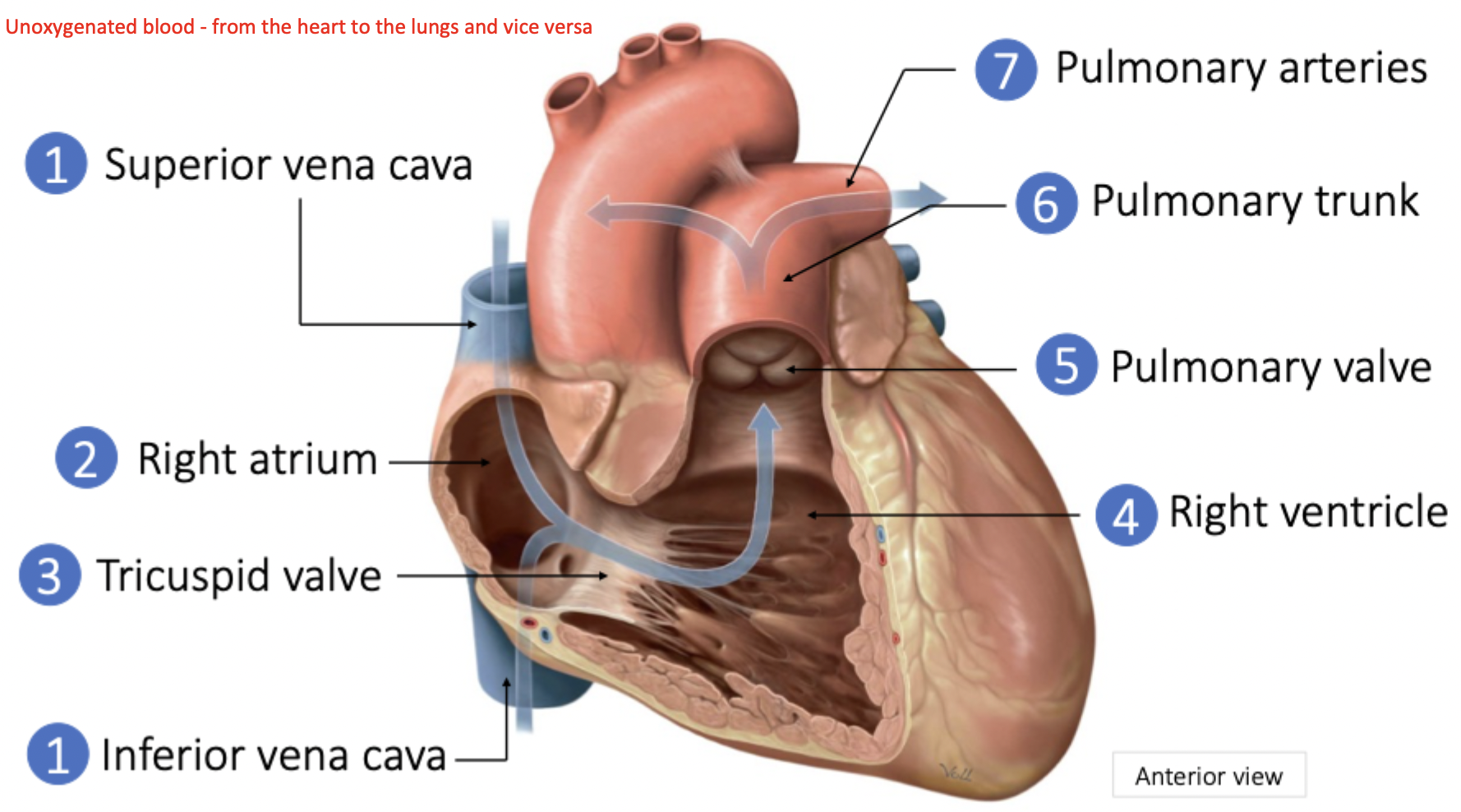
Pulmonary Circulation
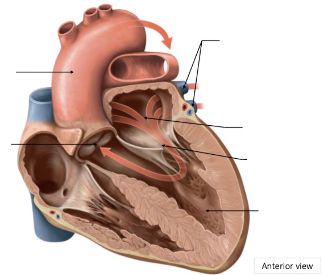
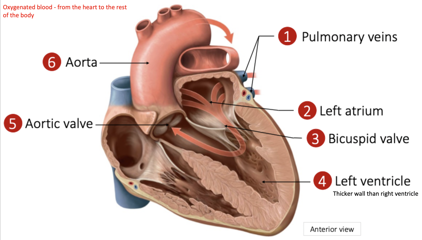
Systemic Circulation
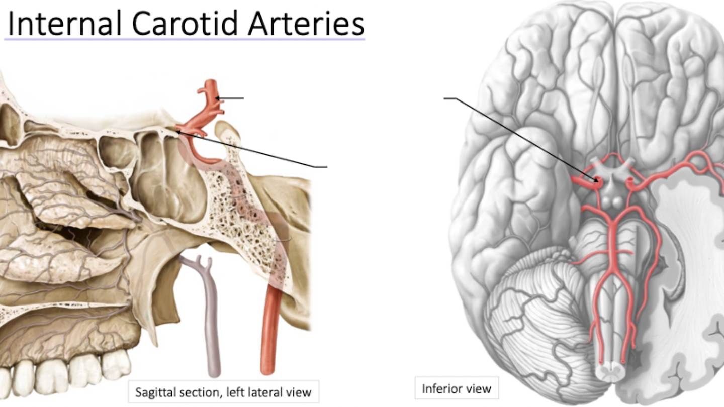
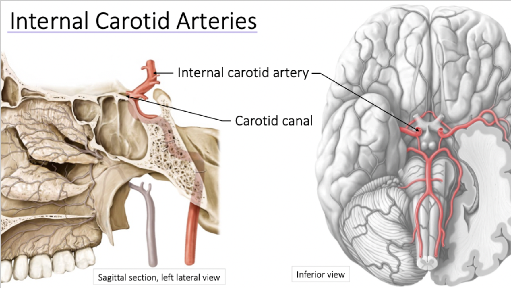
Contributes to circle of Willis
Passes through carotid canal
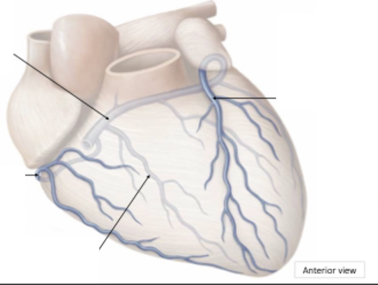
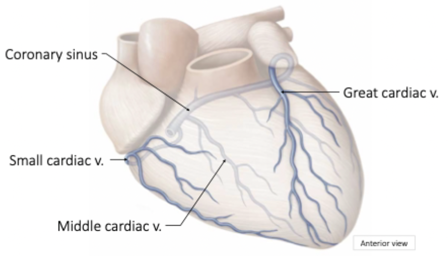
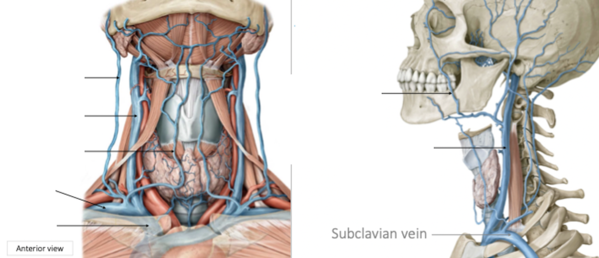
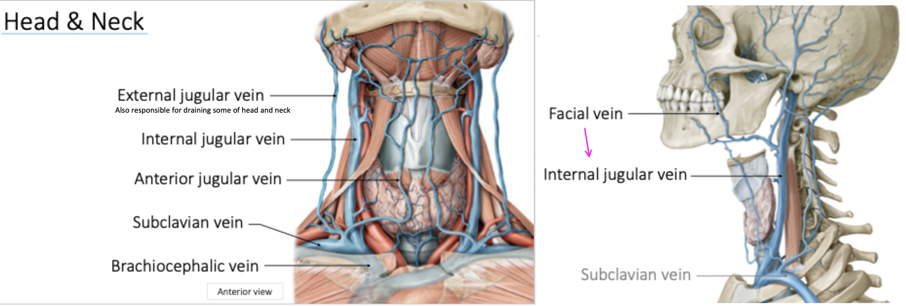
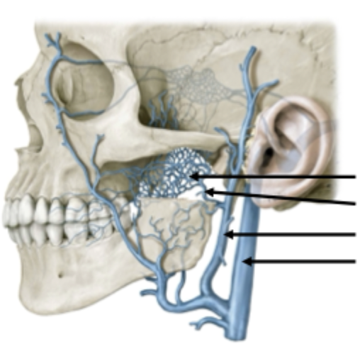
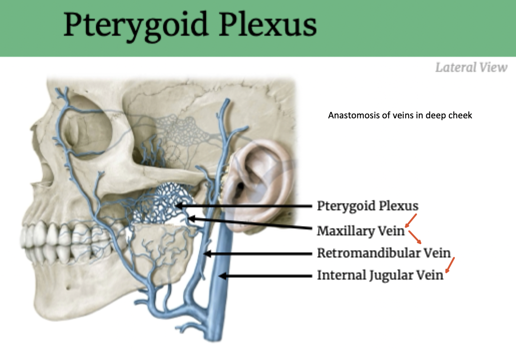
Head and Neck Venous Drainage
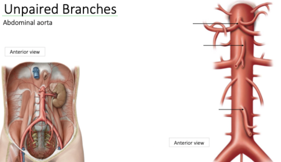
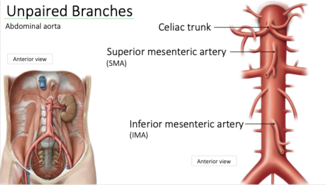
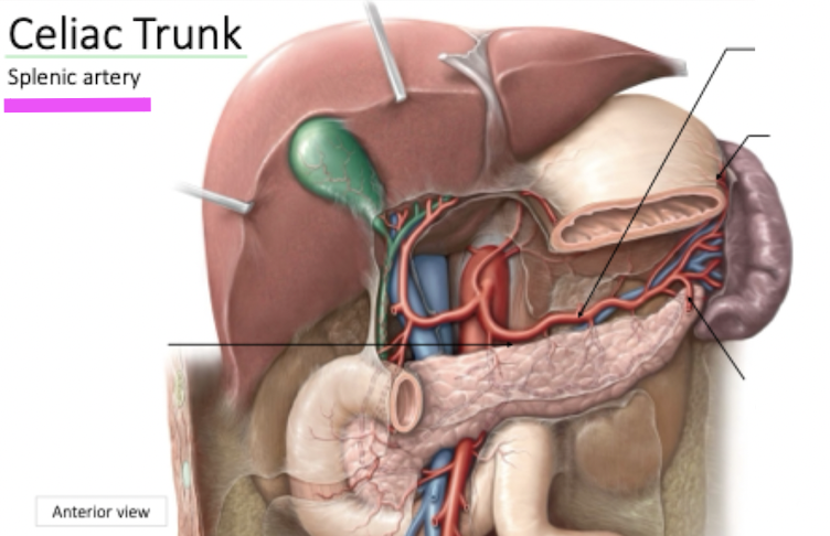
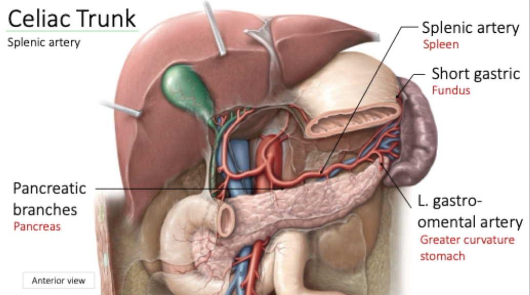
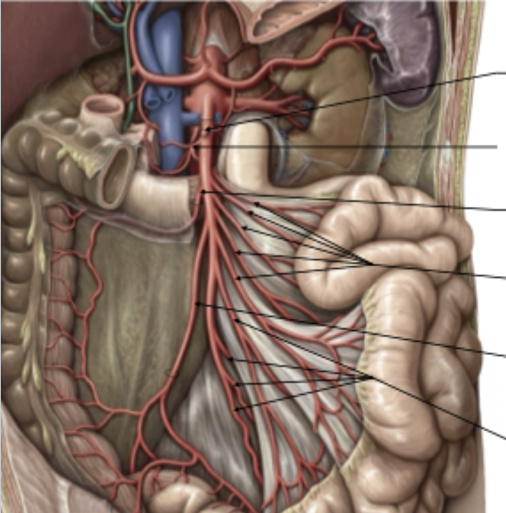
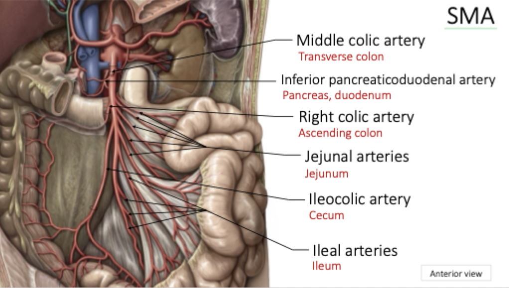
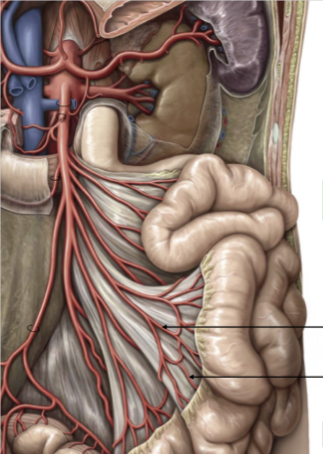
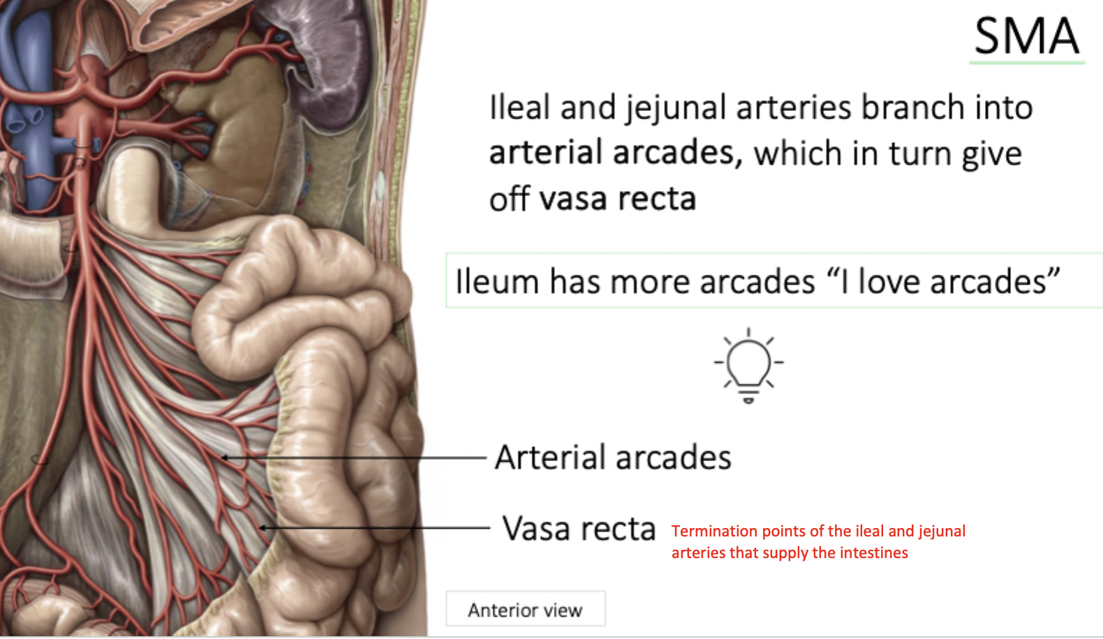
Superior Mesenteric Artery
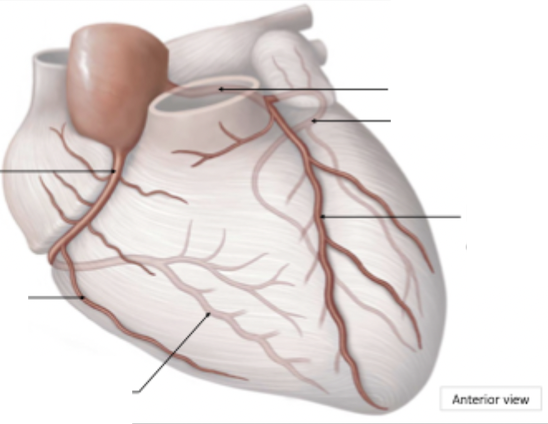
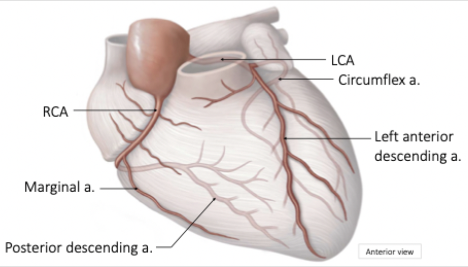
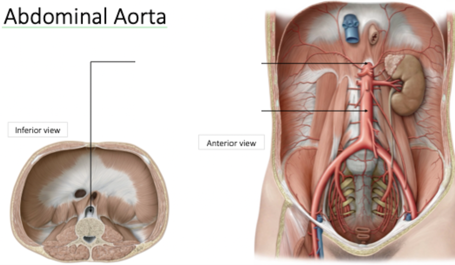
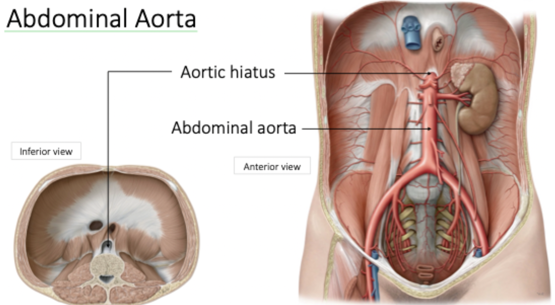
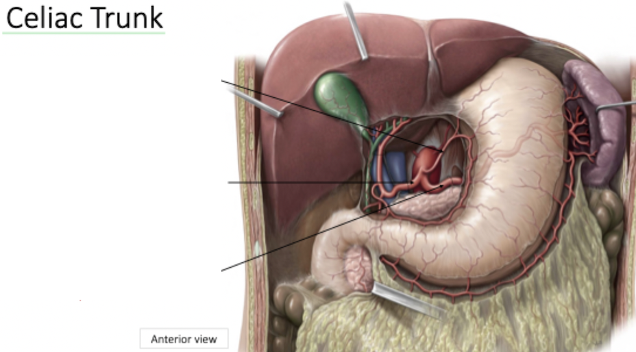
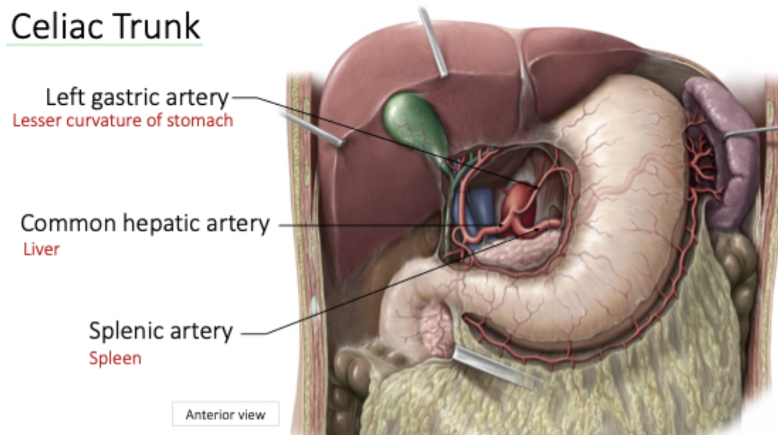
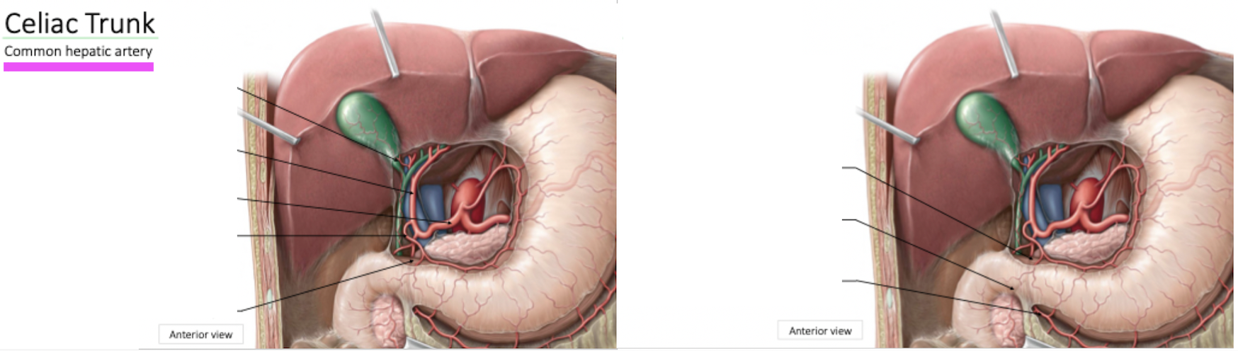
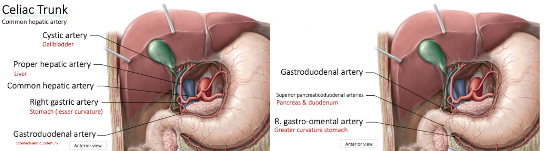
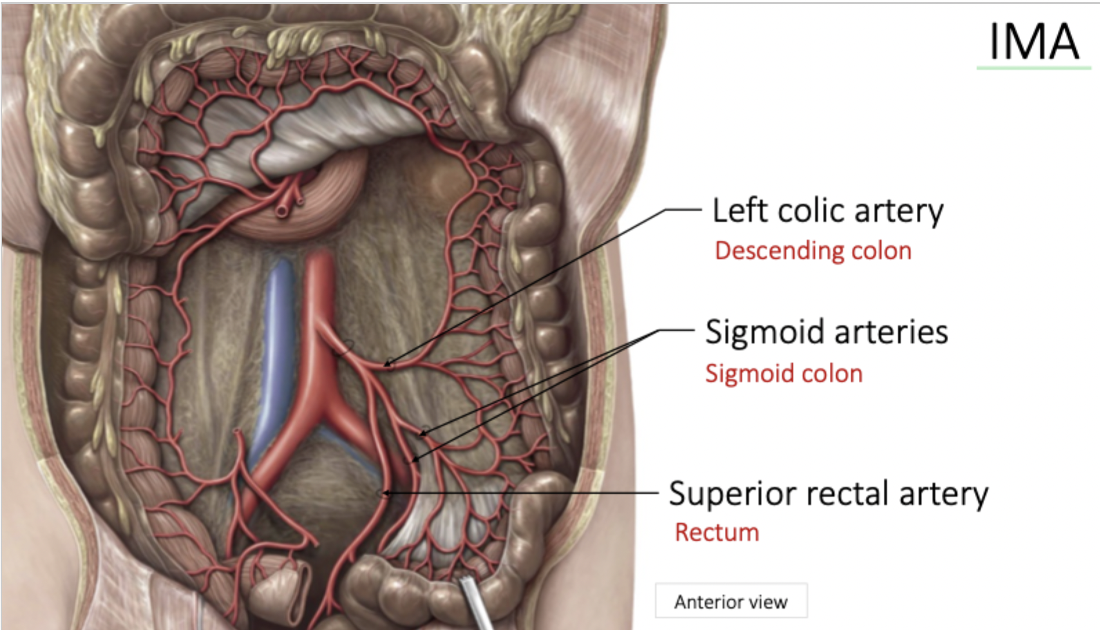
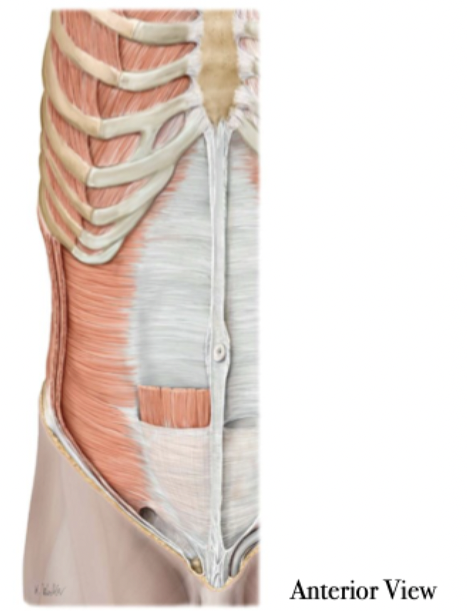
Anterior view
SMA: Ileal and Jejunal Arteries continued
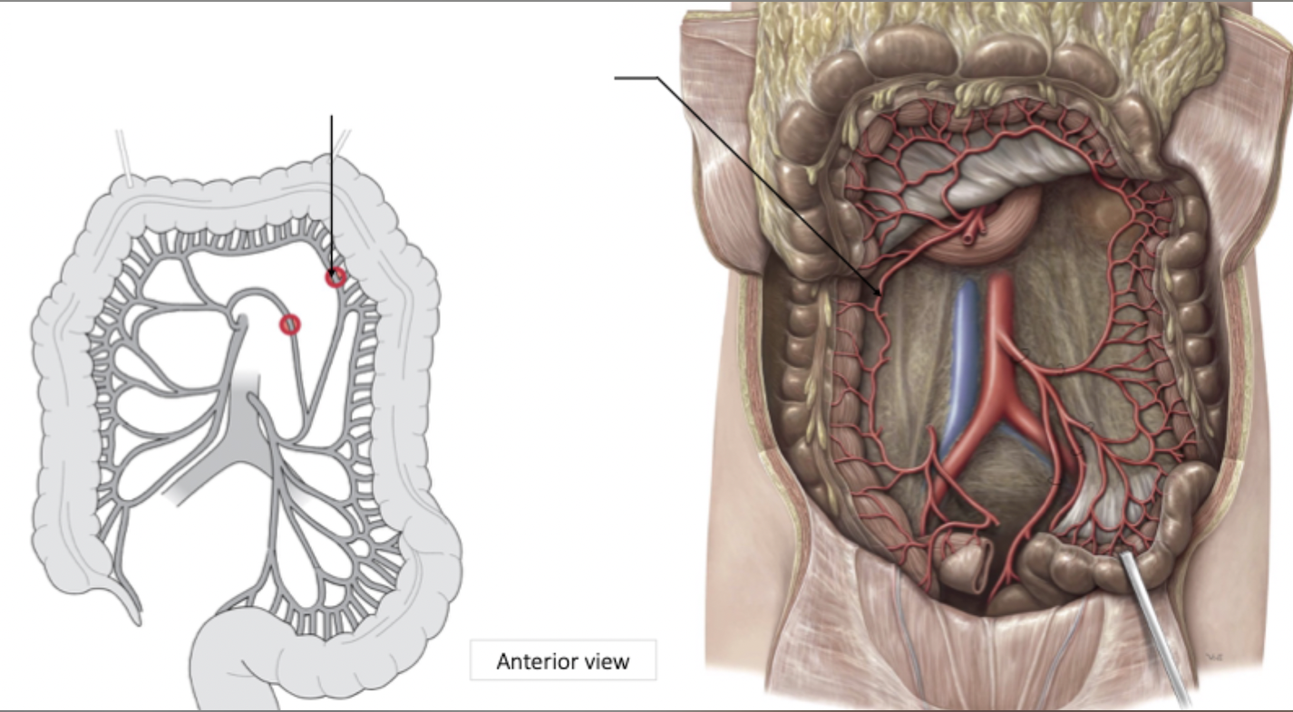
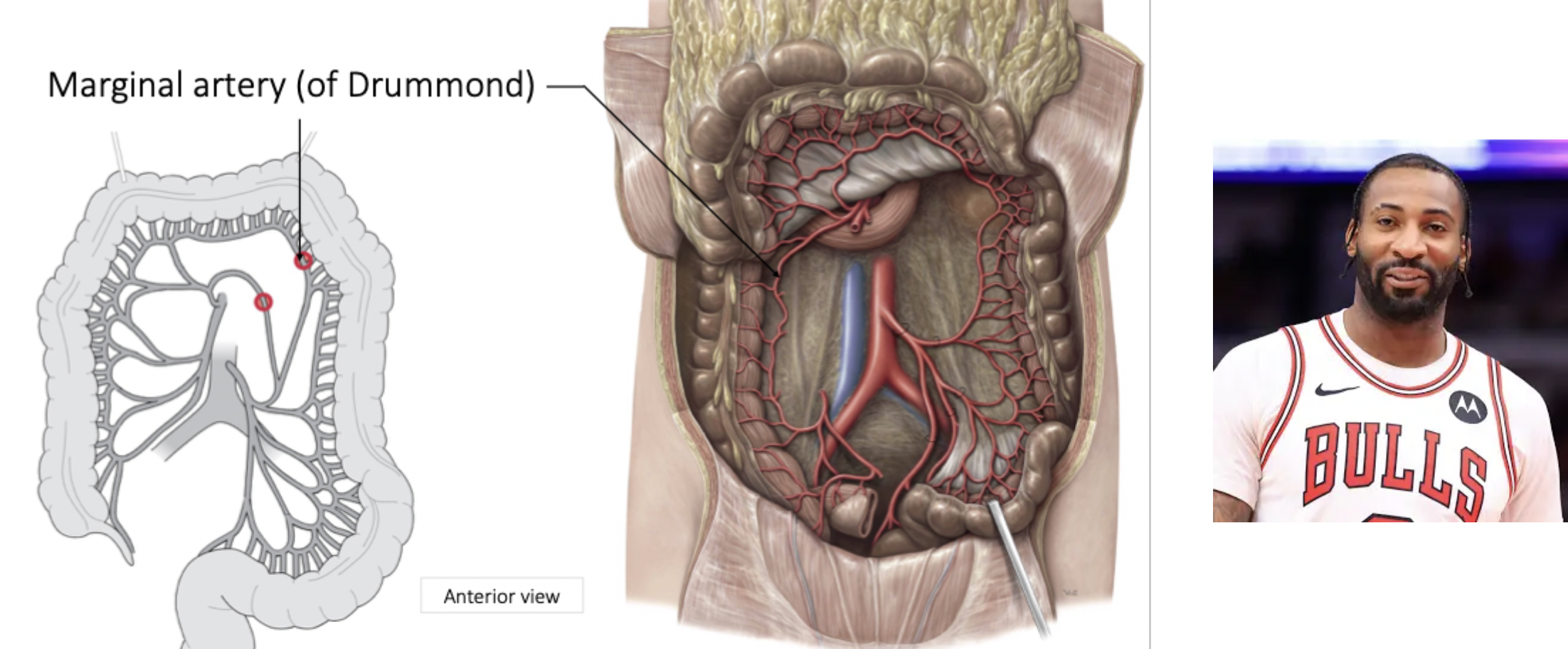
Inferior Mesenteric Artery
Anastomoses between right, middle and left colic arteries - Creates circumference artery around entire large intestine
Key for collateral circulation or alternative supply (compensatory measure)
A pathogen can turn the marginal artery into a hazard as it can spread across whole area
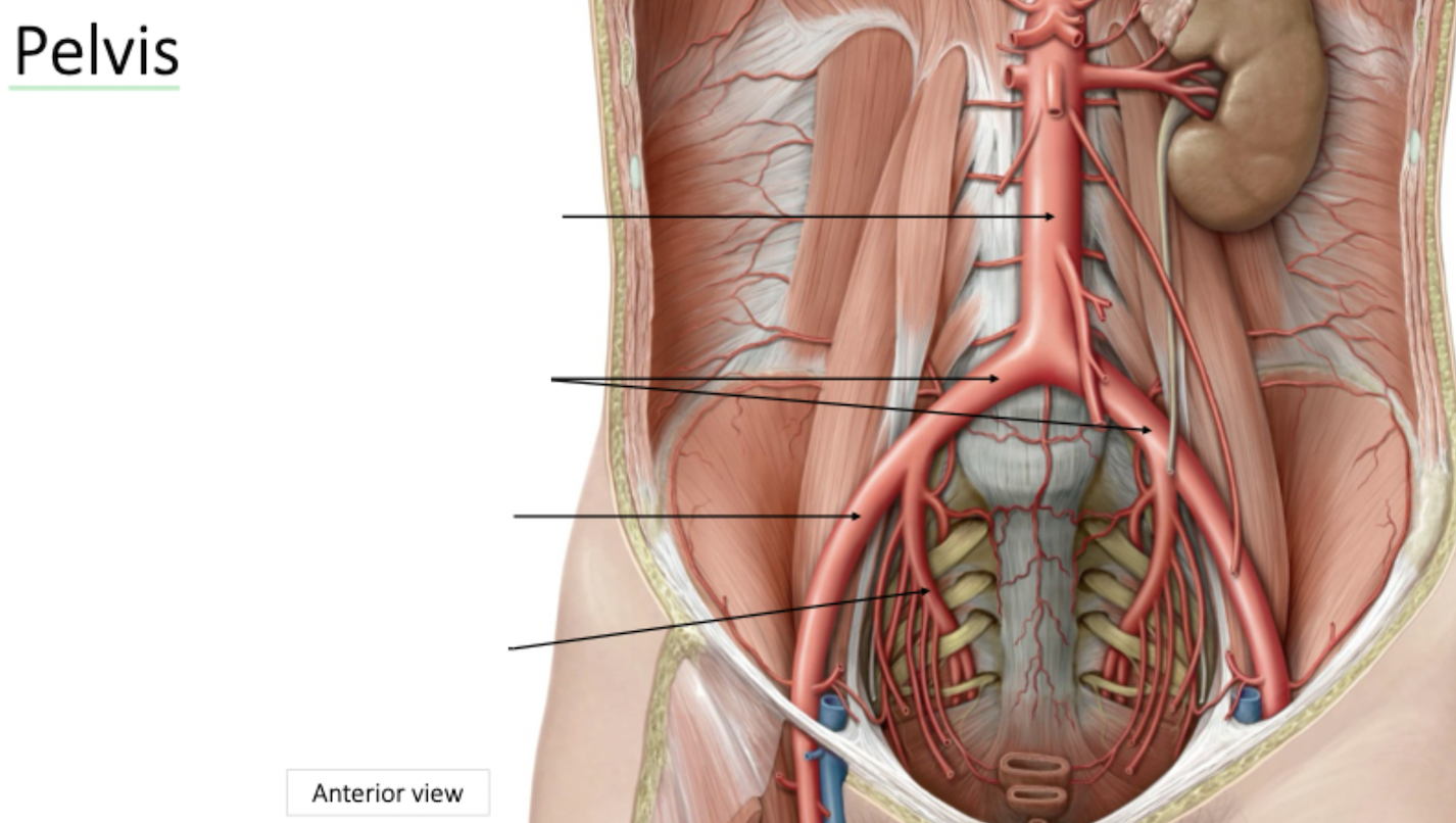
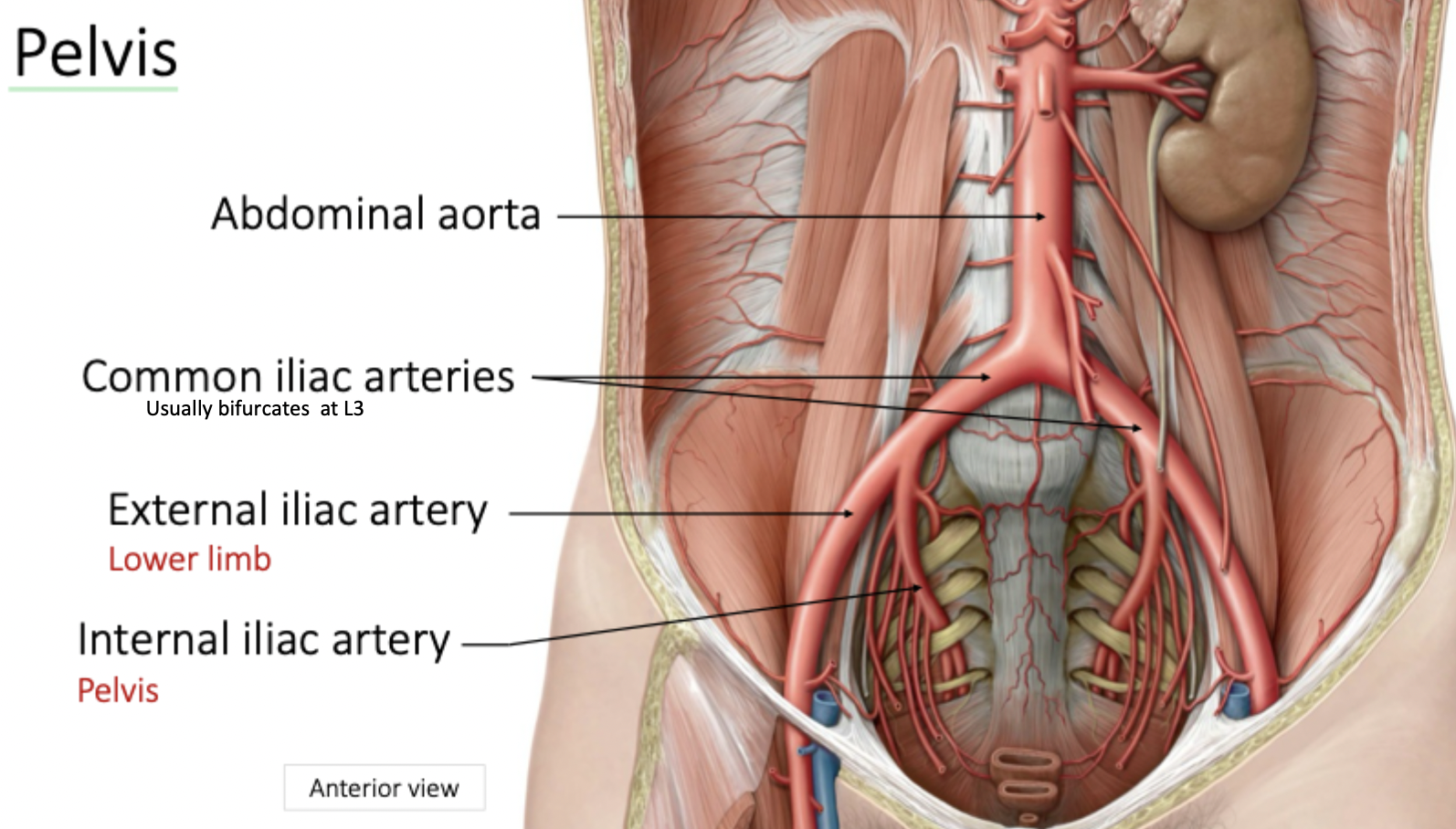
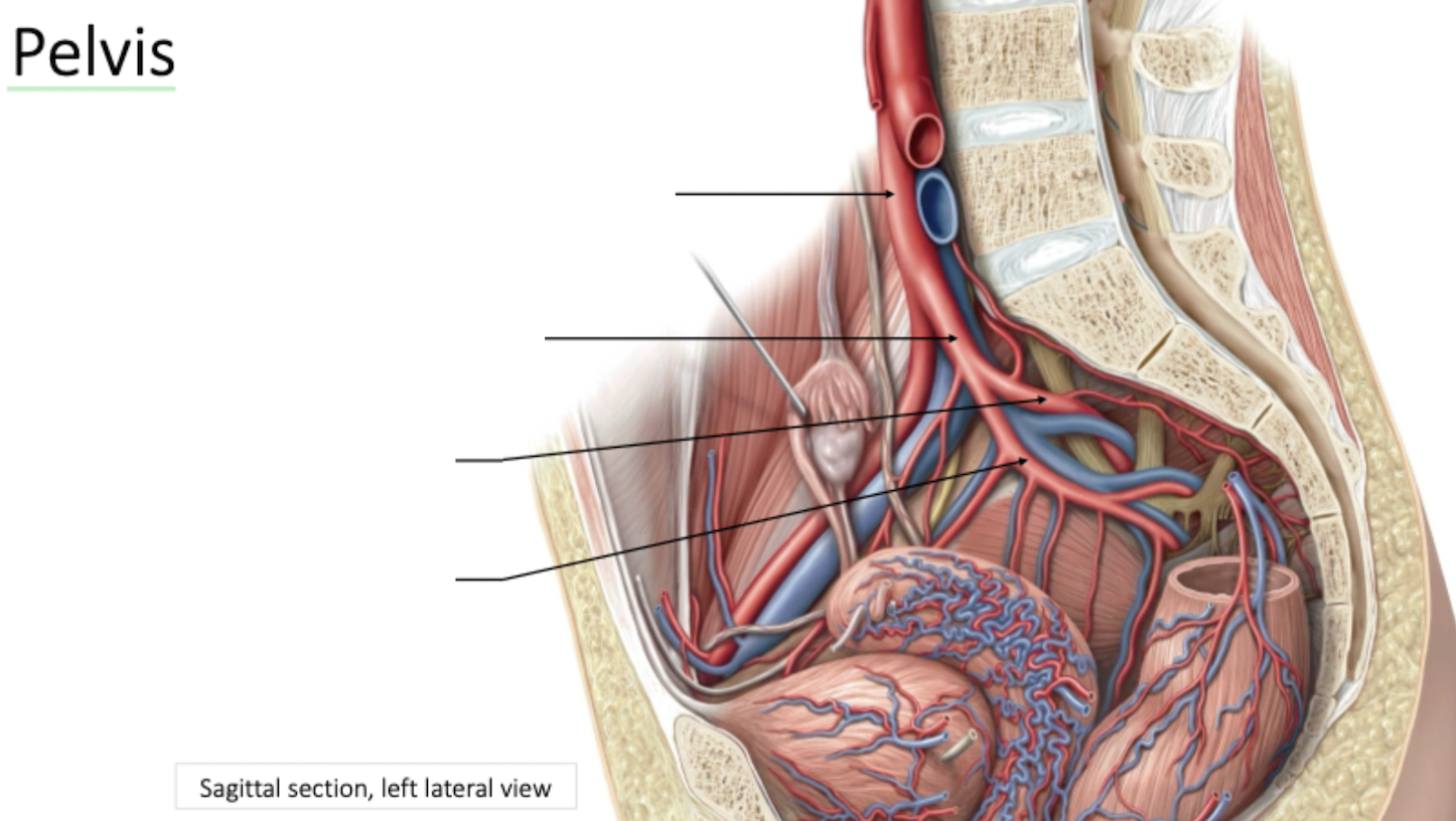
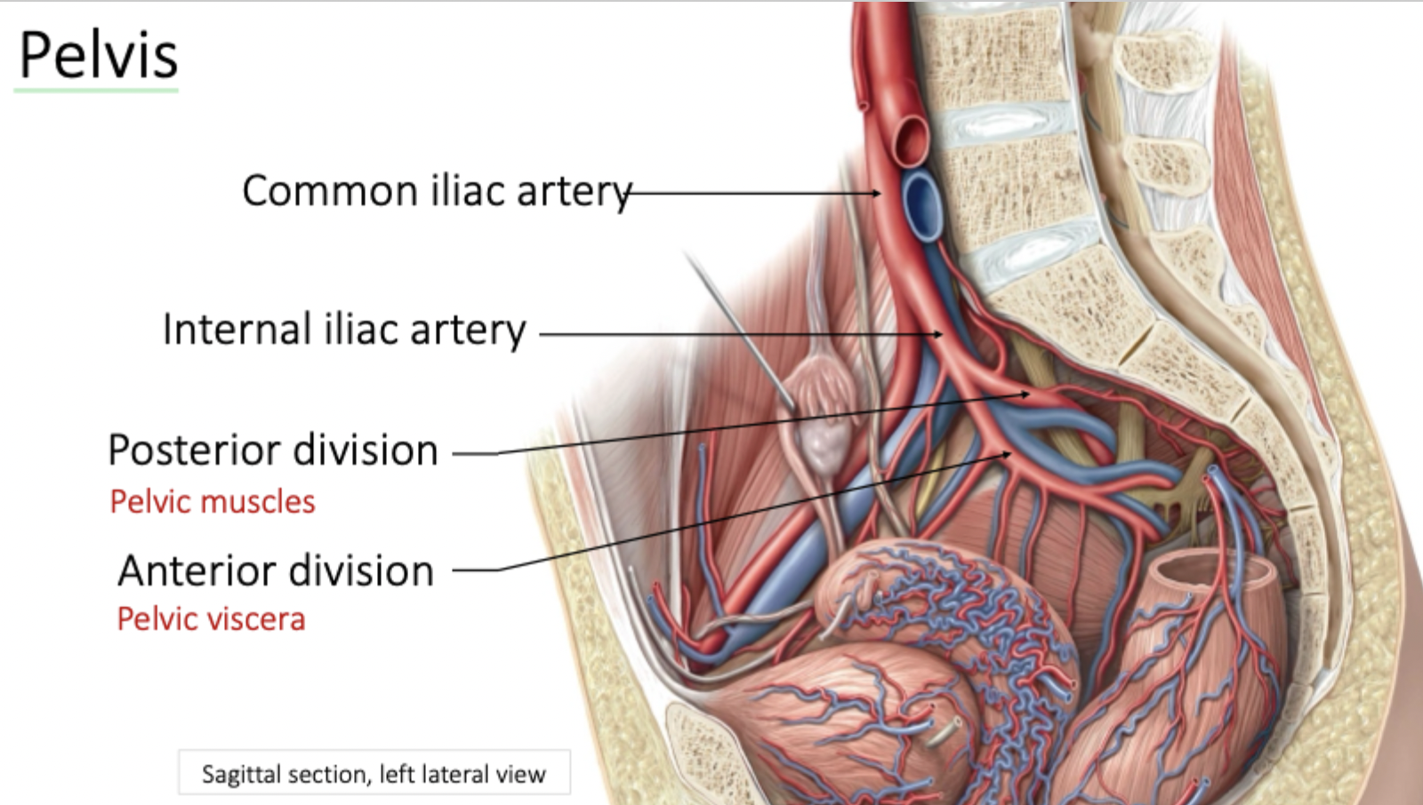
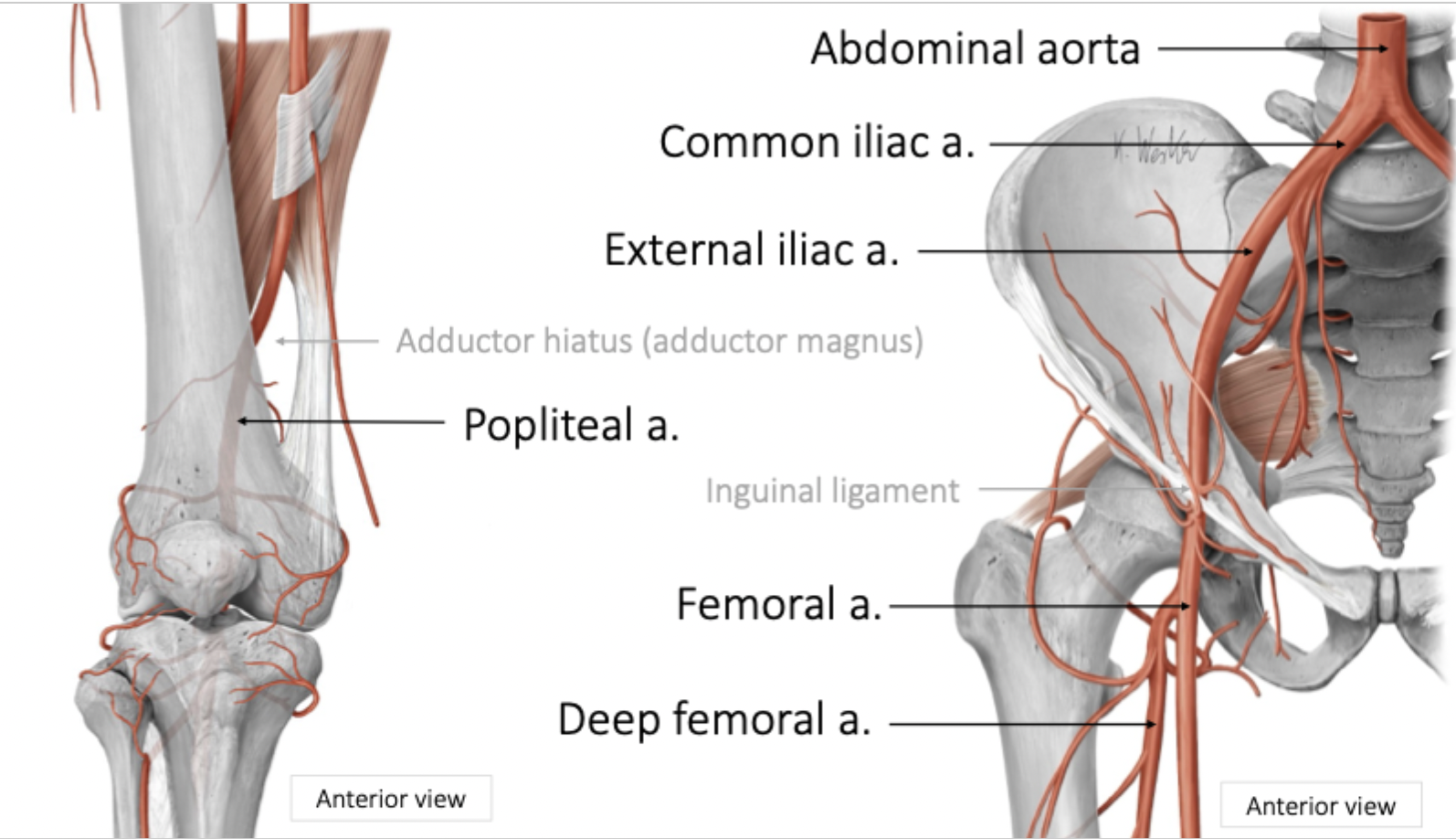
Pelvis Arterial Blood Supply
Pelvis Arterial Blood Supply
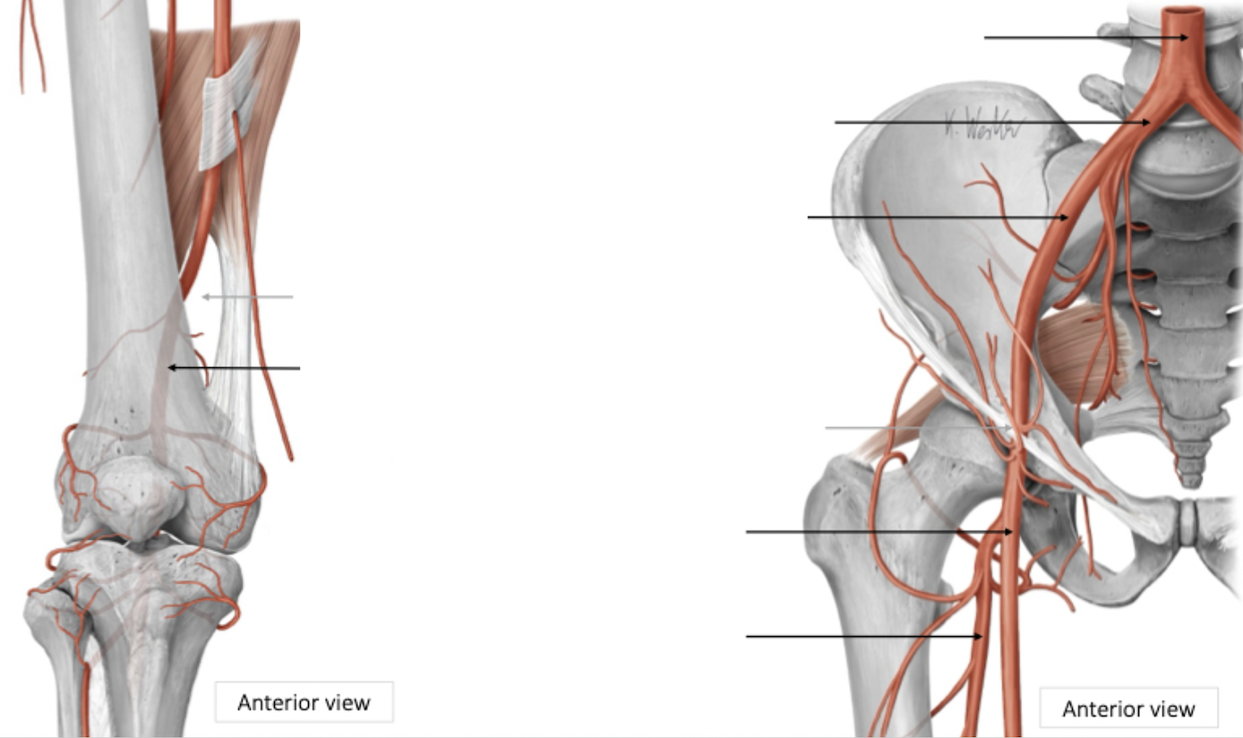
Pelvis → Upper Leg Arterial Blood Supply
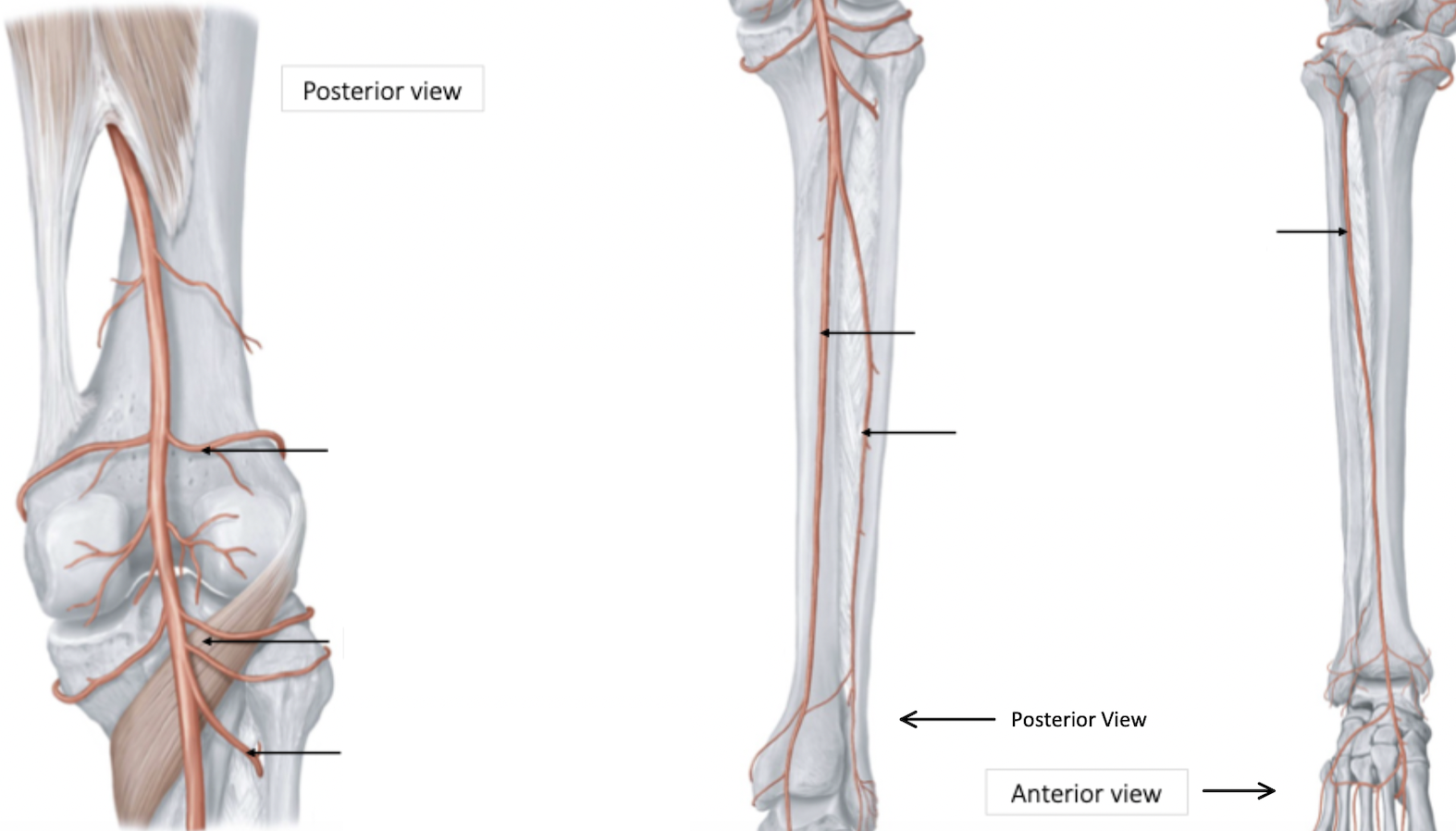
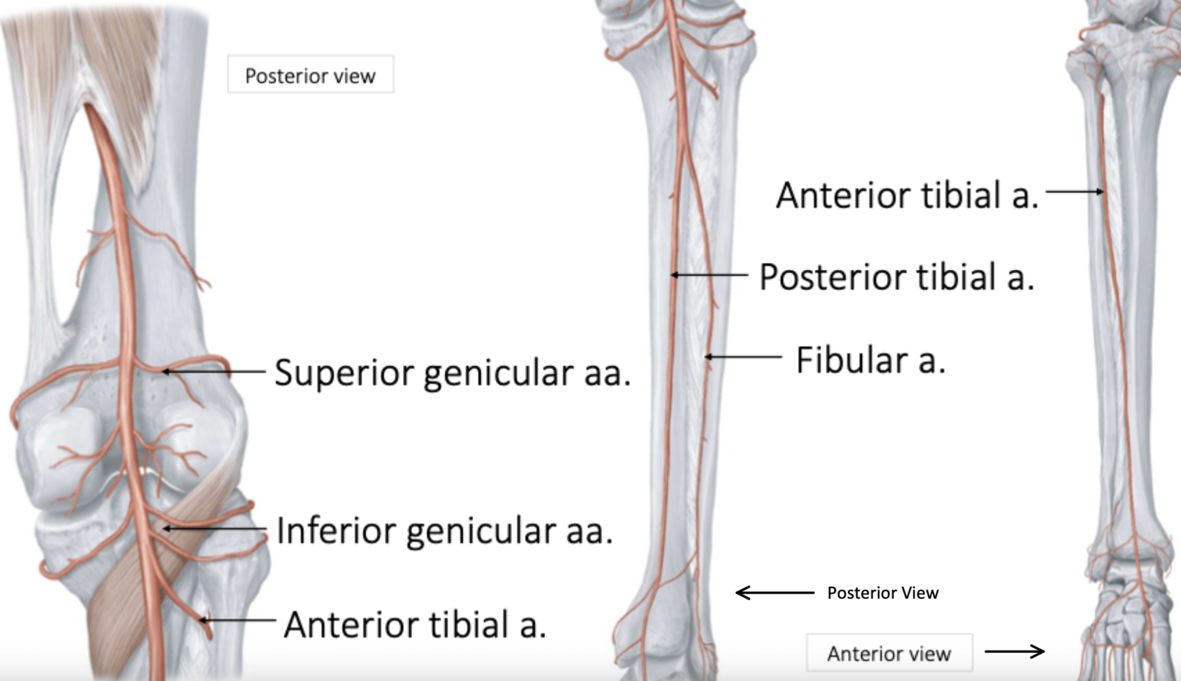
Lower Leg Arterial Blood Supply
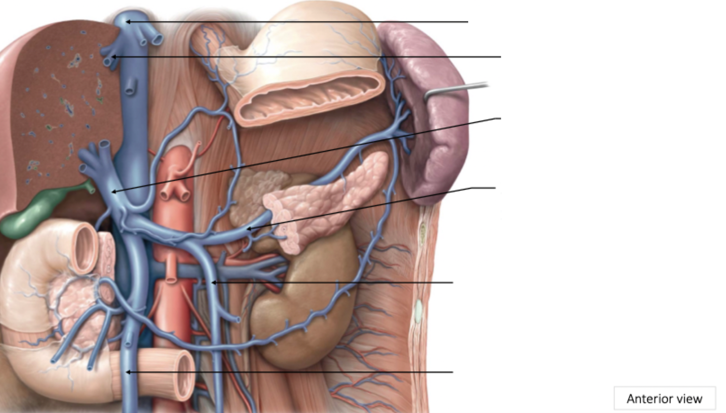
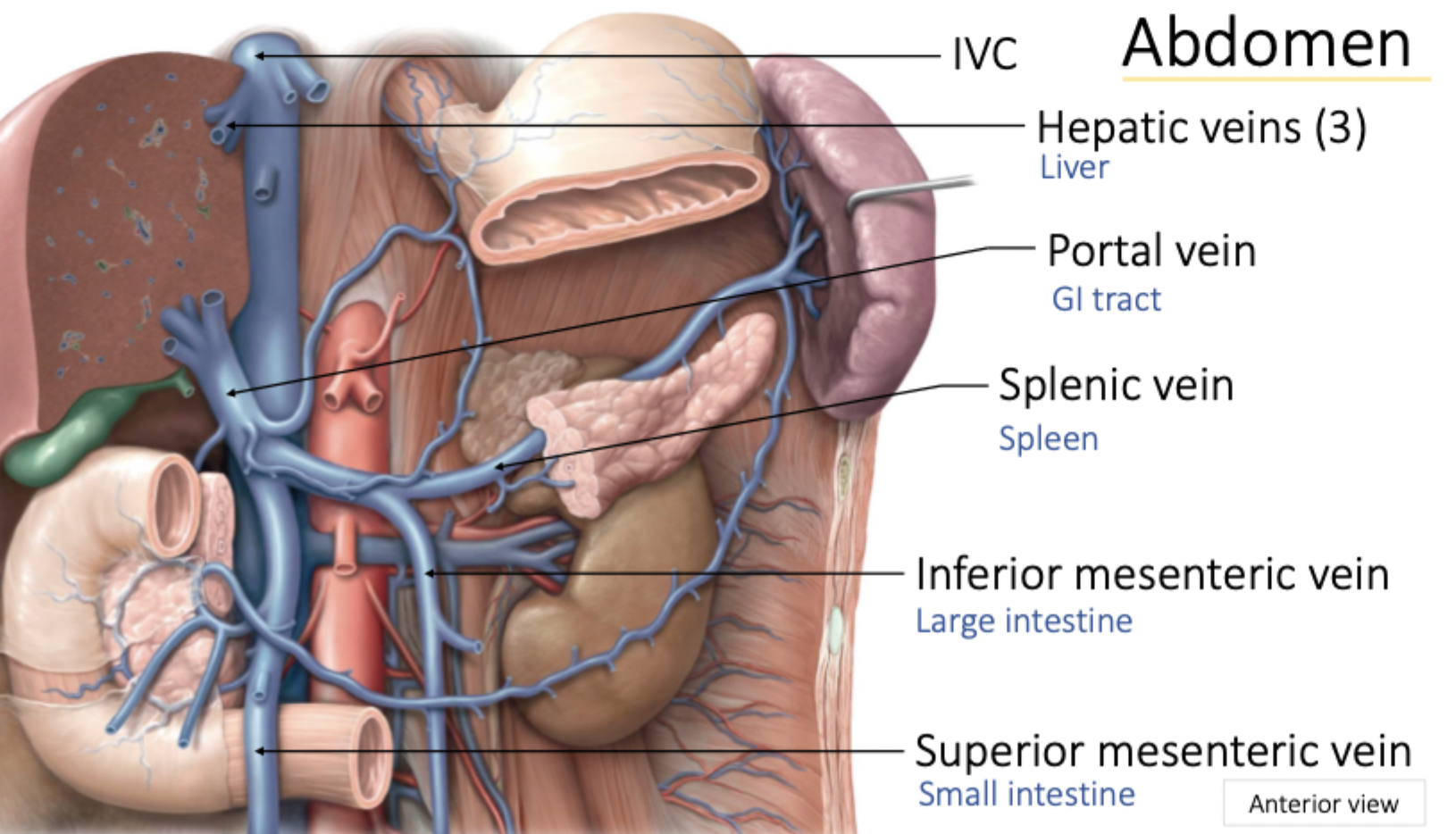
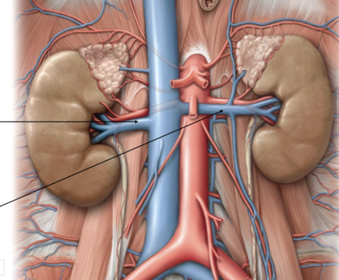
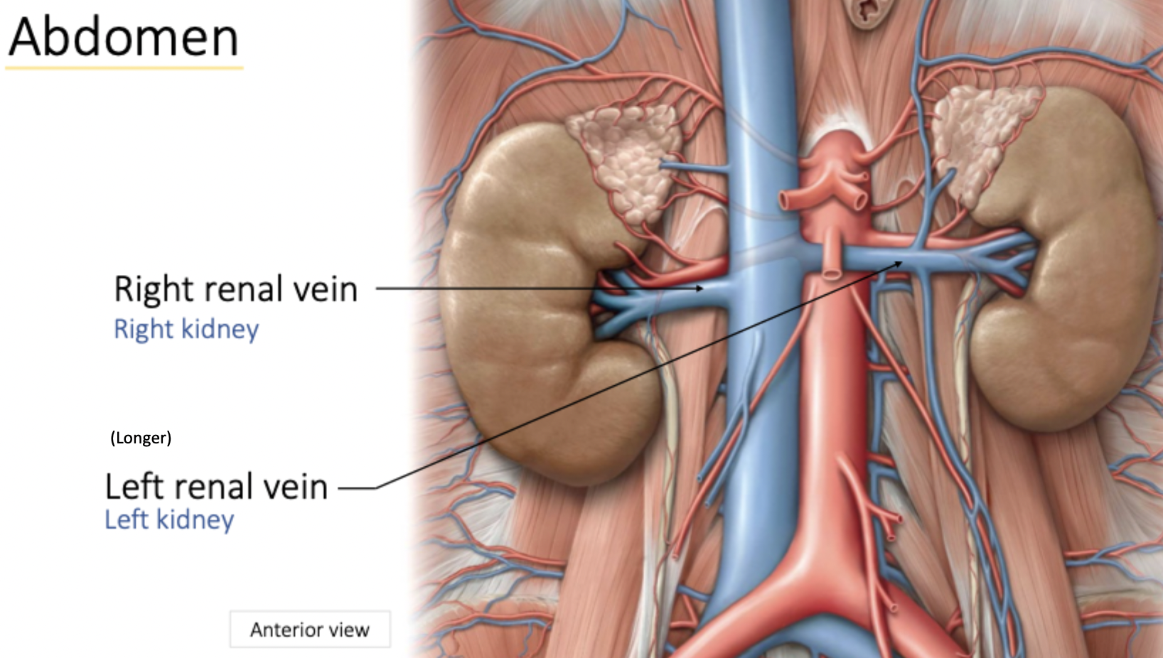
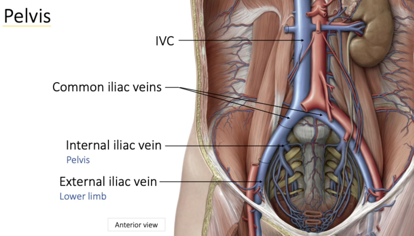
Abdomen Venous Drainage
Abdomen Venous Drainage
Anterior view
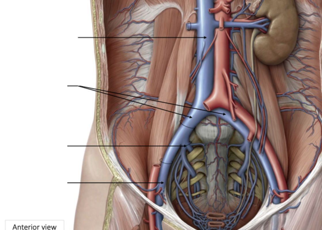
Pelvis Venous Drainage
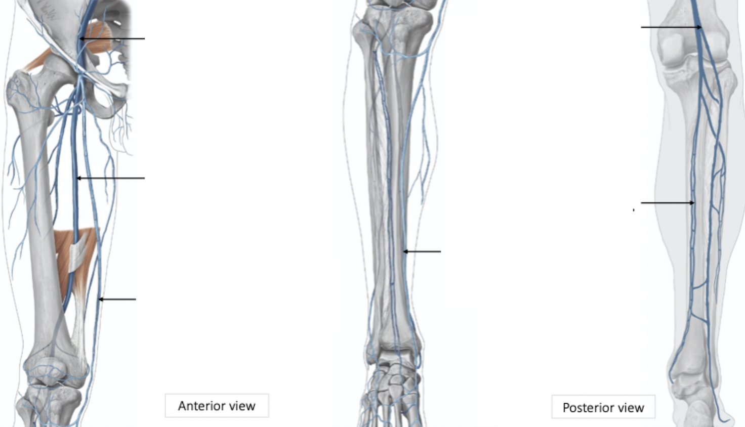
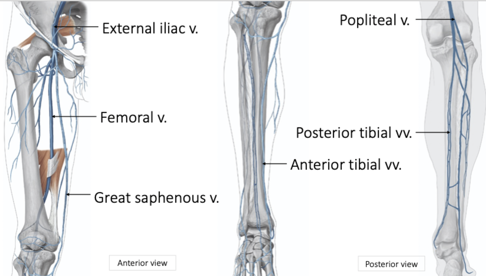
Leg Venous Drainage
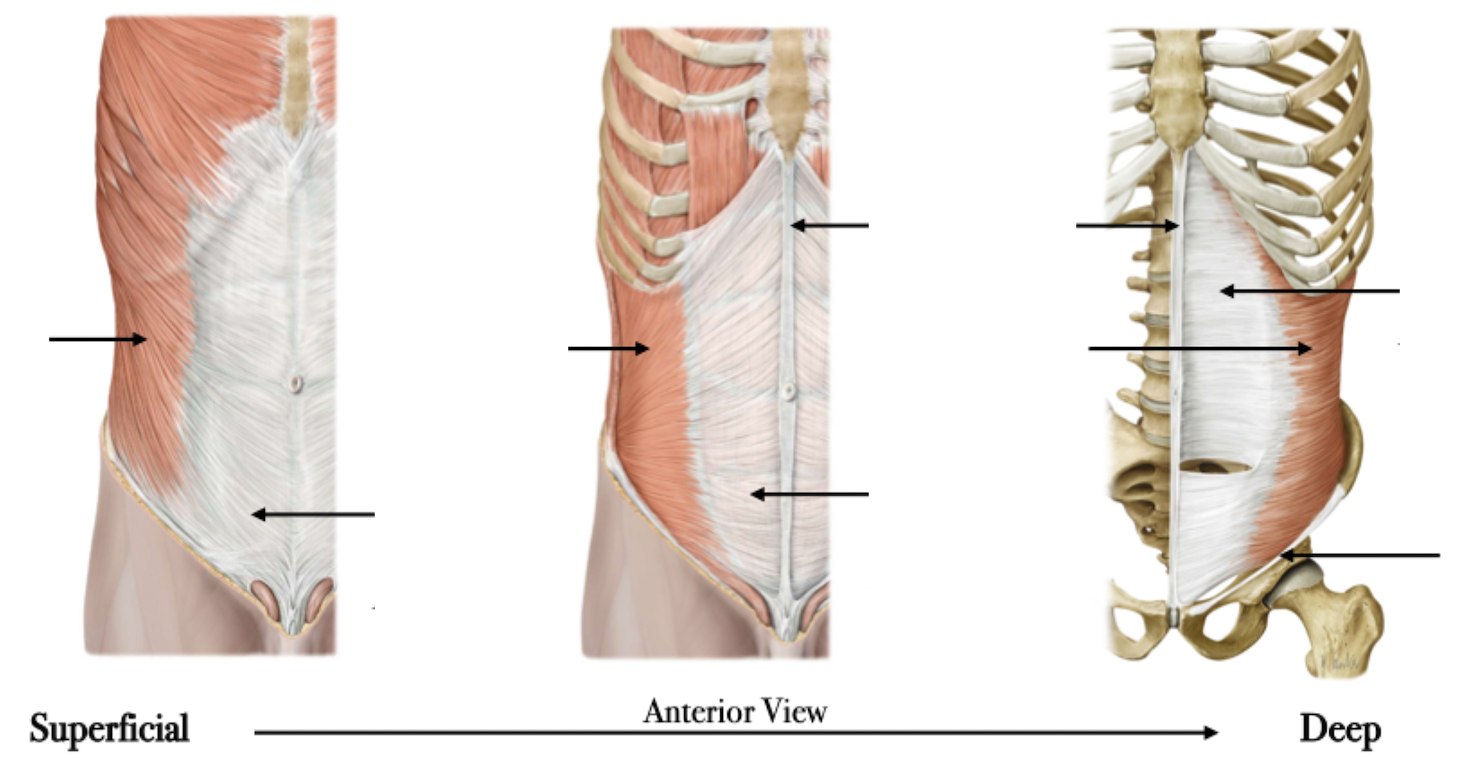
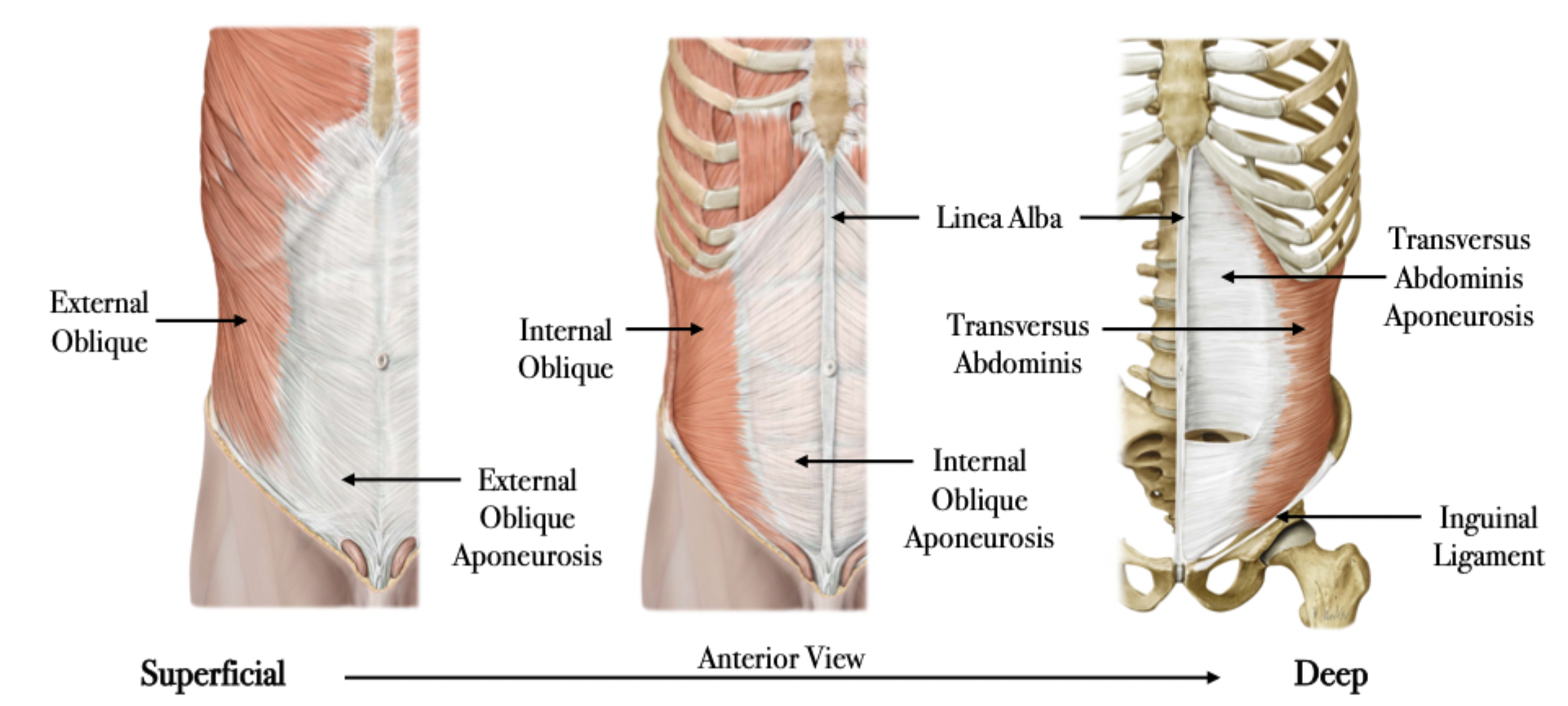
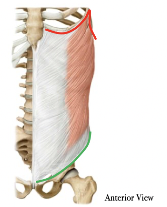
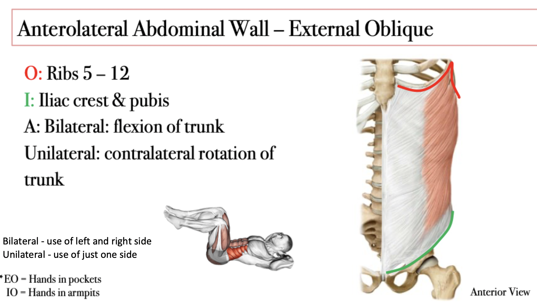
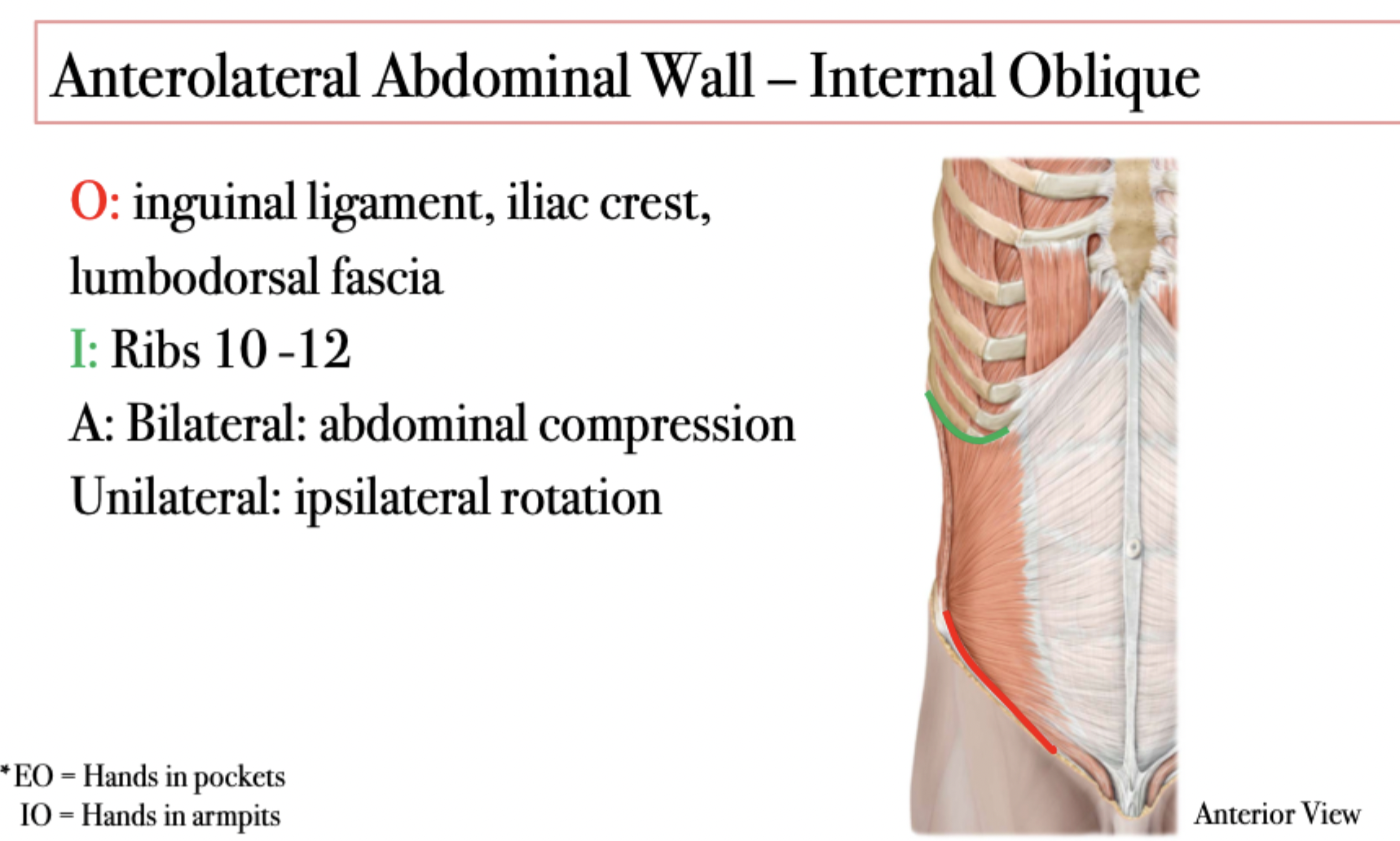
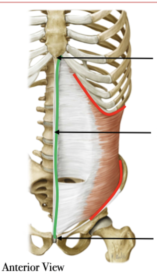
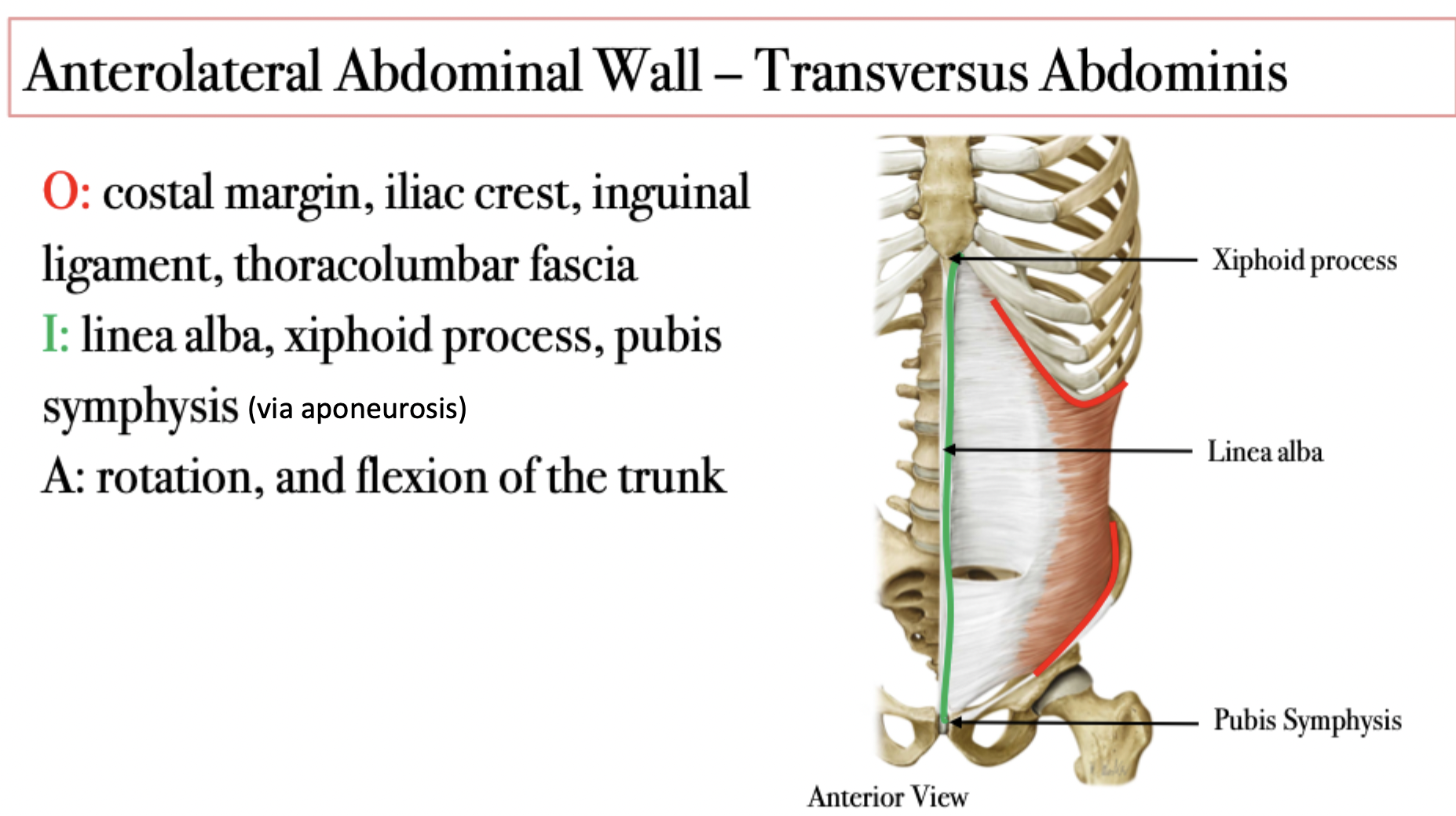
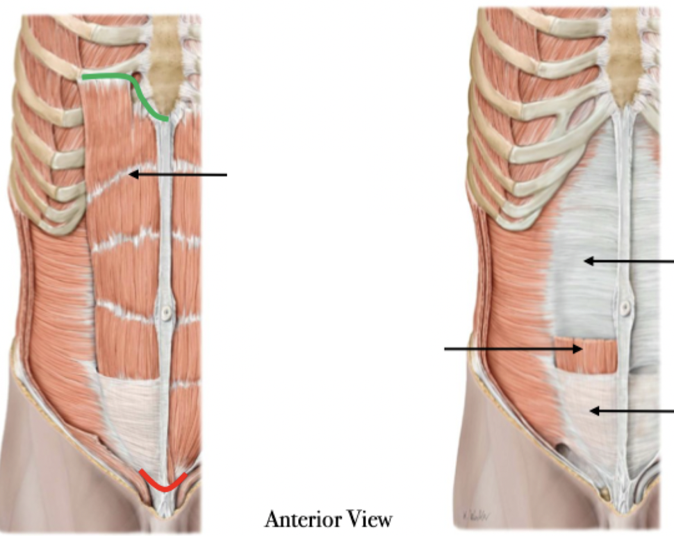
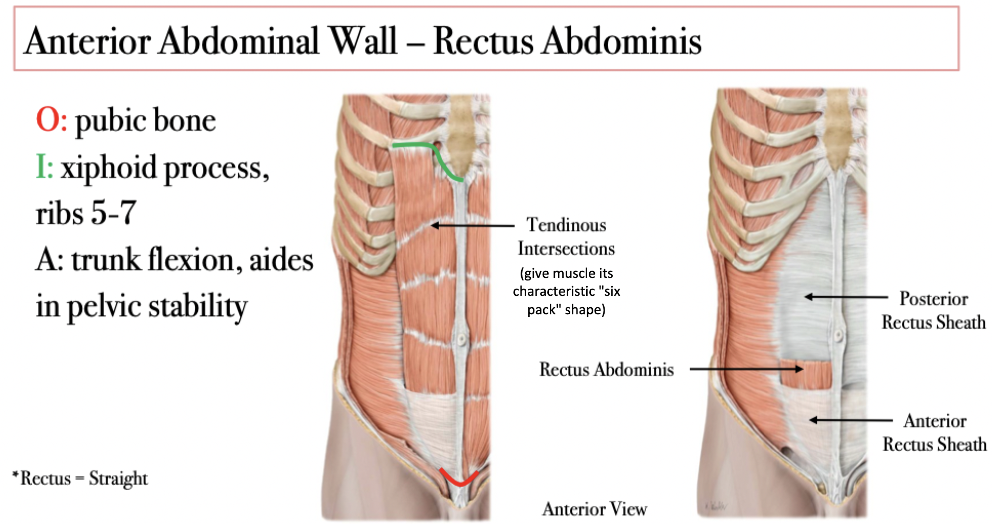
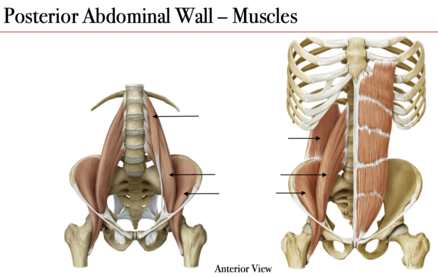
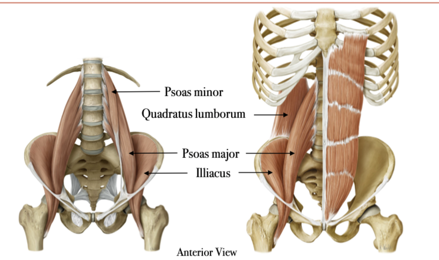
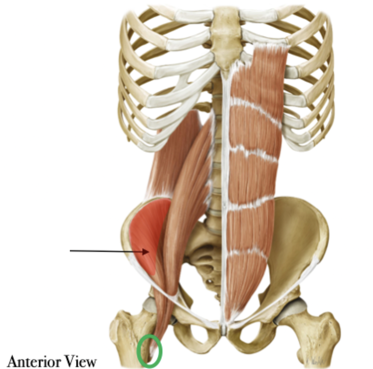
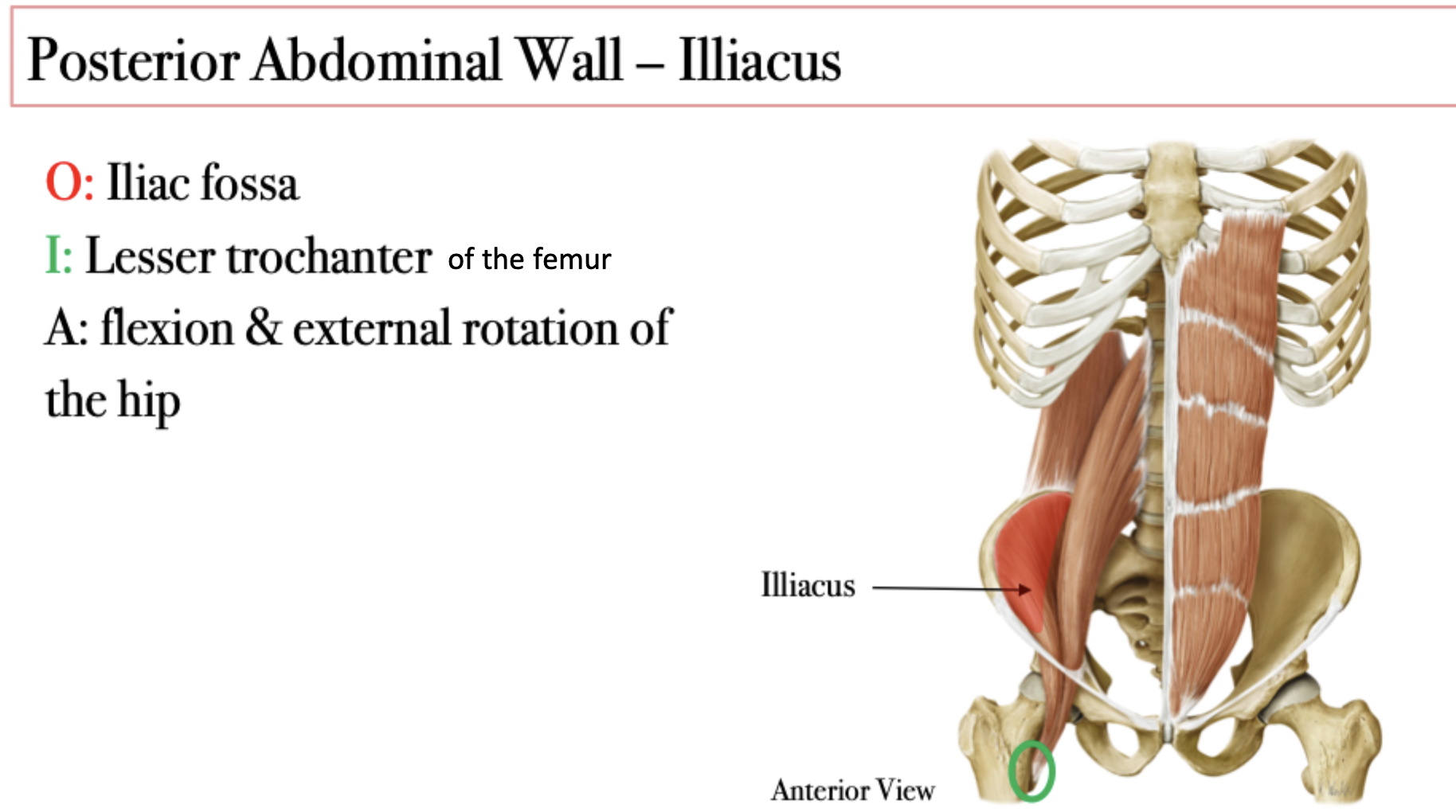
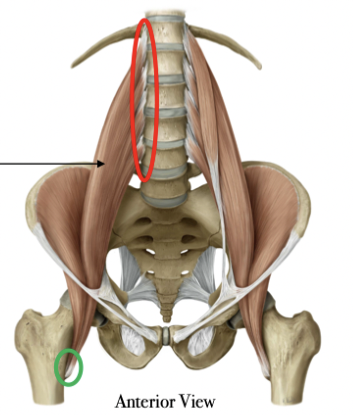
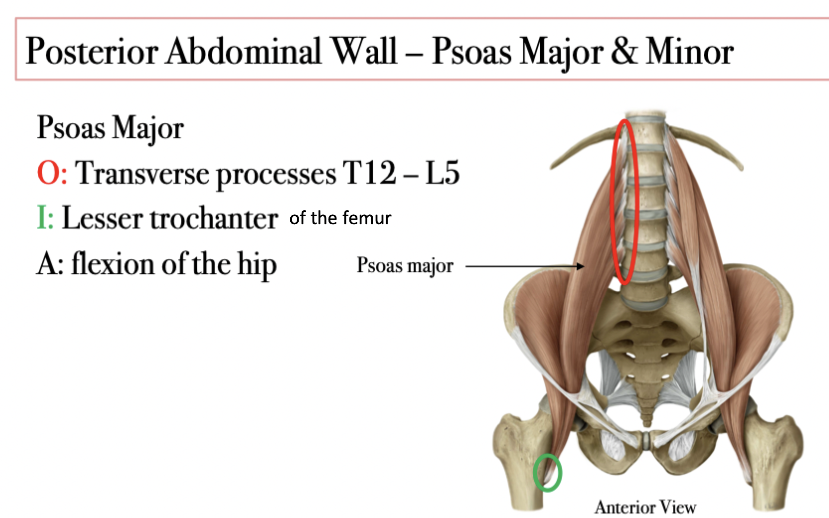
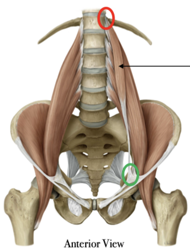
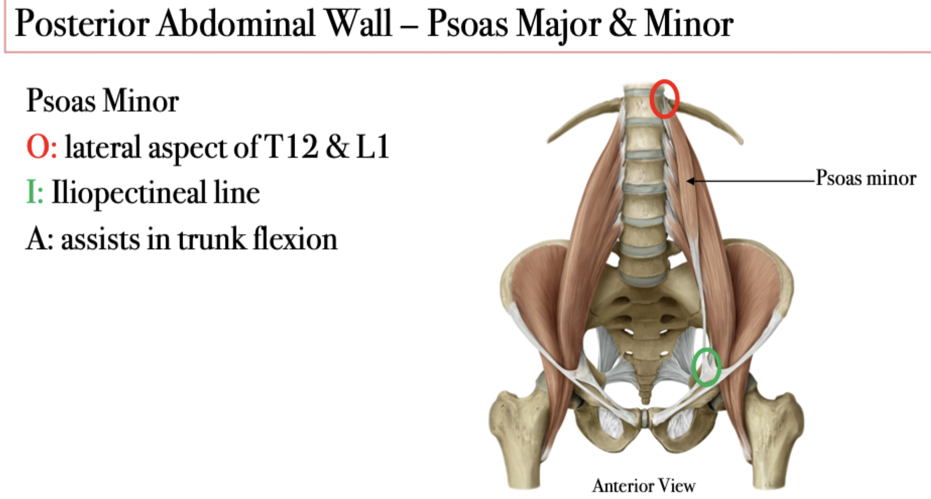
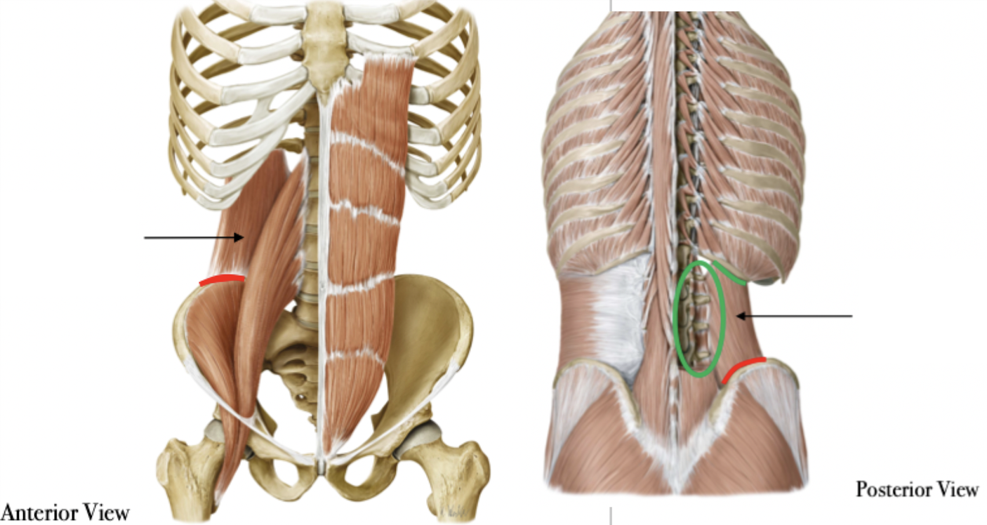
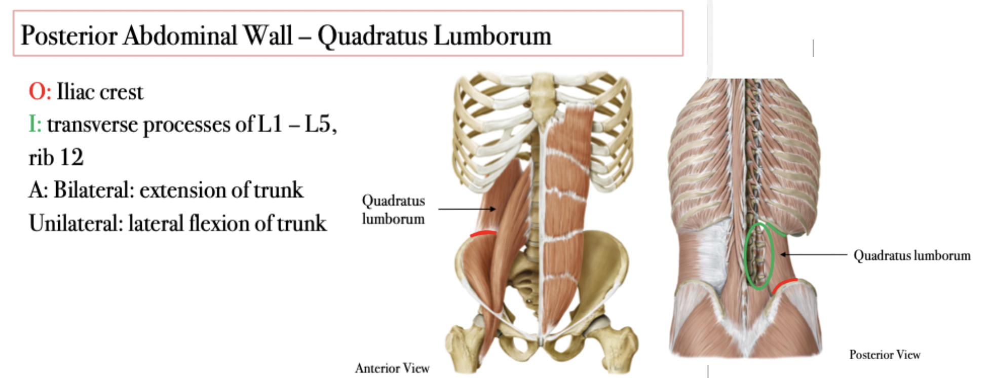
Anterolateral Abdominal Wall - Muscles
External Obliques point inferiorly toward the midline
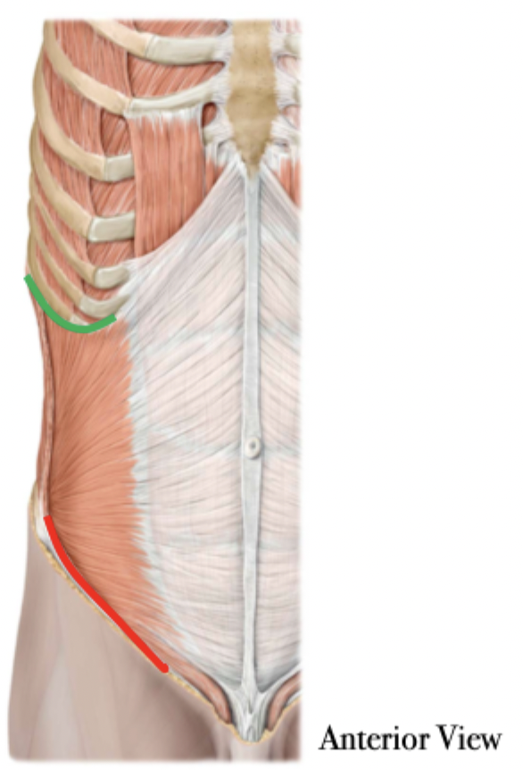
Internal Obliques point superiorly toward the midline
Transversus Abdominus runs horizontally
Housed by the rectus sheathe despite not being part of the anterolateral wall, which is formed by the aponeuroses of the muscles in that group
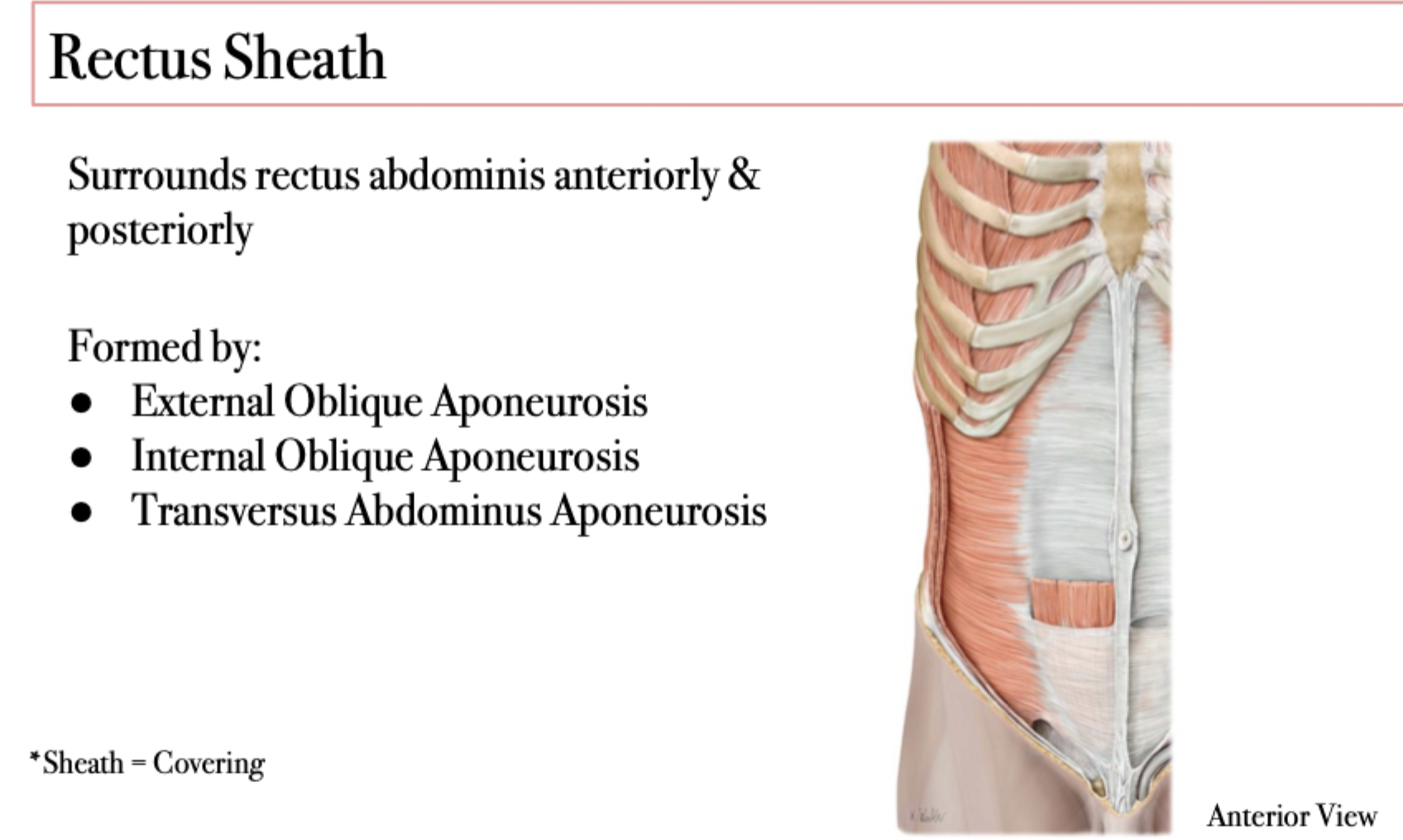
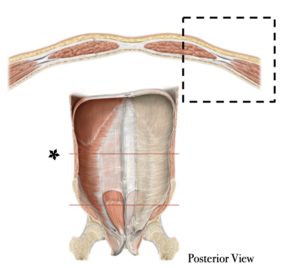
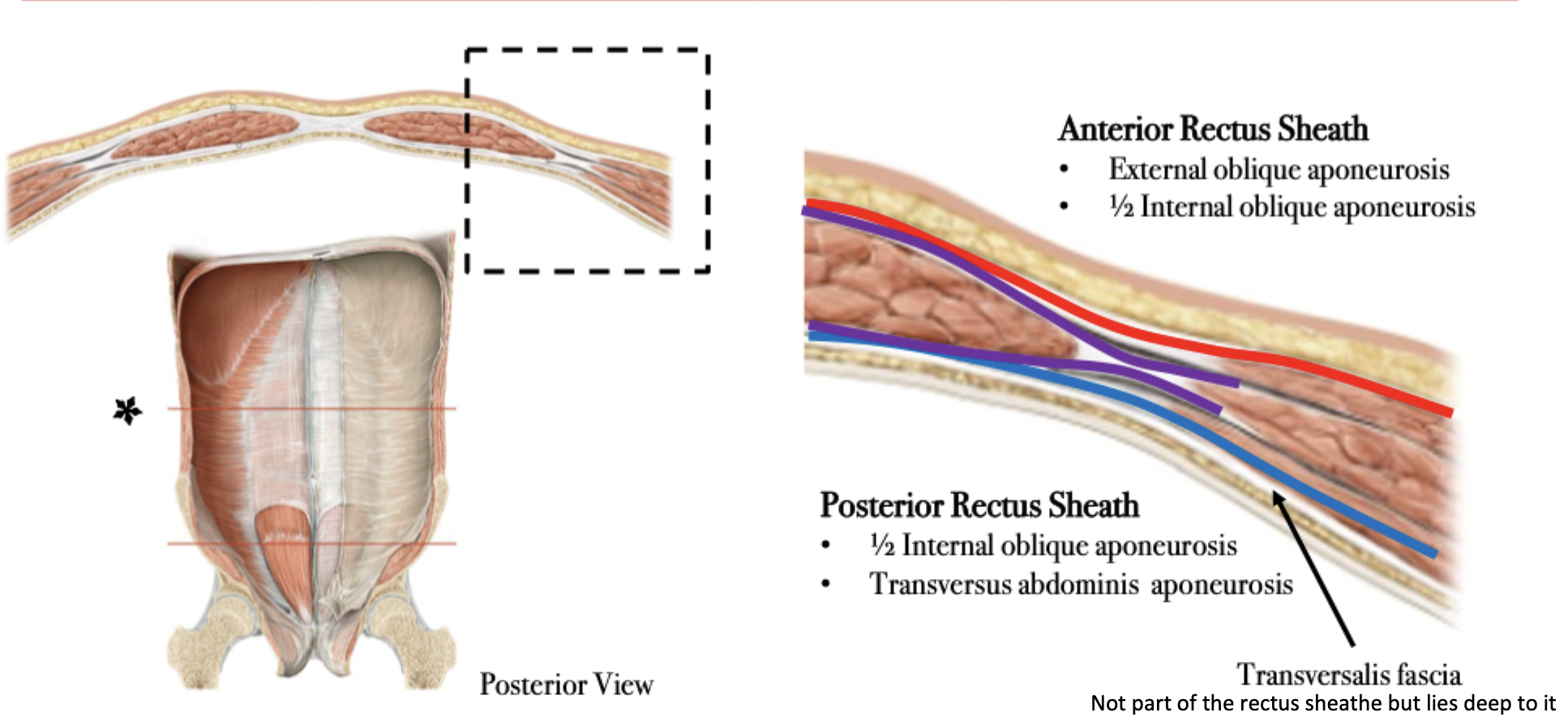
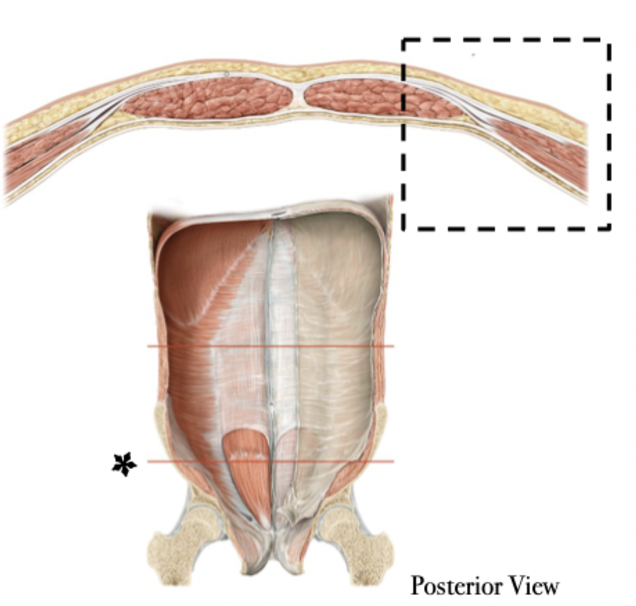
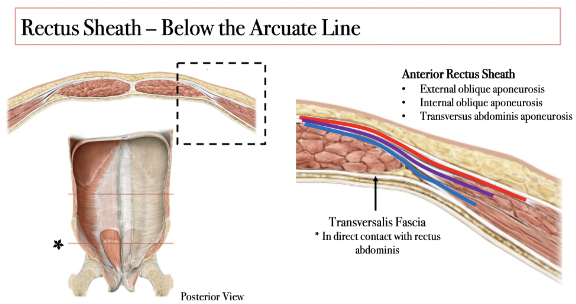
Rectus Sheathe layers depend on…
Location
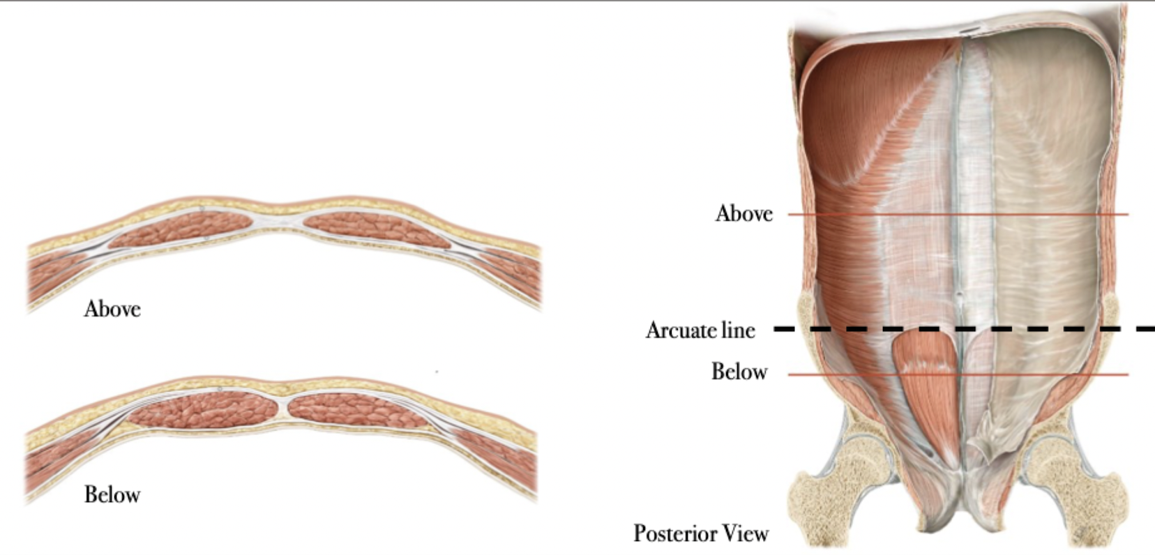
Above or below the arcuate line
Between the umbilicus & pubis
Rectus Sheathe above the arcuate line
Rectus Sheathe below the arcuate line
Inguinal Region Overview
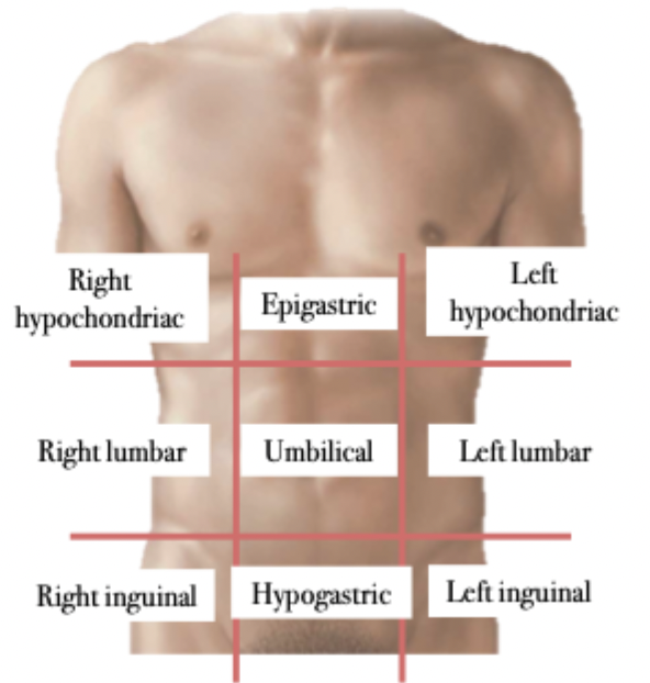
Surface Anatomy
Left & right of the hypogastric region
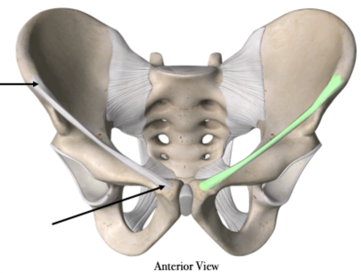
Key Structures
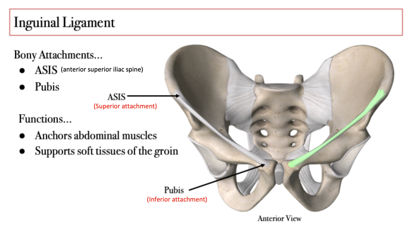
Inguinal Ligament
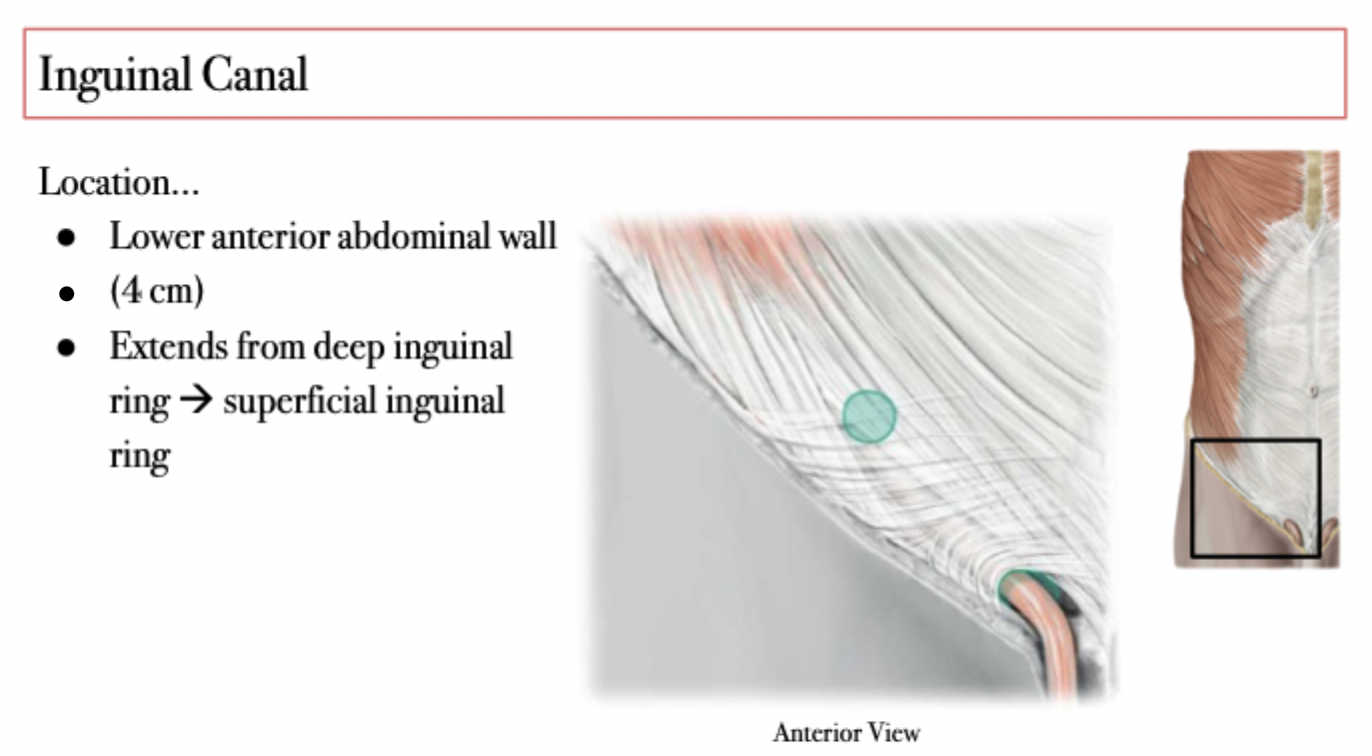
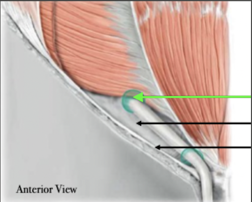
Inguinal Canal
Inguinal Canal - Location
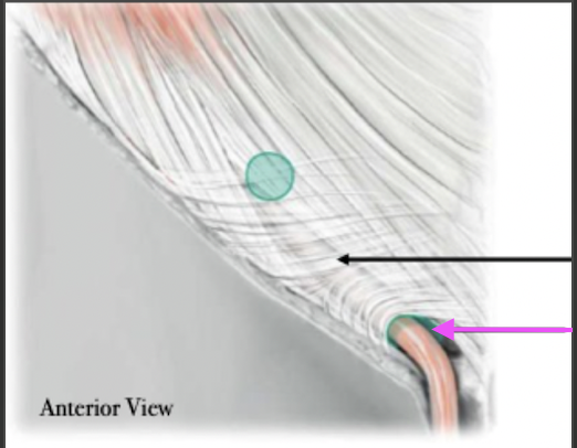
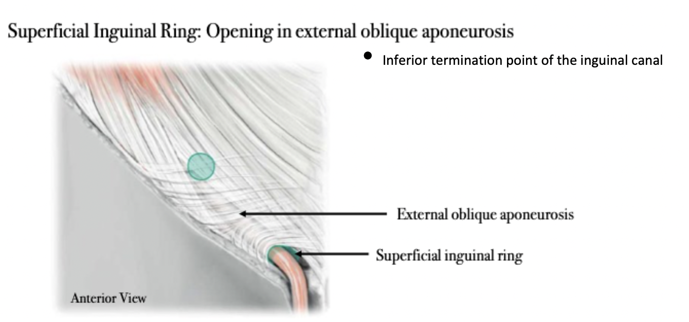
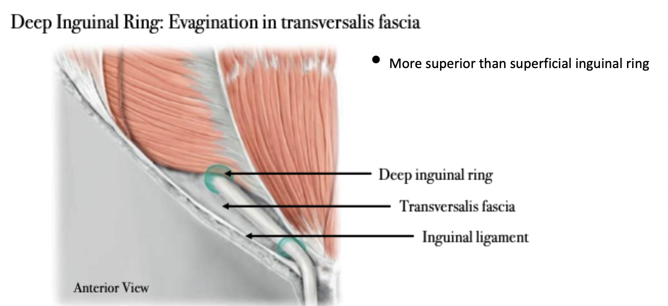
Inguinal Canal Features
Inguinal Canal Features
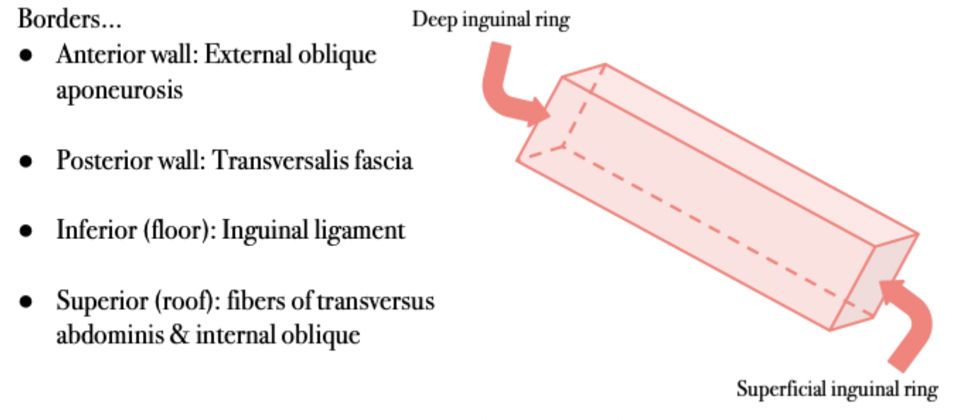
Inguinal Canal Borders
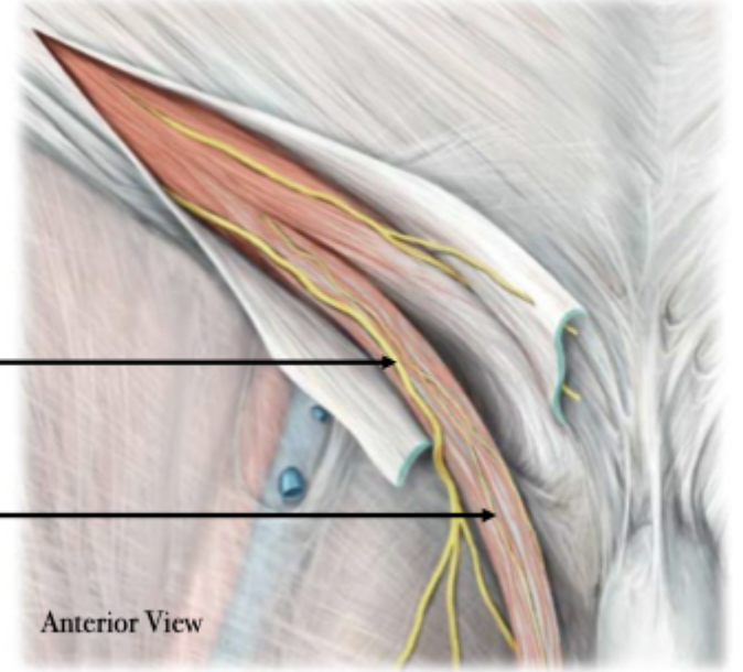
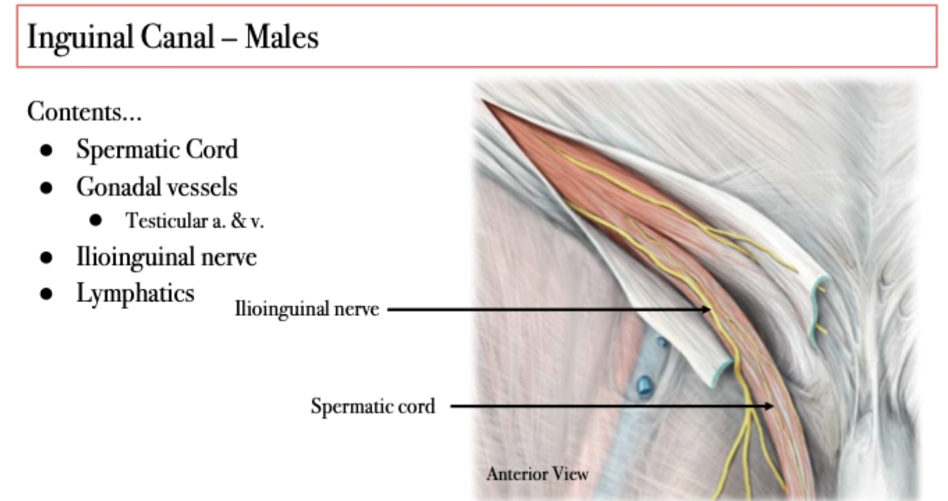
Inguinal Canal - Males
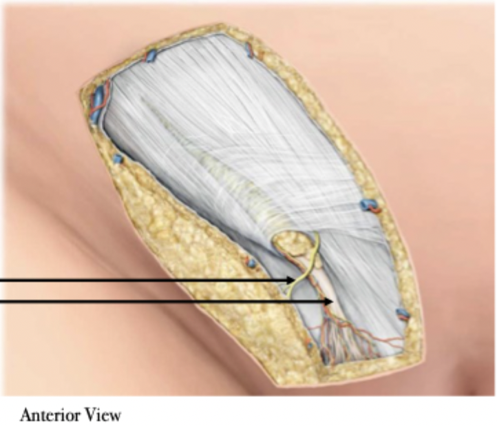
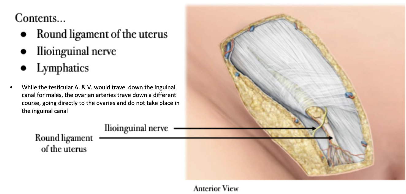
Inguinal Canal - Females
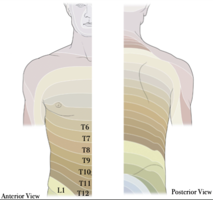
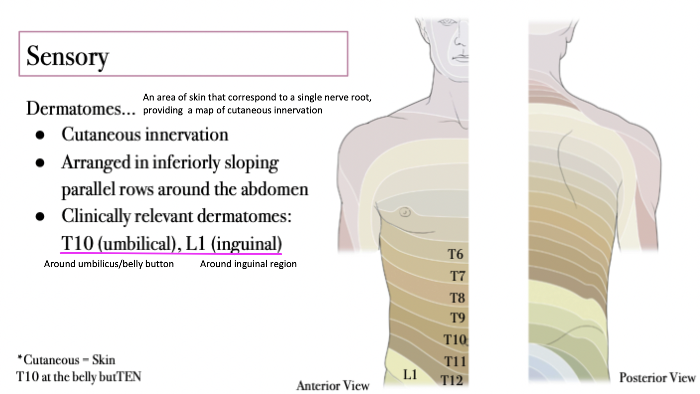
Abdominal Wall Sensory Innervation
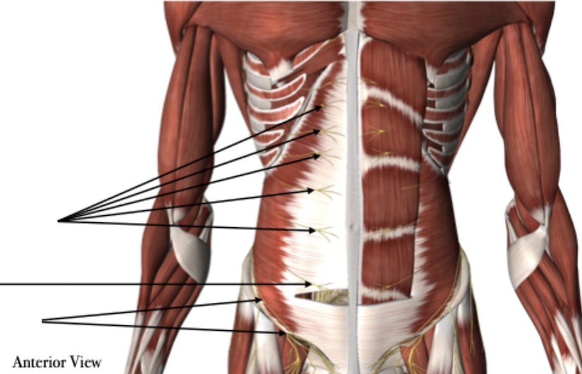
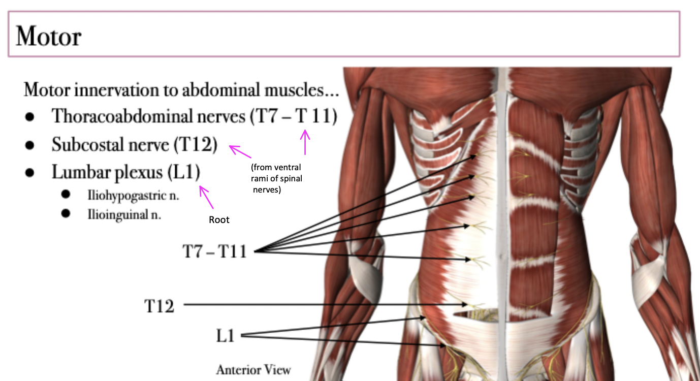
Abdominal Wall Motor Innervation
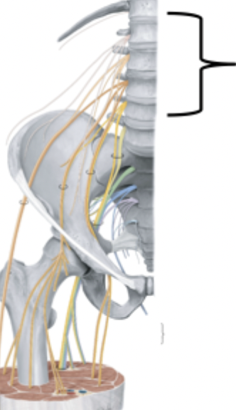
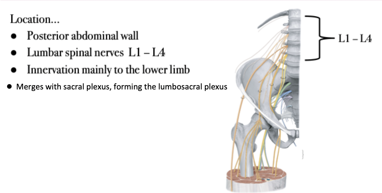
Lumbar Plexus
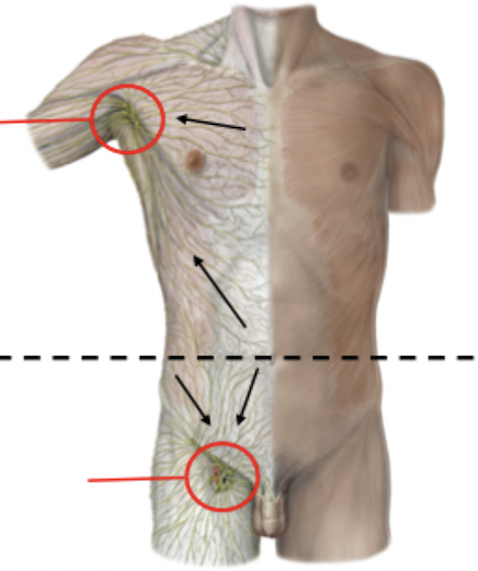
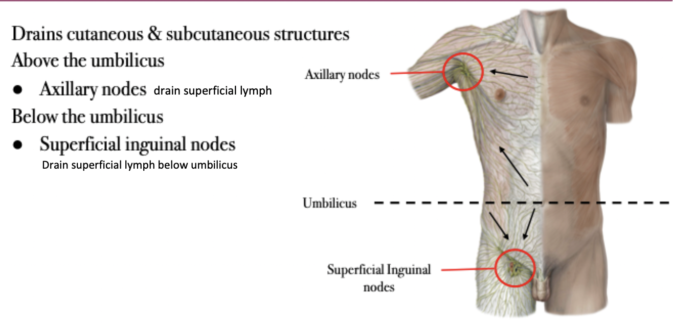
Abdominal Lymphatic Drainage - Superficial
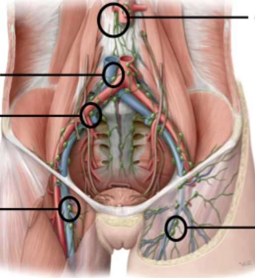
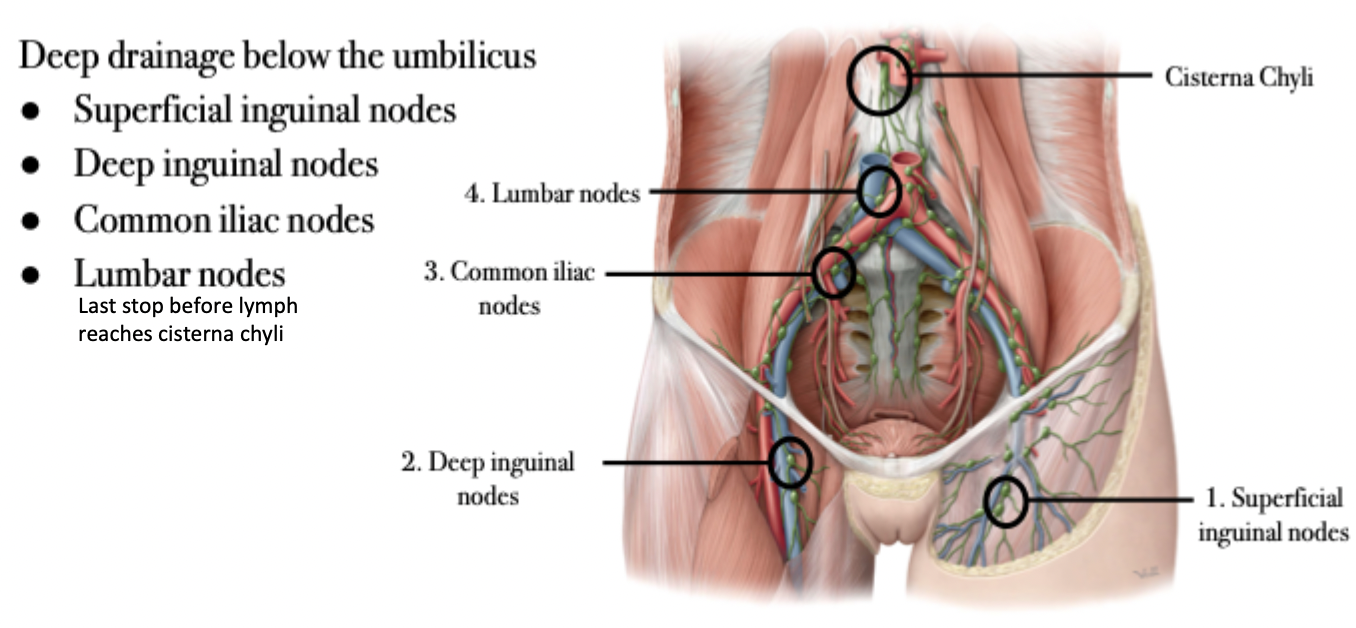
Abdominal Lymphatic Drainage - Deep
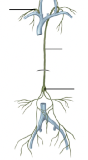
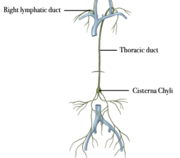
Abdominal Lymphatic Drainage - Return to Venous Circulation
Left abdomen & right abdomen below the umbilicus:
Cisterna chyli → Thoracic duct → venous system
Axillary nodes (left) → Thoracic duct → venous system
Right abdomen above the umbilicus:
Axillary nodes (right) → Right lymphatic duct → venous system
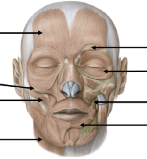
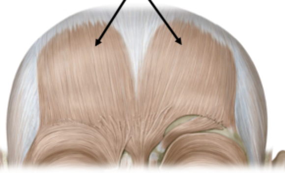
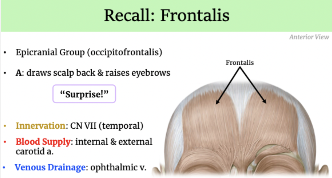
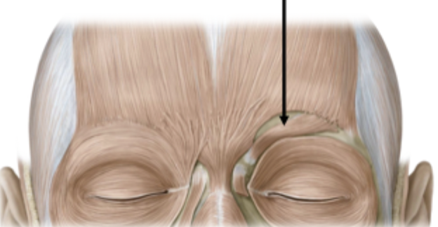
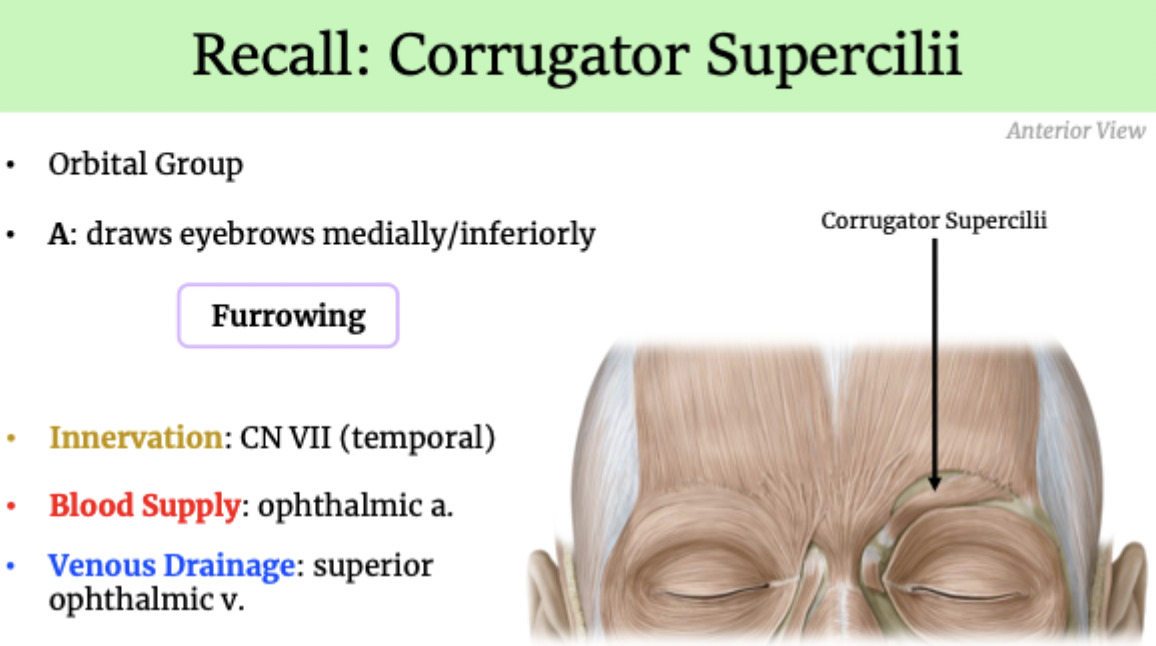
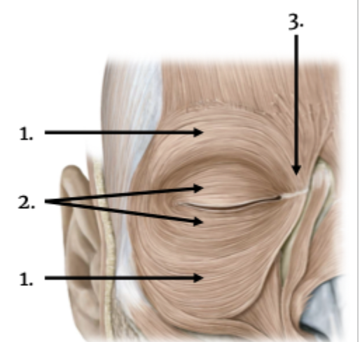
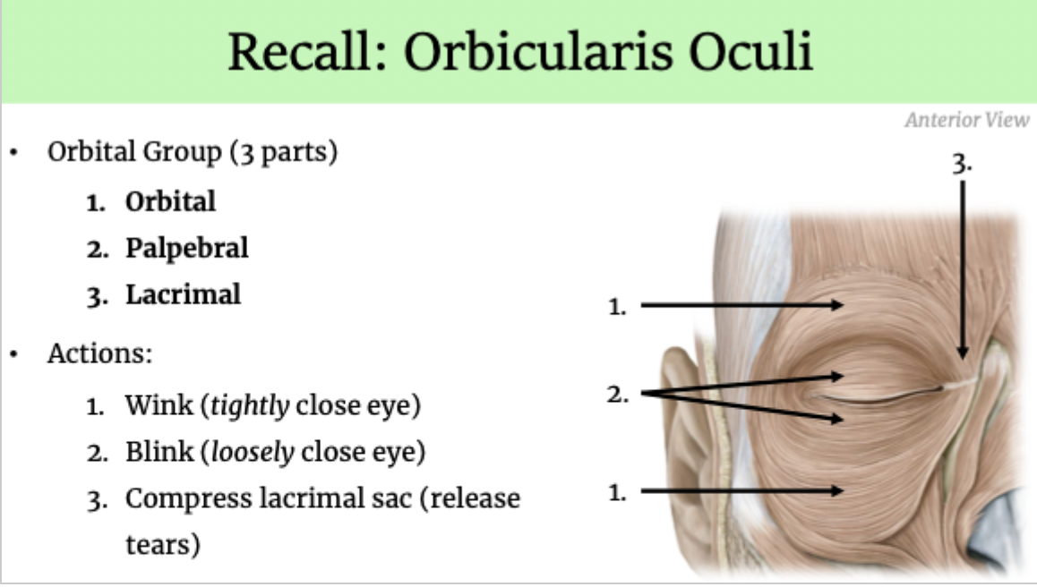
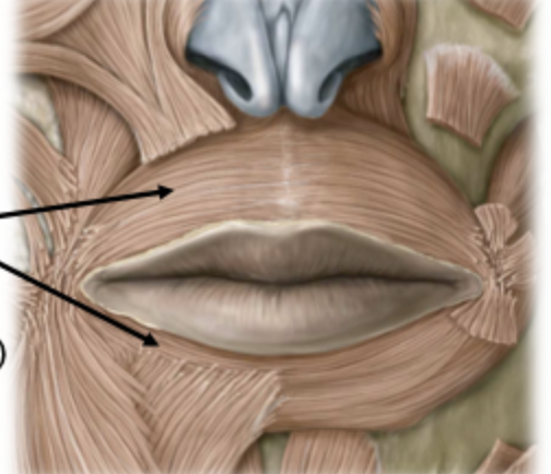
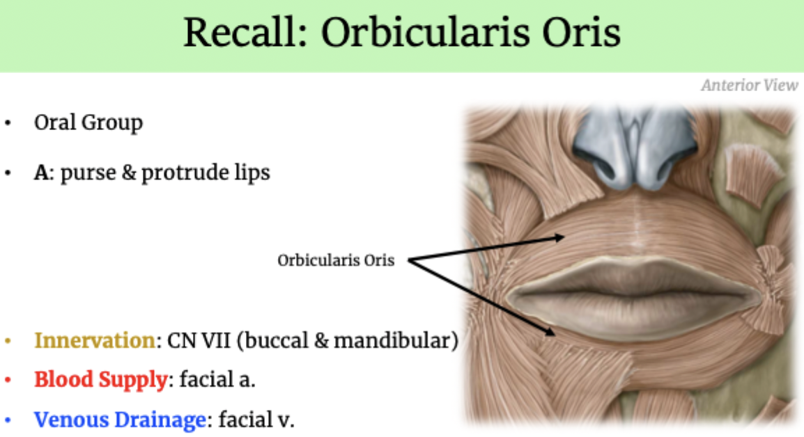
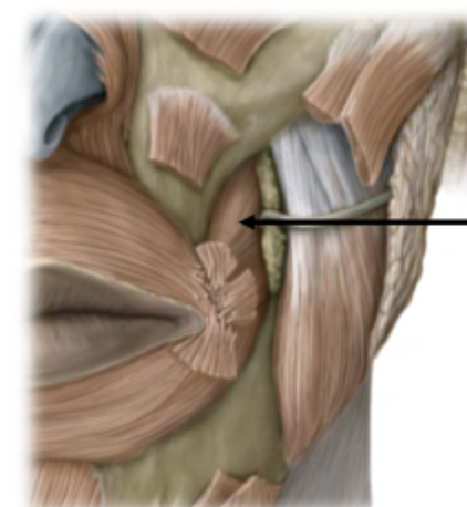
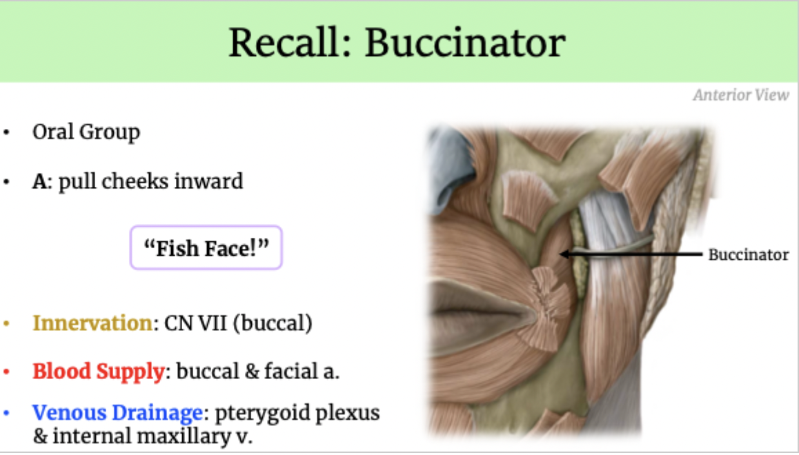
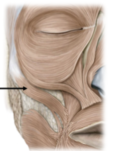
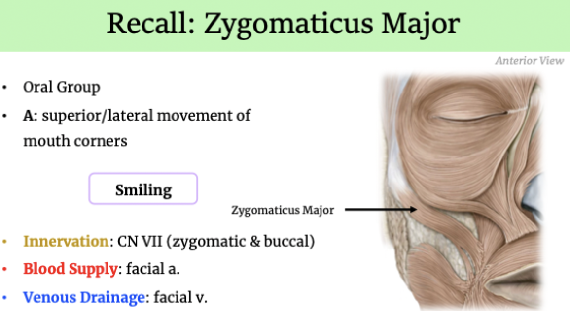
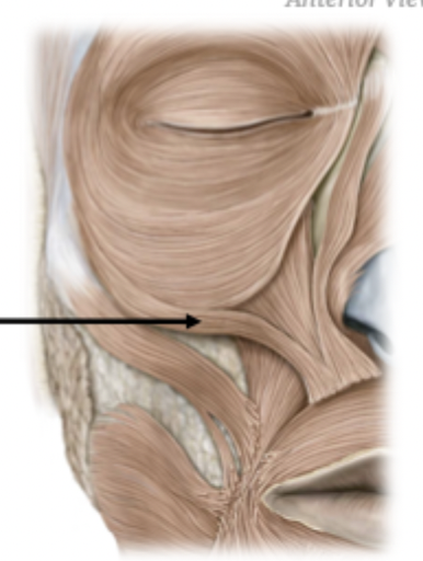
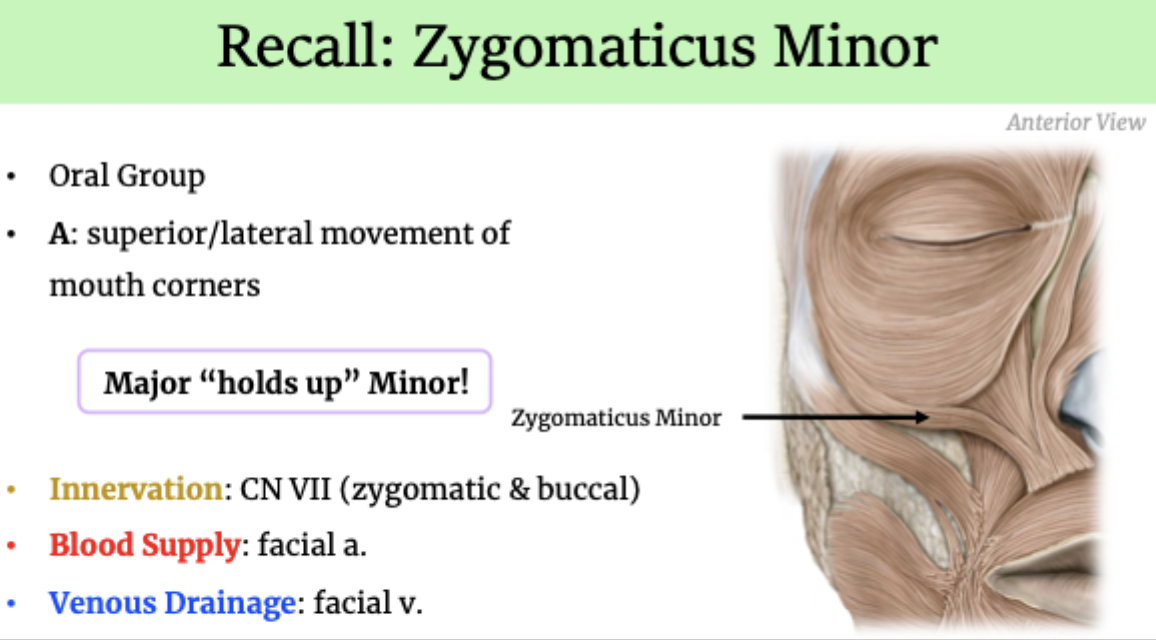
Muscles of Facial Expression
Superficial (subcutaneous) muscles
Origins → facial bones or adjacent muscles
Insertions → skin
Functions:
Sphincters/Dilators
Facial expression alteration
Innervation: CN VII (temporal for the upper half & zygomatic for the lower half)
Blood Supply: facial a., superficial temporal a. & ophthalmic a.
Venous Drainage: facial v.
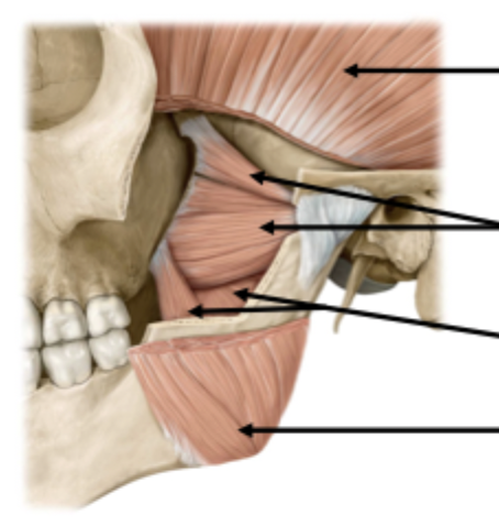
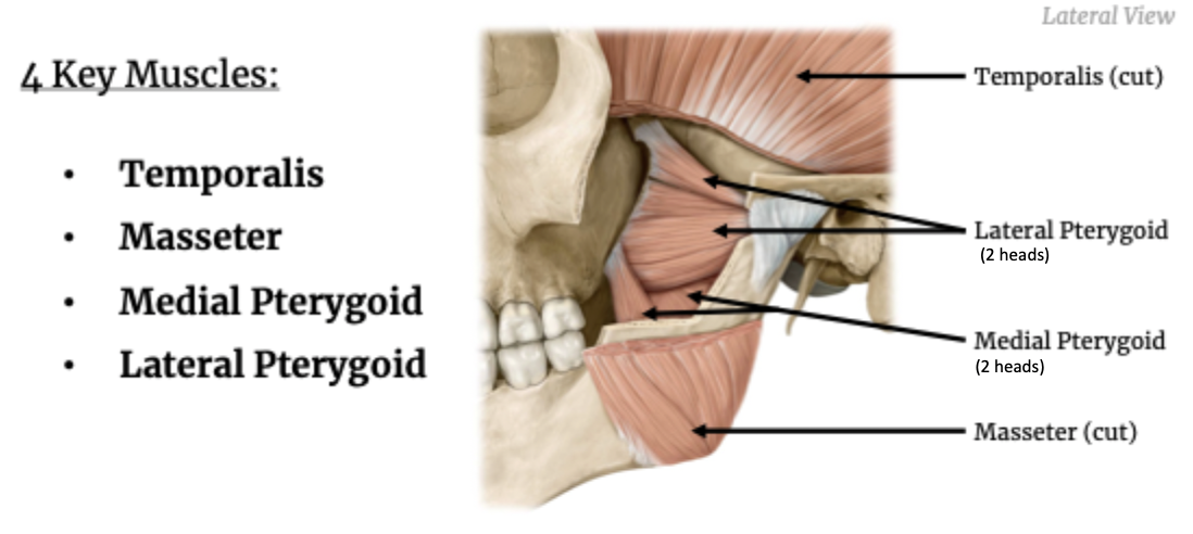
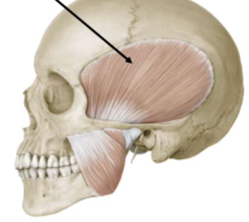
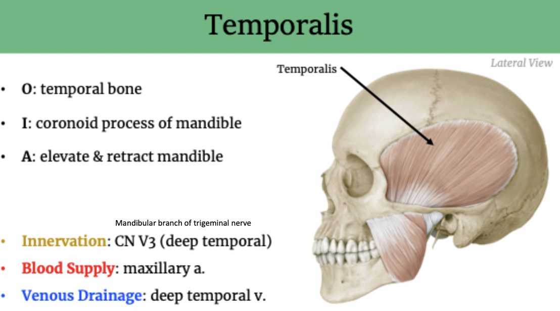
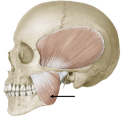
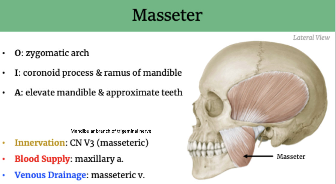
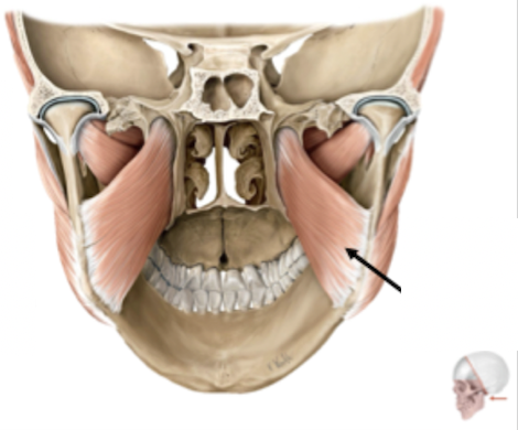
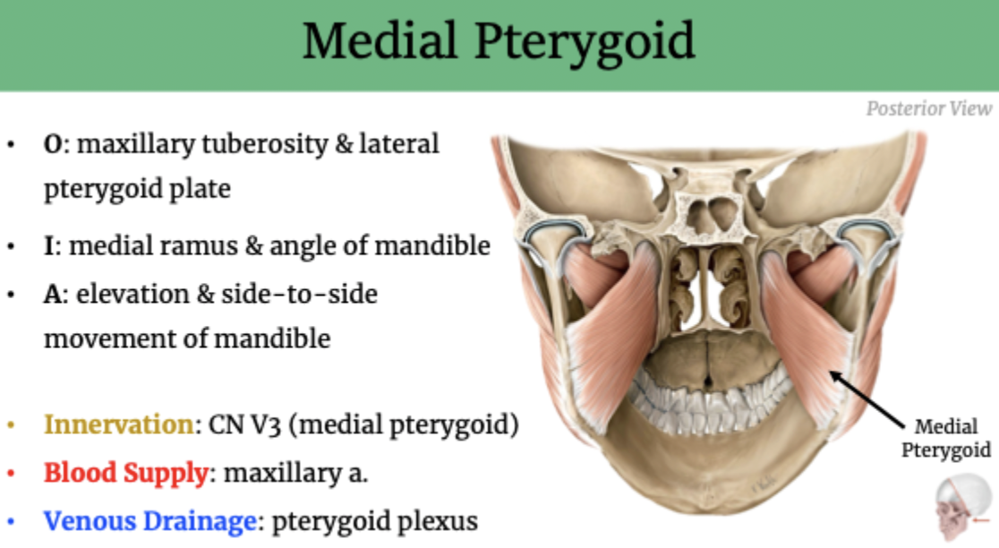
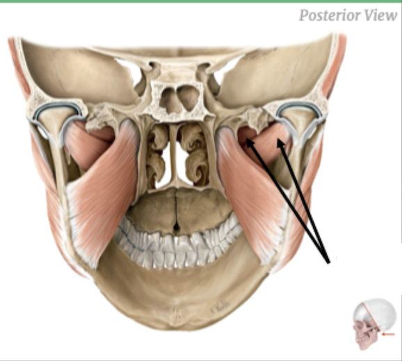
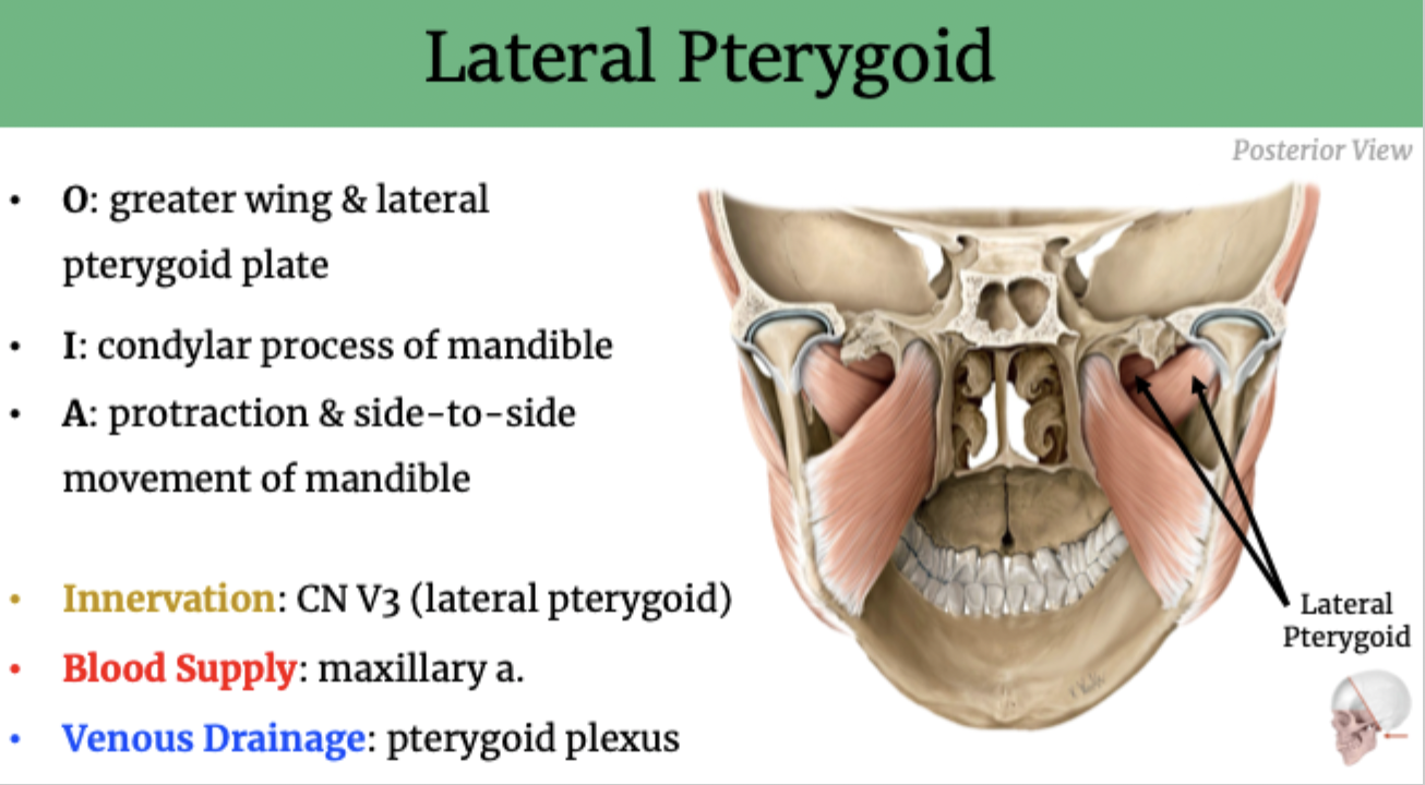
Muscles of Mastication
Both superficial & deep muscles
Functions:
Movement of the mandible
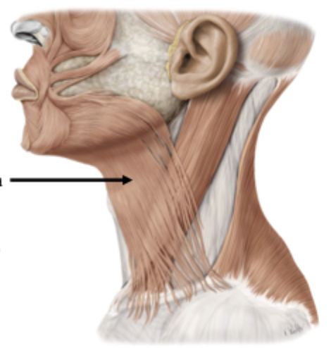
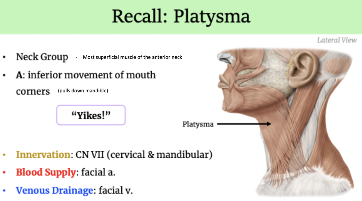
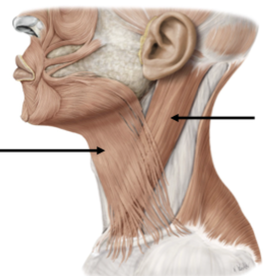
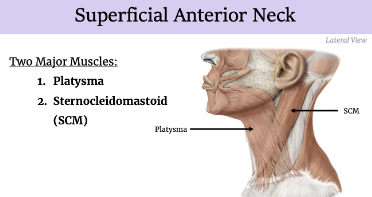
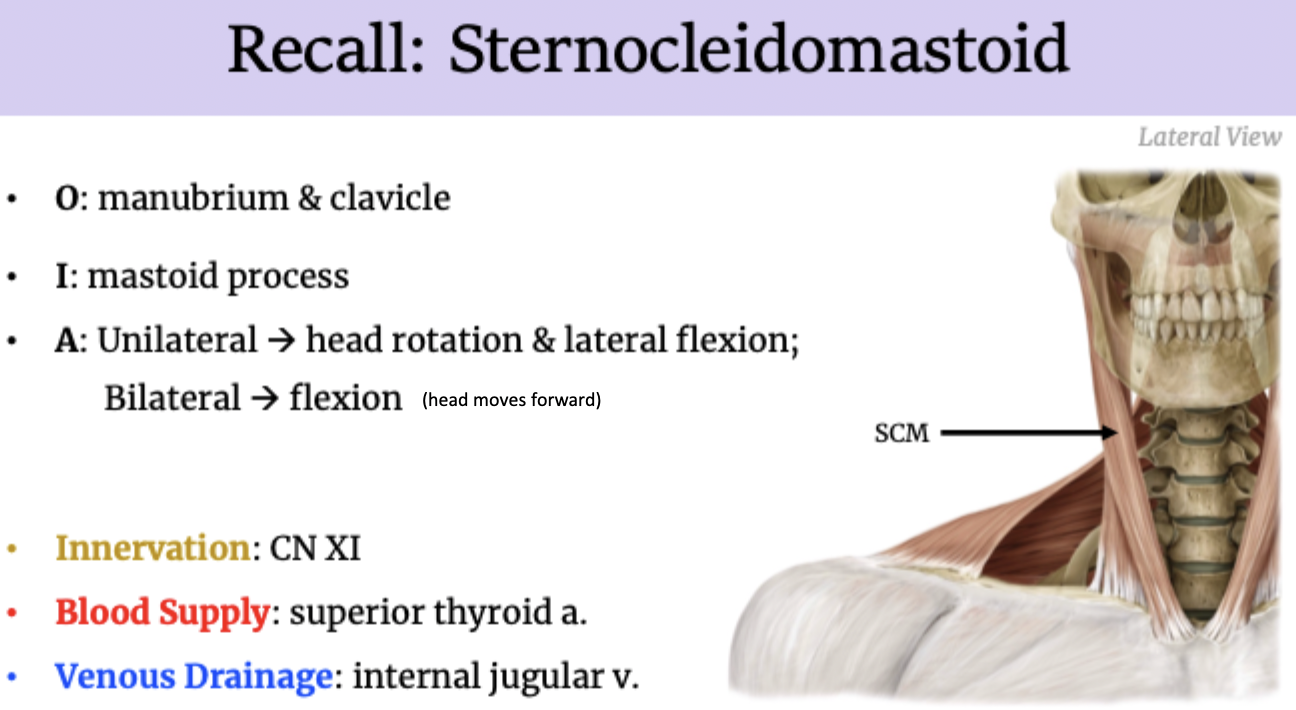
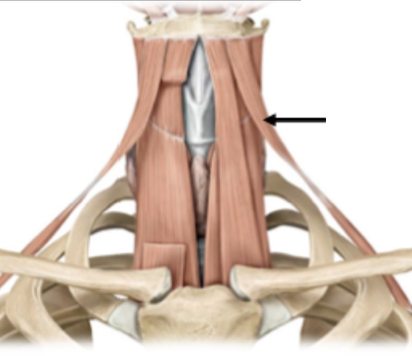
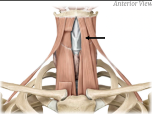
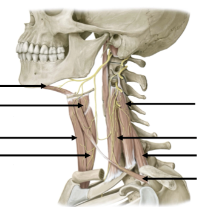
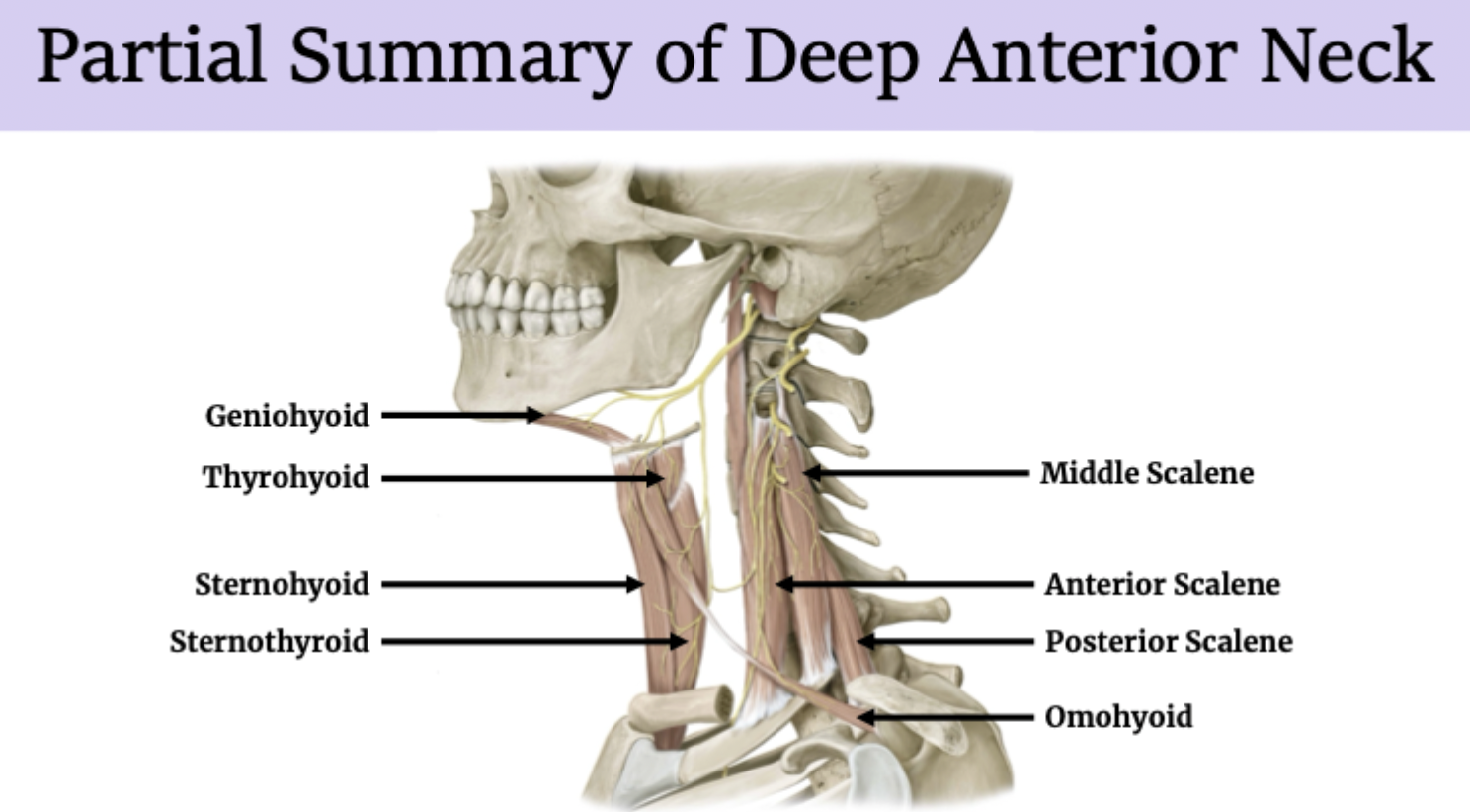
Superficial anterior neck muscles
Deep anterior neck muscles
Two groups
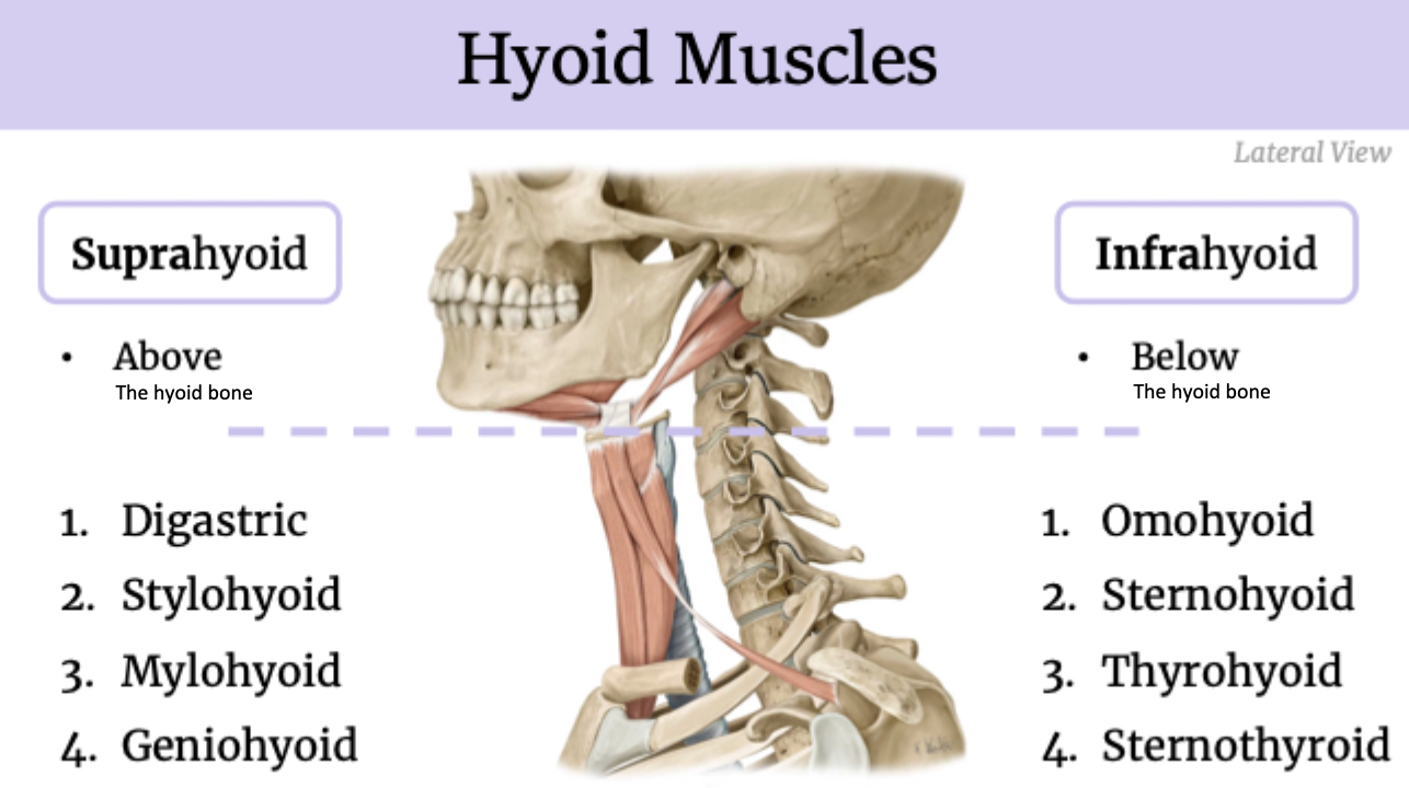
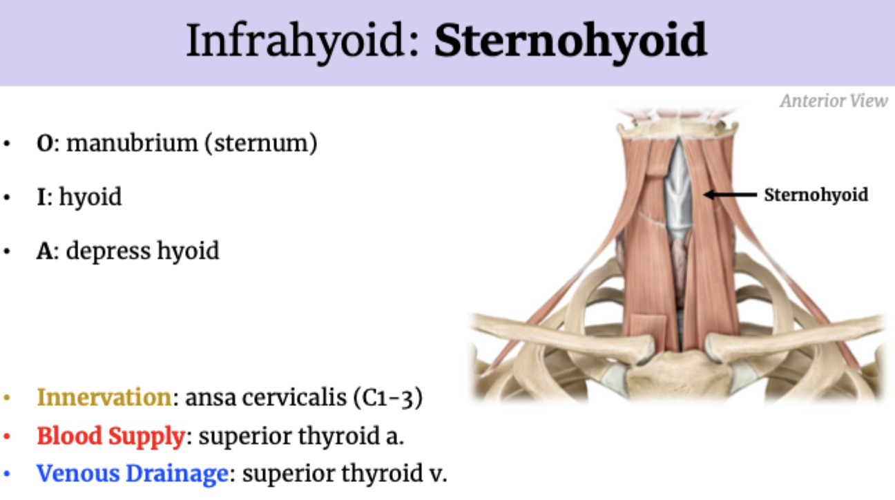
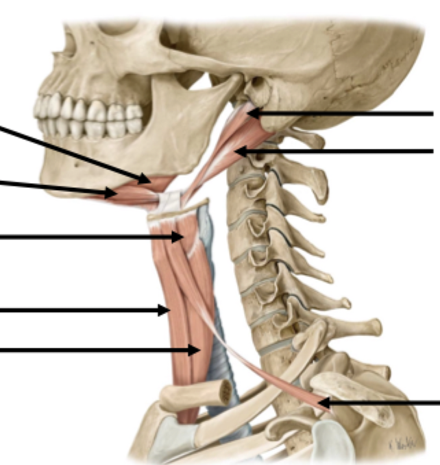
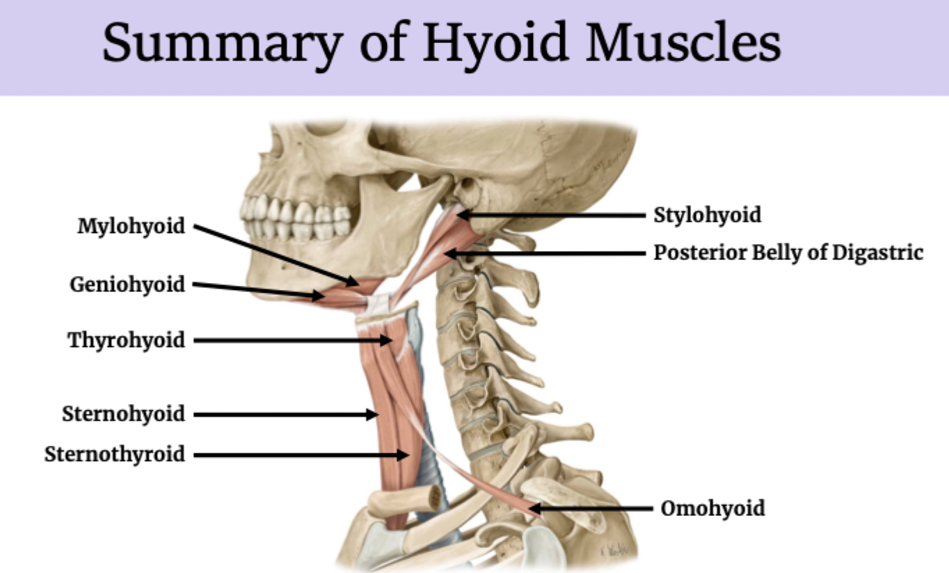
Hyoid muscles
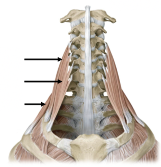
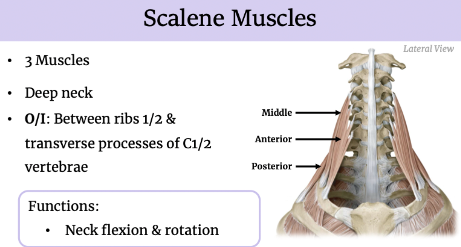
Scalene muscles
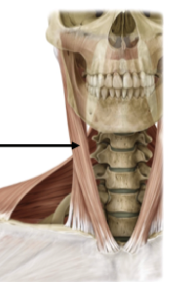
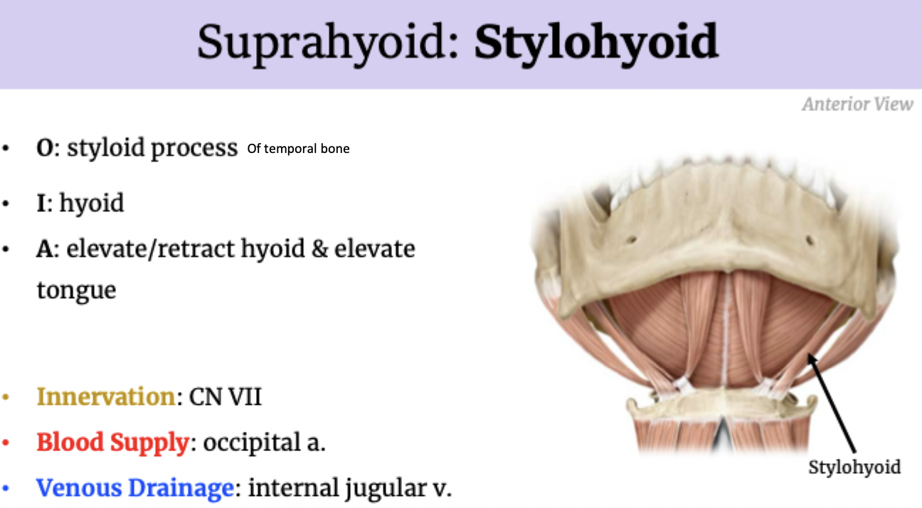
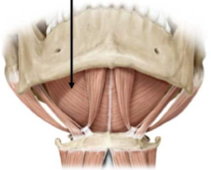
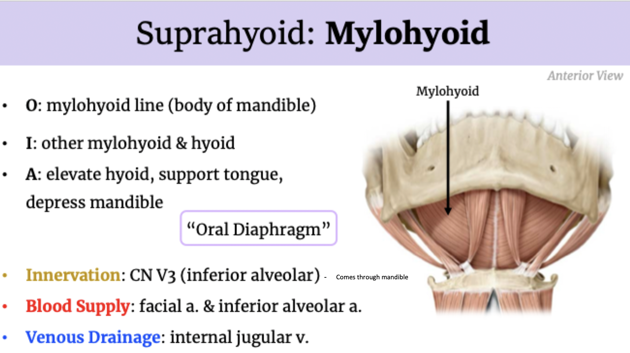
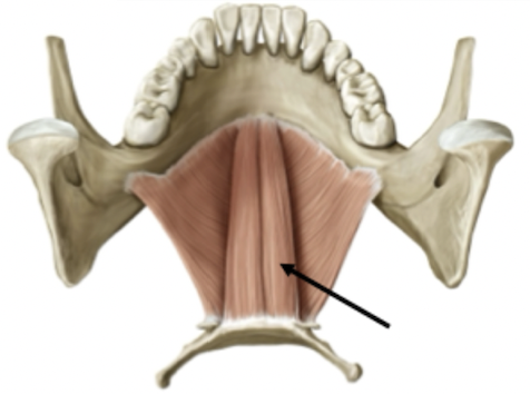
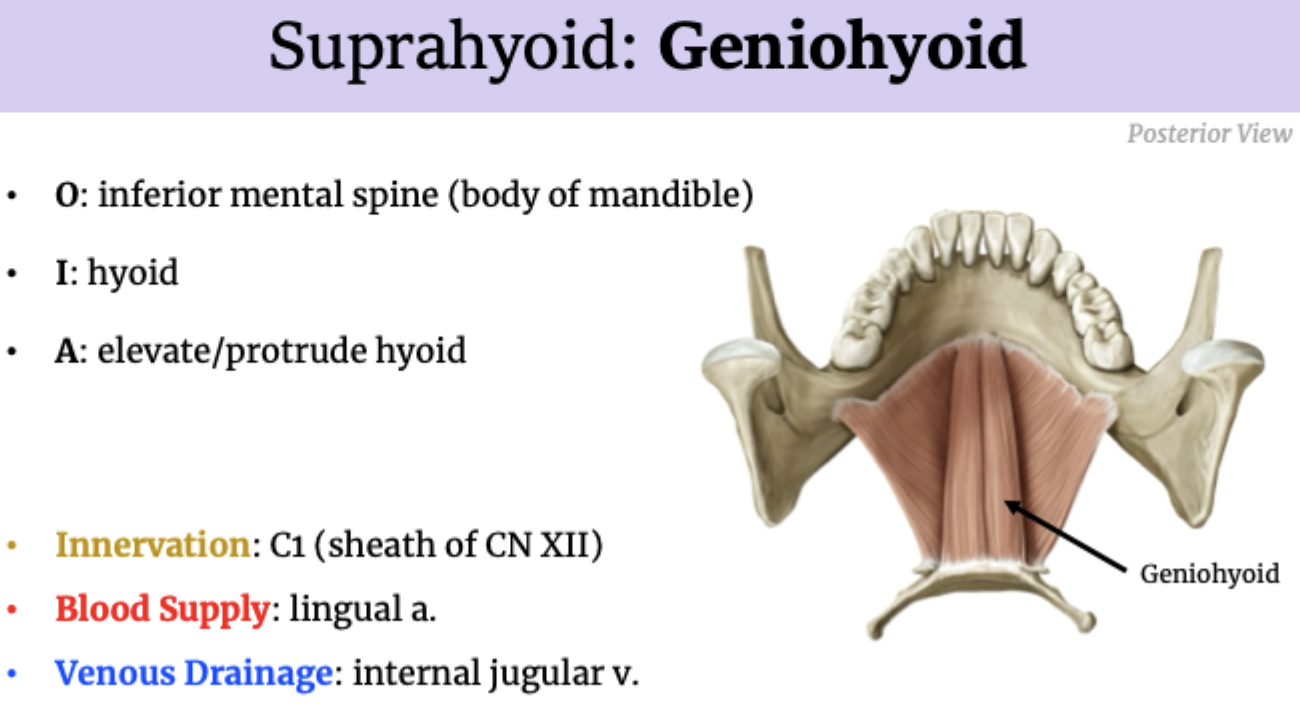
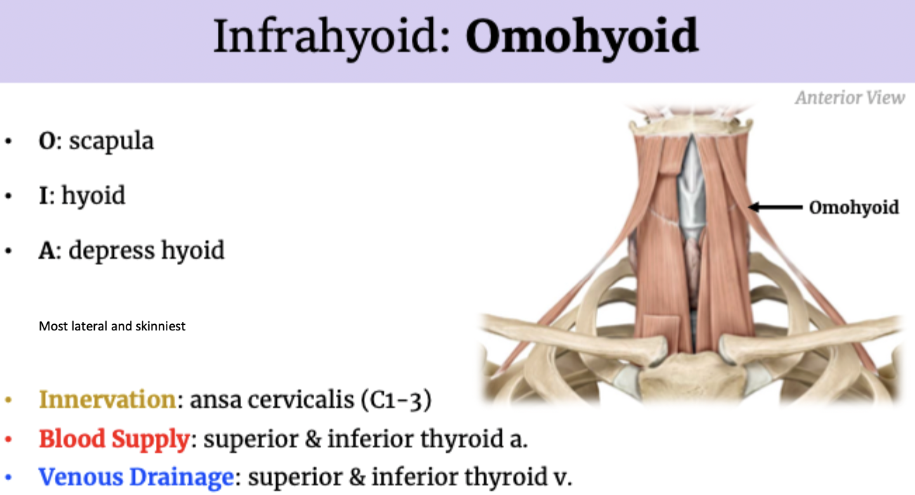
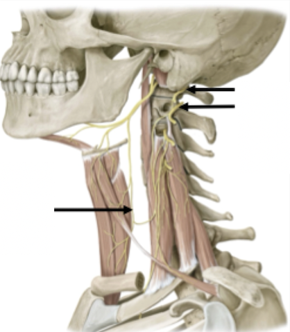
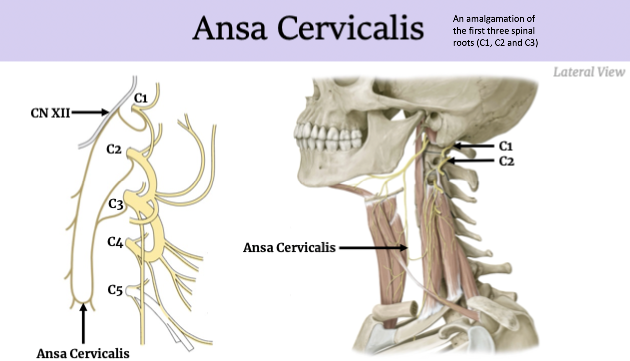
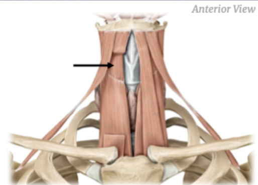
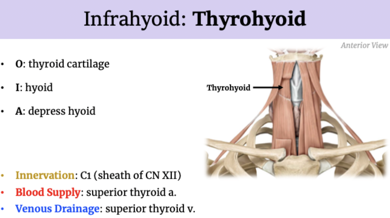
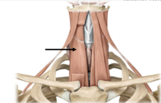
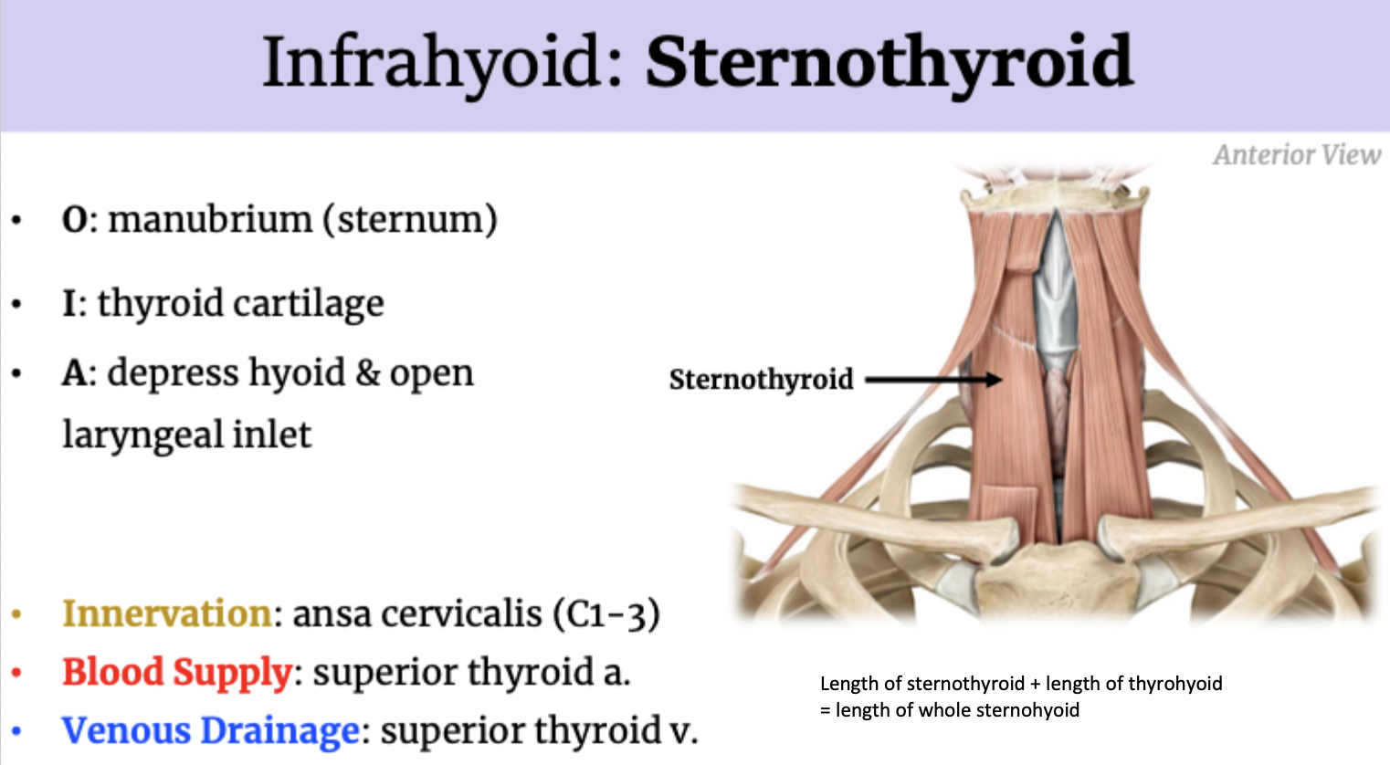
Suprahyoid vs Infrahyoid muscles
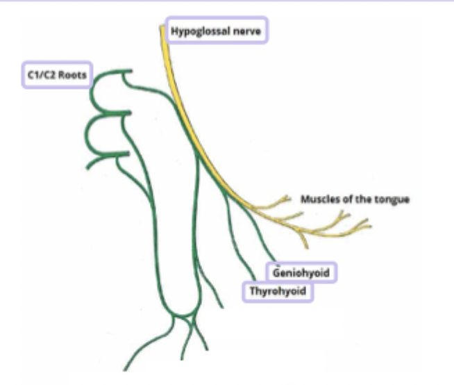
C1 Through CN XII Sheath
C1 travels alongside hypoglossal nerve in the same sheathe and splits off to innervate geniohyoid and thyrohyoid
Innervation: C3-8
Blood Supply: inferior thyroid a.
Venous Drainage: inferior thyroid v.
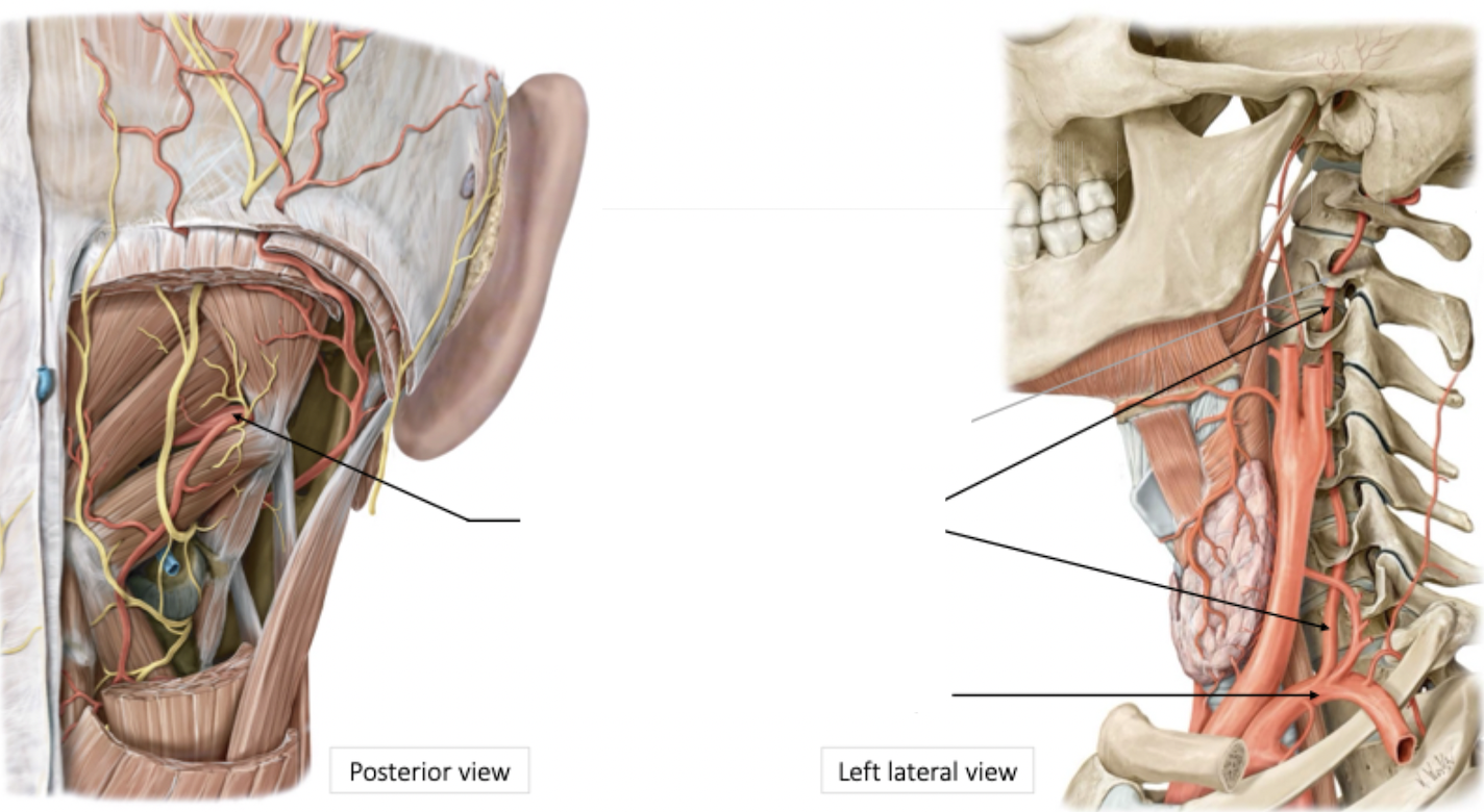
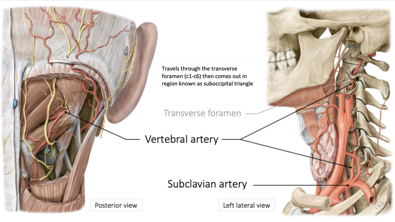
Superficial to deep dissection-based approach to reach the suboccipital triangle!
Remove/reflect trapezius
Remove/reflect splenius capitus
Reflect levator scapulae
Remove/reflect semispinalis capitis
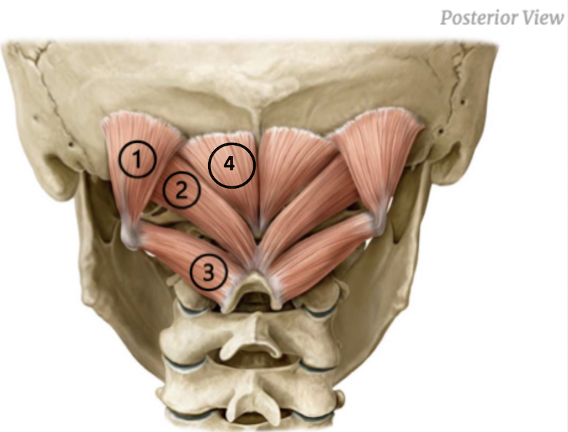
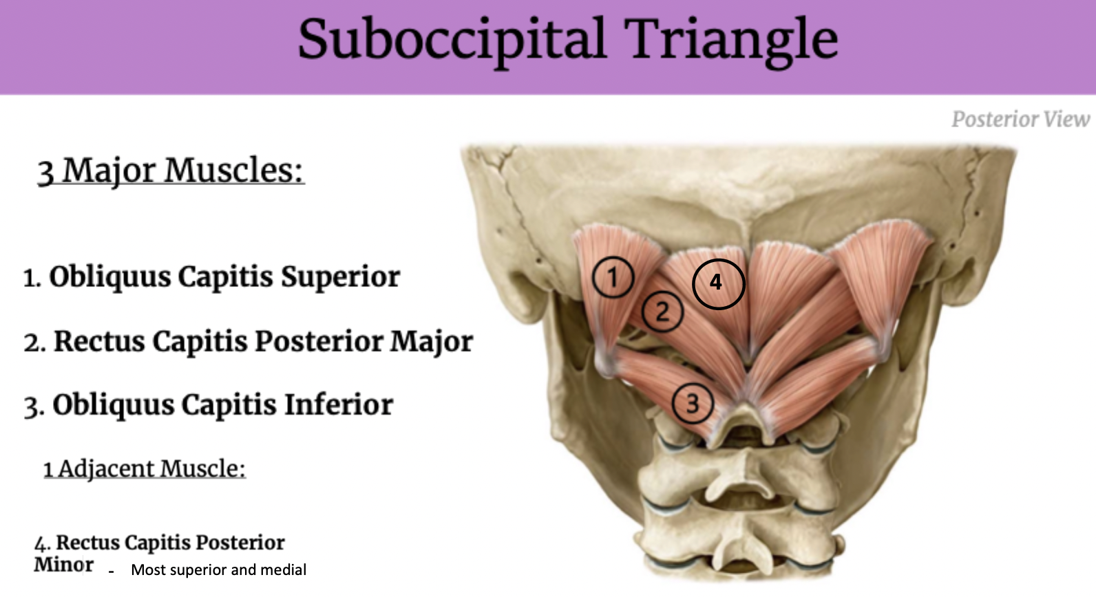
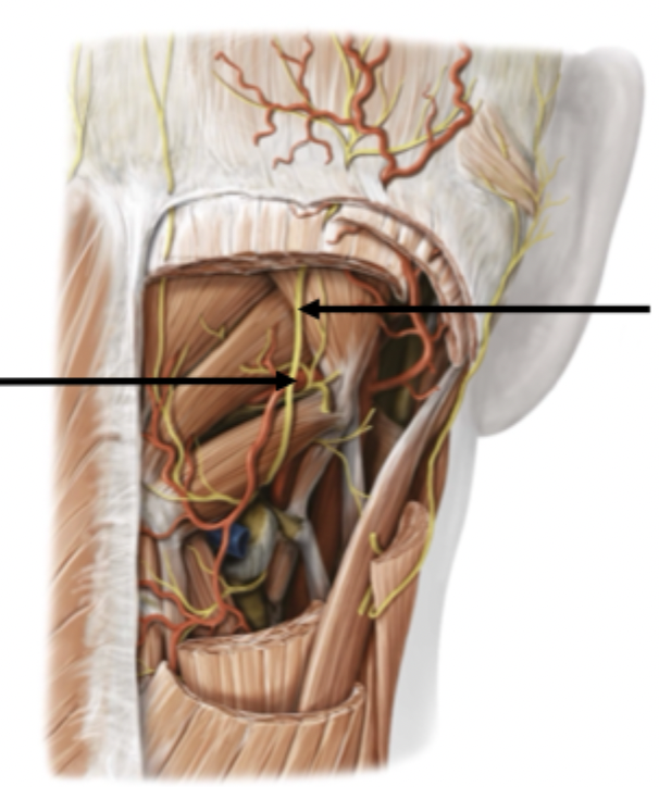
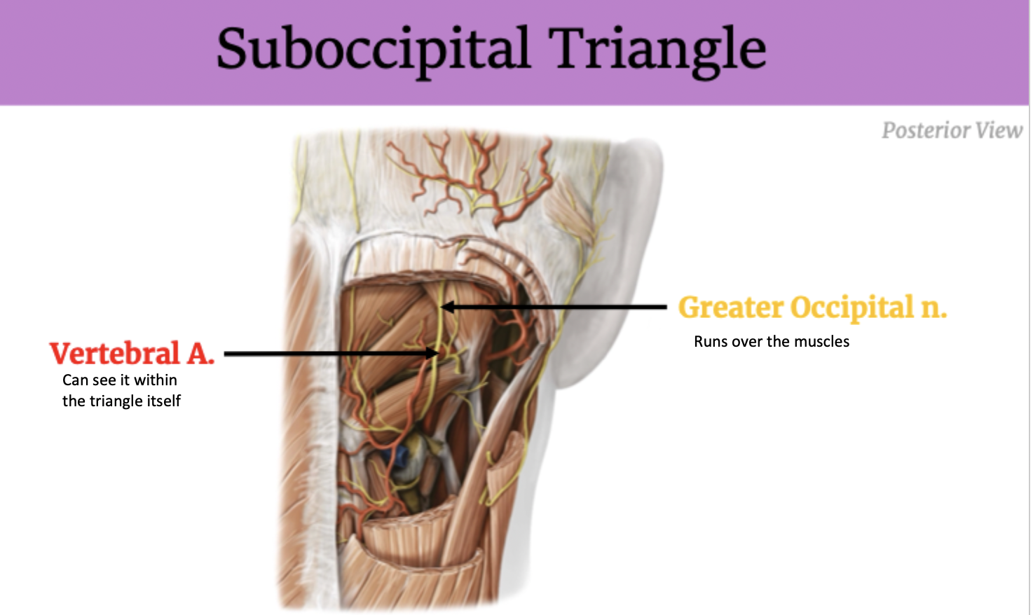
Identify the Suboccipital Triangle
Muscles!
Functions:
Neck extension & rotation
Postural support
Suboccipital Triangle Contents