possible short answer questions for neuroanatomy final
1/25
There's no tags or description
Looks like no tags are added yet.
Name | Mastery | Learn | Test | Matching | Spaced |
|---|
No study sessions yet.
26 Terms
describe the afferent and efferent pathways of the 3 subdivisions of the amygdaloid complex
afferent basolateral
thalamus
visual cortex
orbital cortex
afferent central
hypothalamus
septal nuclei
periaqueductal grey
basolateral amygdala nuclei
afferent corticomedial
olfactory bulb
olfactory cortex
efferent basolateral via amygdalofugal pathway
thalamus (dorsomedial)
ventral striatum (nuc. accumbens)
prefrontal cortex
insula
hippocampus
hypothalamus
efferent basolateral direct output
visual cortex
efferent central via stria terminalis
septal nucleus
hypothalamus
efferent via amygdalofugal pathway
brainstem sites, solitarius, vagal nuclei
efferent corticomedial
anterior olfactory nucleus
olfactory cortex
how do limbic connections contribute to motivational behaviors
Amygdala & limbic cortex project to ventral striatum, dorsomedial nucleus of thalamus, prefrontal cortex. Ventral striatum (VS), which includes nucleus accumbens, receives dense dopamine innervation from the ventral tegmental area. The VS is involved in the neural correlates of reward and the reinforcement of behavior. This enables association between sensory stimuli and behaviors that produce rewards and pleasure
describe and distinguish the layers of the dentate gyrus and the CA fields. include what projections are used in each
dentate gyrus consists of molecular, granular, and polymorphic layer. granular neurons (mossy fibers) provide the main output for the dentate gyrus and project to the apical dendrites of pyramidal cells in the CA3 and CA4 fields.
the CA fields consist of molecular, pyramidal, and polymorphic layers. pyramidal neurons provide the main output for the CA fields and use glutamate as a neurotransmitter.
describe the efferent and afferent pathways of the hippocampus formation
afferent cortical inputs via perforant pathway
entorhinal cortex
posterior cingulate
orbital cortex (via unicate fasc.)
other afferent inputs via fimbria/fornix
septal nuclei (cholinergic inputs)
hippocampus commissure fibers
efferent diencephalic targets
thalamus (anterior nucleus)
mammillary bodies
other hypothalamic sites
efferent telencephalic sites
septal nuclei
ventral striatum
amygdala
neocortex
distinguish between the precommissural and postcommissural fibers of the hippocampus
precommissural
originate in CA1 and CA3
project to septal nuclei and ventral striatum
postcommissural
originate in subiculum
project to mammilary body and thalamus
describe the trisynaptic pathway of the hippocampus
entorhinal cortex to dentate gyrus via perforant pathway
dentate gyrus to CA3 via mossy fibers
CA3 to CA1 via Schaffer collaterals
the trisynaptic pathway uses glutamate as a neurotransmitter
describe the similarities and differences between the sympathetic and parasympathetic subdivisions of the ANS
sympathetic
mediates stress responses (increased heart rate, pupil dilation, suppression of GI tract)
uses cholinergic preganglionic neurons and noradrenergic postganglionic neurons
has a diffuse organization
preganglionic neurons are found in lateral horn of the thoracic-lumbar spinal cord
postganglionic neurons are found in the paravertebral ganglia (sympathetic trunk), prevertebral ganglia in viscera, and chromaffin cells of adrenal medula
parasympathetic
maintains body functions (reduces heart rate, pupil constriction, GI tract activation)
uses cholinergic preganglionic and postganglionic neurons
has a discrete organization
preganglionic neurons are found in the cranio-sacral spinal cord and cranial nerve nuclei
postganglionic neurons are found in ganglia of the head and neck and ganglia close to target organs
describe the four pathways of the sympathetic system
Postganglionic efferent fibers depart through the spinal nerve via the grey communicating ramus to innervate blood vessels and the skin. The ganglia at T1–T5 innervate the heart and lungs.
Ascending efferents often synapse on postganglionic neurons in the cervical ganglia. The superior cervical ganglion innervate the head, eyes, salivary gland, and heart (pupillary dilation). The middle cervical and stellate ganglia innervate the arms, lungs, and heart (bronchial dilation, tachycardia).
Descending efferent fibers synapse on postganglionic neurons in lumbar and sacral ganglia. The postganglionic fibers innervate the lower extremity (skin, blood vessels) via the lumbar and sacral plexuses (vascular dilation, sweating).
Autonomic efferents from the splanchnic nerve synapse on postganglionic neurons in the prevertebral ganglia that lie closer to visceral organs.
describe the ANS descending control pathways
visceral sensory afferents of cortex and amygdala → hypothalamus → brainstem → spinal cord (preganglionic neurons of ANS)
describe the cell proliferation and migration pattern of the myelencephalon, metencephalon, cerebellum, and mesencephalon
myelencephalon (medulla)
alar plate migrate to form inferior olive
roof plate enlarges for 4th ventricle
alar and basal plates form sensory and motor nuclei, respectively
metencephalon (pons)
alar plate migrate to form pontine nuclei
floor plate is thickened by pontine fibers
alar and basal plates form sensory and motor nuclei, respectively
cerebellum
alar plate forms the rhombic lips, which form the cerebellar cortex
mesencephalon (midbrain)
alar plates form the superior colliculi
alar plate migrates to form the red nucleus and substantia nigra
floor plate forms crus cerebri
basal plate forms motor nuclei
describe three congenital effects that can occur during neural development
anencephaly: failure to close cranial (anterior) neuropore
spina bifida: failure to close dorsal vertabrae
hydrocephaly: blockage of ventricular systems
describe the segmentation of CNS from primary vesicles to secondary vesicles to fully formed brain structures
primary vesicles
prosencephalon → telencephalon + diencephalon
mesencephalon → mesencephalon
rhombencephalon → metencephalon + myelencephalon
secondary vesicles
telencephalon → cerebral hemispheres
diencephalon → thalamus + hypothalamus
mesencephalon → midbrain (pontine nuclei)
metencephalon → pons (red nucleus, substantia nigra, crus cerebri)
myelencephalon → medulla (inferior colliculi)
what are the steps of neural development?
neurulation: induction of neural ectoderm
segmentation and pattern formation
cell proliferation
cell migration and differentiation
axonal growth and synapse formation
synaptic stabilization
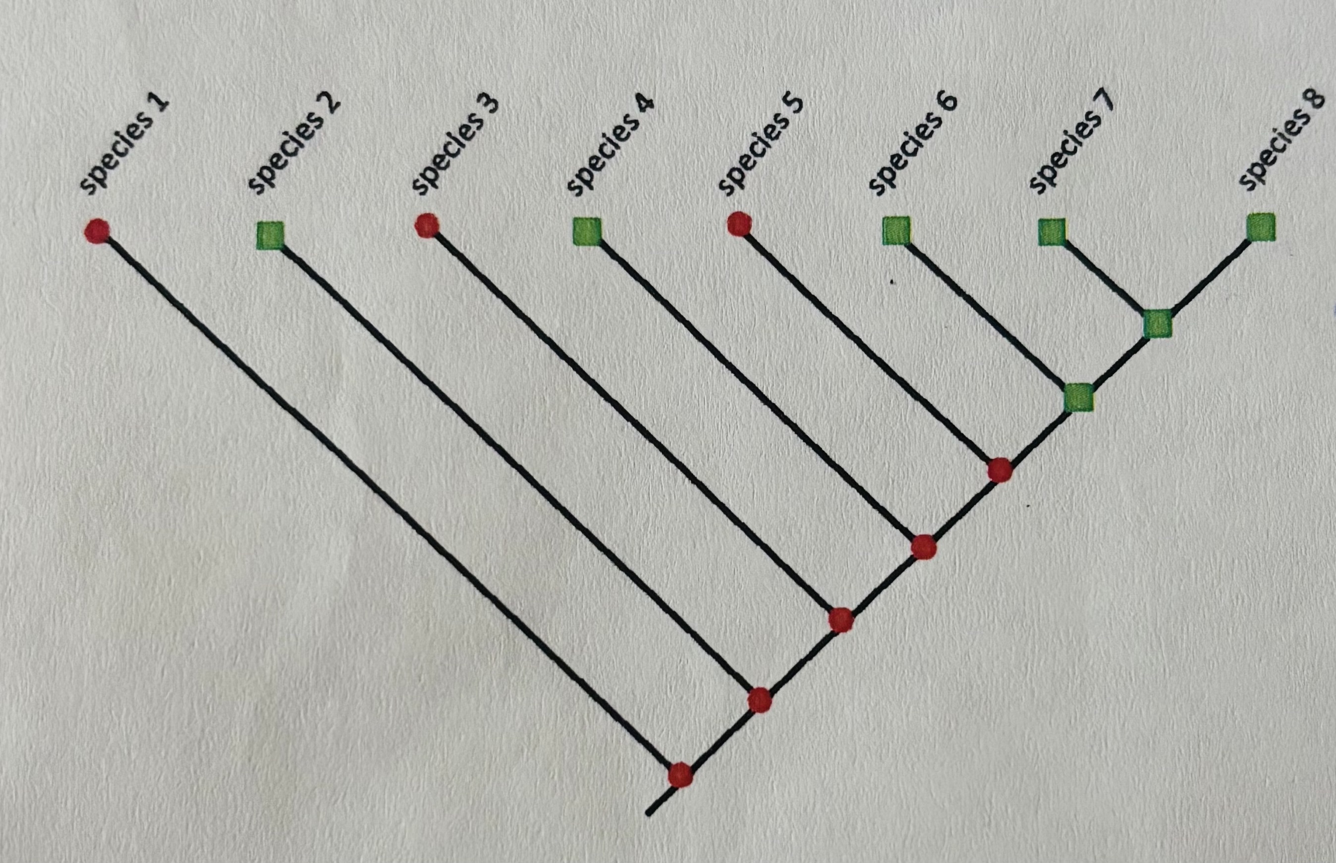
using the cladogram, describe the difference between shared primitive features, shared derived features, and homoplasous features
shared primitive features
old traits from distant ancestors
not monophyletic
example: centralized nerve cords
cladogram: 1 and 3 or 1 and 5
shared derived features
newer traits that evolved in the most recent common ancestor of a group and is unique to that group
monophyletic
example: neocortex in mammals
cladogram: 6 and 8 or 7 and 8
homoplasous features
features that appear similar but arose independently
example: gyri
cladogram: 2 and 4 or 4 and 7
describe the criteria for determining phyletic homology of brain structures
Topological similarity
Topographical organization
embryological development similarity
Neuronal subtype/morphology similarity
Neurochemical/neurotransmitter similarity
Neurophysiological properties
Gene Expression
describe the neuromeric model and its criticisms
CNS has a long RC axis ending at the lamina terminalis
Embryonic CNS is subdivided by neuromeres, each forming a ring around the RC axis
Each neuromere has two longitudinal domains: an alar plate and a basal plate
major criticism: the forebrain does not possess well-defined segments
what characters were present in the first nervous system?
Minimal number of cellular specializations
Reactive to external stimuli
Transduce external information into electrochemical signals
Produce an effector response
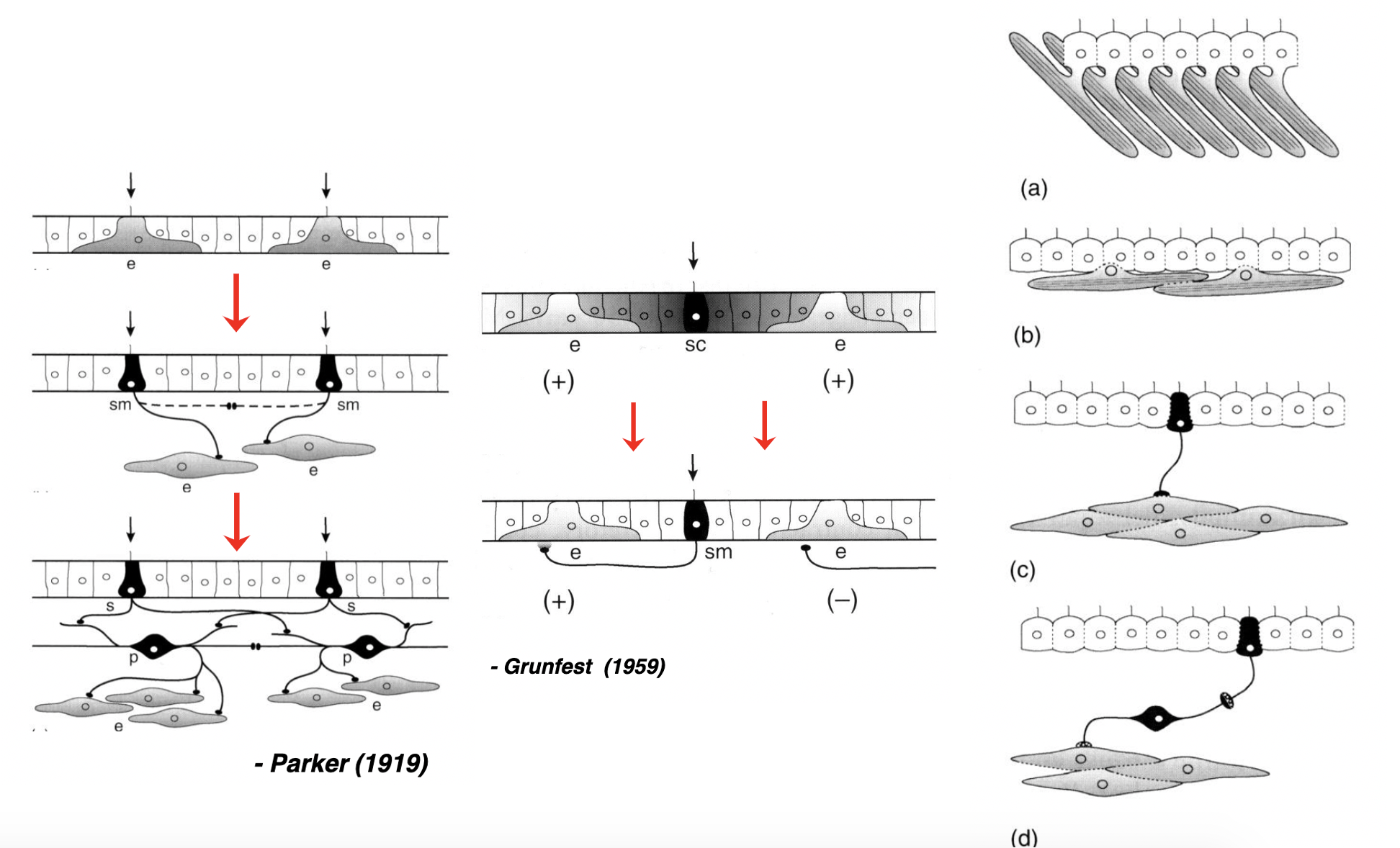
compare the three proposed methods in which the nervous system evolved
describe the differences between a fully-connected network and a regular network, explain which network is optimal for mammals and why
fully-connected (proportional) network
each neuron is directly connected to every other neuron
not possible because he membrane area of a neuron is not large enough to accommodate the synapses, and the metabolic costs of the extra axons would too high
regularly-connected (absolute) network
neurons primarily connect to a small, localized region of adjacent neurons
easy to maintain, but increase the transmission time between brain regions that are widely-separated
describe the three different ways in which brain regions may have evolved
purely mosaic: if the size of a brain region changes independently of other regions
example: visual cortes grows for better sight but other regions are unchanged)
purely concerted: if homologous brain regions show constant proportional changes
example: the forebrain, cerebellum, and brainstem all grow in concert
mildly mosaic: some brain systems show coordinated (concerted) changes, while other regions show independent (mosaic) variation, allowing for specialized adaptations
example: mosaic changes in song-control nuclei of songbird but concerted changes in basic brain structures
describe two species that exhibit examples of mosaic evolution
in mormyrid electric fish, the valvula (part of the cerebellum) is much larger in size compared to other teleocasts, exceeding allometric expectations
olfactory bulbs are smaller in simians than in prosimians, but simian olfactory cortex is much larger than predicted from their small olfactory bulbs
describe and compare the nuclear-to-layered hypothesis and nuclear-to-claustrum/amygdala hypothesis
nuclear-to-layered hypothesis
states that a common ancestor of birds and mammals had a nuclear-based pallium, which evolved into a laminar (layered) structure in the mammalian lineage
direct evolution of ancestral nuclear structures to cortical layers (shared ancestry)
ectostriatum → layer iv, neostriatum → layer ii & iii, archistriatum → layer v & vi
nuclear-to-claustrum hypothesis
states that the avian dorsal ventricular ridge (DVR) represents an elaboration of the mammalian amygdala and claustrum, and that the connectivity that the DVR shares with the neocortex evolved independently
convergent evolution
both avian DVR and mammalian amygdala have nuclear organization and similar connections
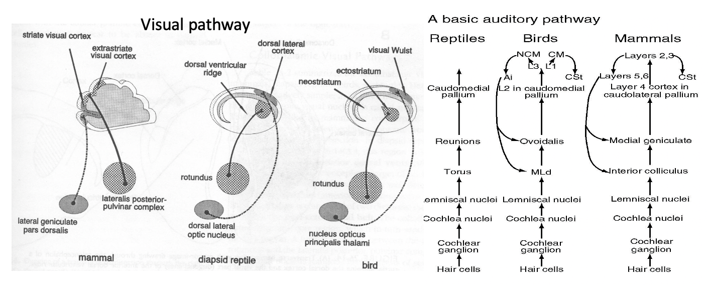
compare two systems between mammals, birds, and reptiles
visual and auditory
describe how the central autonomic network integrates autonomic, endocrine, motor activities
Visceral sensory inputs ascend to the brain via pathways to the NTS and parabrachial nucleus, which relay visceral data to the hypothalamus & amygdala
Descending pathways:
cortex, amygdala → hypothalamus → brainstem → spinal cord
describe the projections of visceral afferent fibers in the ANS. why is visceral pain perceived on the body wall or extremity rather than the affected organ (referred pain)?
Visceral afferent fibers synapse on preganglionic neurons to form an autonomic reflex arc. Second order sensory neurons project to the brain via the spinothalamic tract to convey visceral pain sensations. Visceral pain is perceived on the body wall or extremity rather than at the affected organ (referred pain) because of the convergence of visceral and somatic fibers on second-order neurons in the dorsal horn.
explain why Edward Lewis proposed that duplicate genes play a critical role in evolution
homeotic genes are duplicates of a primordial gene that regulates development of the body.
the original gene of a duplicate gene still performs the old function.
duplicated genes provide extra genes from which new ones arise.
mutations of the duplicated genes enable the performance of new functions