2.1.3 Methods of studying cells
1/30
There's no tags or description
Looks like no tags are added yet.
Name | Mastery | Learn | Test | Matching | Spaced | Call with Kai |
|---|
No analytics yet
Send a link to your students to track their progress
31 Terms
What is magnification and how is it calculated?
Magnification is the number of times greater an image is than the size of the real object.
Magnification equals size of image divided by size of real object.
What is resolution?
The minimum distance apart two objects can be to be distinguished as separate objects, also known as the level of detail.
How does each type of microscope form an image?
Optical: light passes through the specimen; different structures absorb different amounts and wavelengths of light.
TEM: electrons pass through the specimen; denser parts absorb more electrons and appear darker.
SEM: electrons are deflected or bounce off the specimen surface.
What type of image does each microscope generate?
Optical: 2D image of a cross-section.
TEM: 2D image of a cross-section.
SEM: 3D image of the surface.
How is the image focused in each type of microscope?
Optical: light focused using glass lenses.
TEM: electrons focused using electromagnets.
SEM: electrons focused using electromagnets.
What is the resolution quality of each microscope and why?
Optical: low resolution due to the long wavelength of light.
TEM: very high resolution due to the short wavelength of electrons.
SEM: high resolution due to the short wavelength of electrons.
What internal structures can each microscope see?
Optical: cannot see internal structures of organelles or ribosomes.
TEM: can see internal structures of organelles and ribosomes.
SEM: cannot see internal structures, only surface details.
What type of specimen does each microscope require?
Optical: specimen must be thin.
TEM: specimen must be very thin.
SEM: specimen does not need to be thin.
What is the maximum magnification of each microscope?
Optical: low magnification, up to x1500.
TEM: high magnification, up to x1,000,000.
SEM: high magnification, up to x1,000,000.
Can each microscope view living specimens?
Optical: yes, can view living organisms.
TEM: no, can only view dead or dehydrated specimens as it uses a vacuum.
SEM: no, can only view dead or dehydrated specimens as it uses a vacuum.
How complex is the preparation for each microscope?
Optical: simple preparation.
TEM: complex preparation, so artefacts are often present.
SEM: complex preparation, so artefacts are often present.
Can each microscope detect or show colour?
Optical: yes, can detect and show colour.
TEM: no, does not detect or show colour.
SEM: no, does not detect or show colour.
How did scientists distinguish between artefacts and cell organelles?
Scientists prepared specimens in different ways.
If an object was seen with one technique but not another, it was more likely to be an artefact than an organelle.

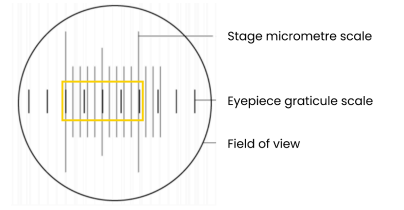
What is the first step in measuring the size of an object viewed with an optical microscope?
Line up the scale of the eyepiece graticule with the scale of the stage micrometre.

What is the second step in measuring the size of an object?
Calibrate the eyepiece graticule by using the stage micrometre to calculate the size of divisions on the eyepiece graticule.

What is the third step in measuring the size of an object?
Remove the stage micrometre and use the eyepiece graticule to measure how many divisions make up the object.
What is the fourth step in measuring the size of an object?
Calculate the size of the object by multiplying the number of divisions by the size of each division.

What is the fifth step in measuring the size of an object?
Recalibrate the eyepiece graticule at different magnifications.
In the worked example, how many eyepiece graticule divisions aligned with 10 stage micrometre divisions?
4 eyepiece graticule divisions equal 10 stage micrometre divisions.
In the worked example, what is the value of 1 subdivision on the stage micrometre?
1 subdivision equals 10 μm.
In the worked example, what is the total value of 10 stage micrometre divisions?
10 μm multiplied by 10 equals 100 μm.
In the worked example, what is the value of 1 eyepiece graticule division?
100 μm divided by 4 equals 25 μm.
What is the first step in cell fractionation and what does it do?
Homogenise tissue or use a blender, which disrupts the cell membrane, breaking open cells to release contents and organelles.
Why is the homogenate placed in a cold solution?
To reduce enzyme activity, so organelles are not broken down or damaged.
Why is the homogenate placed in an isotonic solution?
So water does not move in or out of organelles by osmosis, preventing them from bursting or shrivelling.
Why is the homogenate placed in a buffered solution?
To keep pH constant, so enzymes do not denature.
What is the third step in cell fractionation?
Filter the homogenate to remove large, unwanted debris such as whole cells and connective tissue.
What is the purpose of ultracentrifugation?
To separate organelles in order of density or mass.
What happens during the first spin in ultracentrifugation?
Centrifuge the homogenate in a tube at a low speed, then remove the pellet of the heaviest organelle.
What is done after removing the pellet?
Re-spin the supernatant at a higher speed.
What happens when the process is repeated at increasing speeds?
Each time, the pellet is made of lighter organelles, separating in the order: nuclei, then chloroplasts or mitochondria, then lysosomes, then endoplasmic reticulum, then ribosomes.