Exam 1 : WBC
1/56
There's no tags or description
Looks like no tags are added yet.
Name | Mastery | Learn | Test | Matching | Spaced | Call with Kai |
|---|
No analytics yet
Send a link to your students to track their progress
57 Terms
myelopoiesis (granulopoiesis)
growth and development of granulocytic and monotocytic cells in the bone marrow (WBCs)
WBC maturation takes
7-11 days
granulocytes vs agranulocytes
granulocytes can digest microorganisms; agranulaocytes cannot
Granulocytes
neutrophils, eosinophils, basophils
Agranulocytes
monocytes and lymphocytes
lymphocytes
immune response
granulocytes and monocytes are responsible for
phagocytosis
phagocytosis
ingest bacteria and dispose of dead matter
chemotaxis
movement by a cell or organism in reaction to a chemical stimulus
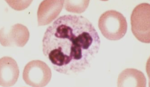
neutrophils
most abundant; phagocytosis; bands/segments; typically 3-5 segments
-increased in bacterial infection!!!

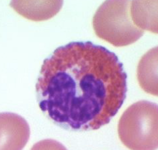
eosinophils
phagocytic; pink; two nuclei; 12-17um
destroy parasitic worms!!

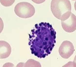
basophils
phagocytic; all purple; granular; one nuclei
response in allergic reactions!!
-increase inflammation

lymphocytes
immune response; B and T cells
-increased in viral infections!!
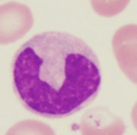
monocytes
can differentiate into macrophages and phagocytize debris; >20um; largest; large monocyte

platelets features
made from megakaryocytes; tiny pink; no nucleus

erythrocyte features
round pink; light middle
lymphocyte features
light purple with dark purple middle
-B all purple no bands/segments
-T smaller all purple; mostly nuclei
neutrophil maturation
myeloblast, promyelocyte, myelocyte, metamyelocyte, band, seg
erythrocyte maturation
rubriblast-->prorubicyte-->rubricyte-->metarubicyte-->polychromatophilic erythrocyte-->mature erythrocyte
-blast
baby
monocyte maturation
Myeloid stem cells -> monoblasts -> promonocytes -> monocytes
lymphoid: plasma maturation
lymphoid stem cell -> pre B -> B lymphoblast -> B cell -> plasma cell
lymphoid: T cell maturation
lymphoid stem cell -> bone marrow to thymus -> prothymocyte -> t lymphoblast -> T cell
hypersegmention
neutrophils with nuclei that have 6 or more lobes
-B12 and folate deficiencies
Hyposegmentation
(Pelger-Huet anomaly) when neutrophils are bilobed/dumbell shaped
T/F it only takes one cell to diagnose hyper/hypo segmentation
F must be several abnormal cells present
neutrophil cytoplasmic inclusions
-bacterial/acute infections
toxic granulation
accentuation of normal cytoplasmic granules. Granules become more numerous, larger and stain a deeper blue-black color
vacuolization
vacuoles appear as "holes" in the cytoplasm
Dohle bodies
large, gray blue structure is neutrophil cytoplasm
-typically near the border
reactive lymphoctyes
increased size, may be round, elongated, or lobulated
-viral infections!! ex mono
-cytoplasm is "soft" and indented by RBCs
ineffective granulopoiesis
increase number of WBC precursor and decreased mature WBC in peripheral blood
ineffective granulopoiesis: secondary problem
signal is sent but stem stems cannot get to WBC
ineffective granulopoiesis: primary problem
no stem cells
megakaryopoiesis
platelet production
platelet maturation
stem cell, megakaryoblast, promegakaryocyte, megakaryocyte, thrombocyte
megakaryocytic splinter to create
platelets; occurs in the red bone marrow -> enter the peripheral blood circulation
platelet life span
5-9 days
what organs remove old platelets
spleen and liver
ineffective thrombopoiesis
Increased abnormal megakaryocytes in the bone marrow
Thrombocytopenia in peripheral blood
CBC requires
lavender tube : EDTA
WBC differential requires _ stain
Wright's stain
WBC differential reports on
neutrophils, lymphocytes, monocytes, eosinophils, basophils
(Never, Let, Monkeys, East, Bananas)
WBC differential ranges
Neutrophils: 60
Lymph: 40
Monocytes: 10
Eosinophils: 2
Basophils: 0
left shift
increase in the number of immature neutrophils
-typically an early response to infection
-"bandemia"
platelet range
150-400
-worry when it comes to bleeding
-too low: prolonged bleeding
-too high: clots
reticulocyte count
they are seen in peripheral samples!!
von Willebrand's disease
bleeding disorder
factor deficiency anemia
-cancer and sickle cell
erythrocyte sedimentation rate
detects inflammatory conditions
-very non specific!!!
-auto immune diseases
changes in the ESR are
result of changes in plasma protein levels
following an acute inflammatory process or acute injury protein levels will
increase
ESR
measures sedimentation rate of red blood cells; indicates inflammation; how fast it fills
G6PD deficiency
deficient enzyme activity
-whole blood anticoagulated w/ EDTA or heparin
several G6PD enzyme variants exists with what populations
greek, Italian, jewish
bone marrow studies are used to diagnose
polycythemia vera, acute and chronic leukemia, myelodysplastic syndrome, aplastic anemia
bone marrow tissue sample
drawn from the pelvis