VIBS 243 Final Review
1/153
Earn XP
Name | Mastery | Learn | Test | Matching | Spaced | Call with Kai |
|---|
No analytics yet
Send a link to your students to track their progress
154 Terms
The star’s located in which compartment of the lymph node?

A. Medullary Sinus
B. Medullary Cords
C. Cortex sinus
D. Cortex cords
E. Nodule/follicle
F. None of these
A
The arrows are pointing to which cells?

A. Plasma Cells
B. Mast Cells
C. Macrophages
D. Neutrophils
E. Lymphocytes
B
B lymphocytes primarily reside in which compartment?

A
B
A and B
None of the above
A
Which is correct regarding the flow of lymph fluid?
Tissue fluid, Lymphatic capillary, Afferent Lymphatic vessel, medullary sinus subcapsular sinus, trabecular sinus, efferent lymphatics
Tissue fluid, Lymphatic capillary, Efferent Lymphatic vessel, subcapsular sinus, trabecular sinus, medullary sinus, afferent lymphatics
Tissue fluid, Lymphatic capillary, Afferent Lymphatic vessel, subcapsular sinus, trabecular sinus, medullary sinus, efferent lymphatics
Lymphatic capillary, Afferent Lymphatic vessel, Tissue fluid, subcapsular sinus, trabecular sinus, medullary sinus, efferent lymphatics
3
Which of the following are part of the innate immune system?
Skin
pH
Mucosa
Apical modifications such as cilia
All of the above
None of the above
5
Which image of the lymph node represents one that is responding to a vaccine?

A
B
A and B
None of these
A
The arrow is pointing to which cell?

Plasma cell
Macrophage
Neutrophil
Mast Cell
Lymphocyte
2
The arrows are pointing to which cells?

Plasma cell
Macrophage
Neutrophil
Mast Cell
3
Which of the following is true regarding the cell type primarily seen in the cord?

Lymphocytes respond to antigens by binding to their epitopes.
Plasma cells will secrete antibodies in response to an antigen.
Macrophages will engulf bacteria and infected cells.
Mast cells will release their granules filled with histamine.
2
The medullary cords in the image primarily contain which cell type?

Macrophages
Plasma Cells
Neutrophils
Lymphocytes
4
Which of the following cells are considered tissue bound?
Mast cells, macrophages, and lymphocytes
Lymphocytes, plasma cells, and monocytes
Monocytes, neutrophils, and lymphocytes
Mast cells, macrophages, and plasma cells
4
Which of the following is/are true about the organ pictured below?

It is a lymph node
It contains lymphocytes
It filters lymph fluid
All of the above
None of the above
2
What cell is indicated by the arrows below?

Thymocyte
Macrophage
Epithelial Reticular Cell
Dendritic Cell
3
What statement is FALSE about thymocyte development within the thymus?
A. Prothymocytes enter the thymus from the bone marrow through corticomedullary vessels
B. Thymocyte receptors are first tested in the cortex
C. In positive selection, thymocytes are selected for apoptosis if they react strongly with the self
D. Specific CD4 or CD8 T cell receptors are tested in the medulla
C
Which of the following is FALSE about the image below?

A. It is in the thymus cortex
B. It contains more mature thymocytes that are less densely packed
C. It contains Hassall’s Corpuscles that are concentric layers of epithelial reticular cells that are degenerating
D. It contains dendritic antigen-presenting cells
A
Which of the following is true about the image below?

A. It is surrounded by a capsule of connective tissue
B. Septa extensions divide the organ into different lobes
C. Each lobe is divided into a cortex and medulla
D. It is the thymus
E. All of the above
E
Which of the following is true in regard to antigen-antibody interactions?

a. A single antibody can respond to hundreds or thousands of different antigens
b. A single cell will only produce a single type of antigen
c. Antibodies can only respond to the exact antigen they were made for
d. None of the above are true
c
Which of the following is FALSE about the thymus?
a. It is the site of maturation into CD4 or CD8 T Cells
b. It is lobulated
c. It is avascular
d. It has consistent structure across species
c
Which of the following is true regarding the image of the lymph node?

a. The nodules/follicles are secondary, so the lymph node is responding to antigens.
b. The nodules/follicles are secondary, so the lymph node is not responding to antigens.
c. The nodules/follicles are primary , so the lymph node is responding to antigens.
d. The nodules/follicles are primary, so the lymph node is not responding to antigens.
a
Which of the following statements is true regarding the image of the lymph node?

a. The nodules/follicles are secondary, so the lymph node is responding to antigens.
b. The nodules/follicles are secondary, so the lymph node is not responding to antigens.
c. The nodules/follicles are primary, so the lymph node is responding to antigens.
d. The nodules/follicles are primary, so the lymph node is not responding to antigens.
d
What is true about the function of Epithelial Reticular Cells?
a. They provided the structural framework for lymph nodes
b. They create the blood-thymus barrier
c. They help in positive and negative selection of macrophages
d. None of the above
b
The arrow is pointing to which cell?

a. Plasma Cells
b. Mast Cells
c. Macrophages
d. Neutrophils
e. Lymphocytes
a.
True or False. The lymph node filters blood?
False
The star is located in which compartment of the lymph node?

a. Medullary Sinus
b. Cortex Sinus
c. Medullary Cord
d. Cortex Cord
e. Nodule/Follicle
c
What cell in the thymus cortex is pictured below?

A. Thymocyte
B. Dendritic cells
C. Macrophages
D. Epithelial reticular cells
C
Which of the following is true about the cells in the thymus cortex?
thymocytes are very densely packed
macrophages ingest apoptotic thymocytes
epithelial reticular cells are mainly structural and make the blood-thymus barrier
all of the above
4
Which of the following is correct when a lymph node responds to an antigen?
A. T lymphocytes proliferate in the germinal centers, move out of the nodule, differentiate into plasma cells which will then make antibodies.
B. T lymphocytes proliferate in the germinal centers, move out of the nodule, differentiate into macrophages which will then make antibodies.
C. B lymphocytes proliferate in the germinal centers, move out of the nodule, differentiate into plasma cells which will then make antibodies.
D. B lymphocytes proliferate in the germinal centers, move out of the nodule, differentiate into macrophages which will then make antibodies.
C
Which of the following best describes an antigen and the immune system's response?
a. An amino acid sequence specific for the cell, and antibodies will be nonspecifically bind to it.
b. A molecular structure present on cells, and antibodies made to this antigen will be specific.
c. A molecular structure on the inside of cells that is undetectable by the immune system.
d. An amino acid sequence that doesn't provoke an immune response due to the cleverness of the virus.
b
Which of the following organs is the site of T Cell Maturation?
a. Lymph Node
b. Tonsils
c. Thymus
d. Spleen
c
The arrows are pointing to which cells in the lymph node?

a. Plasma Cells
b. Mast cells
c. neutrophils
d. Macrophages
e. None of the above
d
Which of the following are part of the acquired immune system?
a. Plasma cells
b. Neutrophils
c. Macrophages
d. Mast cell
e. All of the above
f. None of the above
a
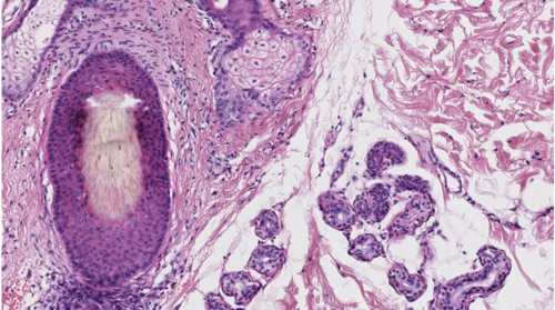
Which of the following structure(s) are located in the dermis?
Hair follicles
Sebaceous glands
Sweat glands
Arrector pili muscle
A, B, and C
A, B, C, and D
6
Which of the following is correct regarding the image below?

Layer A is the stratum basale
Layer B contains melanocytes
Layer C is the stratum corneum
Layer D contains cells that can undergo mitosis
4
This is a cytology image from a mass. The cells with the brown pigment are…

Stratum corneum cells
Stratum basale cells
Melanocytes
Stratum granulosum cells
3
What glandular structures are seen in the image below?

White fat
Sweat glands
Sebaceous glands
Arrector pili muscle
2
What structure is seen in the image below?

A. Meissner’s corpuscle
B. Pacinian corpuscle
C. Sweat gland
D. Sebaceous gland
B
Which structure(s) best match(es) the image(s)?

Stratum spinosum
Sebaceous gland
Eccrine sweat glands
A and B
A, B, and C
3
Melanin granules cap the nuclei of the stratum basale to:

Prevent UV rays of sunlight from damaging the cytoplasm
Enhance UV rays of sunlight to damage DNA in nuclei
Prevent UV rays of sunlight from damaging DNA in nuclei
A and B
A, B, and C
3
The duct in the image below belongs to which gland?

Eccrine sweat gland
Apocrine sweat gland
Sebaceous gland
Messiner’s corpuscle
1
Which is/are characteristic(s) related to melanin or melanocyte?
Freckles are produced by melanin distributed in patches
Melanocytes produce melanin to give away to other cells
Malignant melanomas is a cancer related to melanocytes
A and B
A, B, and C
5
Which of the following is true regarding the image below?

A. There is a hair follicle in the image
B. There are sweat glands in the image
C. Both the dermis and epidermis are visible
D. These structures are located in the subcutaneous region
E. A, B, and C
F. A and C
G. A, B, and D
F
The arrow is pointing to which structure in the TEM?

Desmosome
Basement membrane
Mitotic figure
Dead cell
1
Which of the following is true regarding the image below?

Pacinian corpuscles will be found in the area indicated by the yellow arrow.
Meissner's corpuscles will be found in the area indicated by the yellow arrow.
Capillaries will be found in the area indicated by the yellow arrow.
A and C
B and C
A and C
5
Which adnexa is seen in the image?

A. Dermis
B. Hair follicle
C. Arrector pili muscle
D. Sebaceous glands
E. All of the above
F. B, C, and D
F
The layer indicated by the blue arrow in the picture on the left corresponds to which histological layer in the image on the right?

A
B
C
None of the above
C
What two regions of the skin is visible in this image below?

Epidermis
Dermis
Subcutaneous
A and B
B and C
A, B, and C
4
'Melanin capping,' or the deposit of melanin over the nuclei of the cells of the stratum basale, is designed to-

A) Protect the nuclei of this layer from UV light-induced damage
B) Protect the nuclei of this layer from dividing
C) Encourage UV light to penetrate deep into the nuclei of this layer
D) Provide nutrient to the nuclei of this layer
A
Which image(s) match(es) basic functions of the integument?

A) Fat metabolism in the subcutaneous layer
B) Prevents desiccation and allows terrestrial (land-welling) existence
C)Basis of recognition and yields clues to one's well being
D) A and B
E) A, B, and C
E
Which answer choice below is true regarding the structure in the image?

A) Secretions from this gland functions in water-proofing
B) Secretions from this gland functions in antibacterial properties
C) The duct system from this gland empties directly onto skin surface
D) A and B
E) A and C
F) A, B, and C
D
Which of the following is correct regarding the TEM image below?

A) This is the interface between stratum basale and dermis
B) This area contains adhesion molecules in the basement membrane
C) This area will contain capillaries to supply the epidermis
D) A and B
E) A and C
F) A, B, and C
F
Which of the following is correct regarding the image?

a. Layer labeled A contains dead, flat cells
b. Layer B contains cells that can undergo mitosis
c. Layer C contains cells that have keratohyalin granules
d. Layer D contains cells that are dead
a
The cytology image below came from which layer of the epidermis?

a. Stratum Corneum
b. Stratum Granulosum
c. Stratum Spinosum
d. Stratum Basale
d
The sweat glands in the image are considered......
a. Sebaceous glands
b. Apocrine
c. Eccrine
d. Arrector Pili
b
Which of the below are cells found in the epidermis?
a. Keratinocytes
b. Melanocytes
c. Sebaceous Gland secretory cells
d. A and B
e. A, B, and C
d
Which of the following is not a function/feature of melanin?
a. skin pigmentation
b. produced by keratinocytes
c. passed by cytocrine secretion from one type of cell to another type
d. a & b
e. a, b & c
b
The blue star in the image below is located in which compartment of a tubular organ?
Mucosa
Submucosa
Tunica muscularis
Serosa
2
The black arrow in the image below is indicating a structure. Choose the best answer in regards to the structure indicated by the arrow.

The blood vessel is responsible for providing oxygen to the villi
The goblet cell is responsible for secreting mucus into the lumen
The intestinal crypt is responsible for providing replacement of epithelial cells
The central lacteal is responsible for transporting lipids and lymph
All of the above
None of the above
4
What is the function of the structure indicated by the blue arrow in the image?

Secretion of mucous
Absorption of fats
Absorption of protein
Absorption of carbohydrates
B, C, and D
C and D
A, B, C, and D
5
The image shows an abnormal structure in the intestine. What is the consequence of this abnormal structure?

Loss of lymphocytes
Loss of proteins
Loss of weight
Increase in lipid absorption
A and B
A, B, and C
A, B, C, and D
6
The boxed in area in the image below clearly shows some structures that help to increase the surface area of the small intestine. What does this image show?

A. Short length of the small intestine
B. Plica circularis
C. Villi
D. Microcilia
E. B and C
F. B, C, and D
G. A, B, C, and D
E
Which of the below is/are true?
There are more goblet cells in the small intestine than large intestine
Microvilli is present only in the small intestine and not the large intestine
The large intestine absorbs more than the small intestine
Goblet cells are only found in the large intestine
C and D
B and C
A and C
None of the above
8
The scanning electron image shows the luminal surface of an organ. Which of the following is true regarding the image?

Plica circularis are easily seen in the image
Villi are easily seen in the image
Microvilli are easily seen in the image
B and C
A and B
A, B, and C
2
The image shows an abnormal structure in the intestine. What is the consequence of this abnormal structure.

No replacement of epithelial cells
Exposed lamina propria
Decreased absorption
Bloody diarrhea
C and D
A and B
A, B, C, and D
7
Which of the following are FALSE?
The large intestine does not have villi
The large intestine does not have intestinal crypts
The large intestine does not contain mitotic figure
B and C
A, B, and C
None of the above
4
The yellow arrows in the image are pointing to cells. Please choose the correct response in regard to these cells.

Mitotic figures found in the crypts provide replacement for connective tissue
Mitotic figures found in the crypts provide replacement for epithelial cells
Mitotic figures found in the villi tips provide replacement for epithelial cells
Mitotic figures found in the villi tips provide replacement for connective tissue
A and B
C and D
None of the above
2
Which of the following is TRUE about a central lacteal?
They are lined by endothelium
They transport lymph fluid
They transport absorbed lipids
All of the above
4
The scanning electron image shows the luminal surface of an organ. Which of the following is/are true regarding the image.

Intestinal crypts are easily seen in the image
Villi are easily seen in the image
Microvilli are easily seen in the image
B and C
A and B
A, B, and C
1
Which of the following is TRUE about the organ pictured below?

It is the small intestine
It functions in absorption of lipids, carbohydrates, and proteins
It contains intestinal crypts
It contains intestinal villi
3
Which of the following is true about intestinal villi?
They function to increase absorption
They are a series of permanent folds made of the mucosa and submucosa
They are found in both the small and large intestine
All of the above
1
Which of the following parts of the small intestine is paired correctly with their components?
Mucosa - epithelium and lamina propria
Submucosa - collagenous connective tissue and glands
Tunica muscularis - double layer of muscle
All of the above
4
The blue star in the image below is located in which compartment of a tubular organ.

a. Mucosa
b. Submucosa
c. Tunica Muscularis
d. Serosa
c
Which of the below is true?
a. Microvilli is present in only the small intestine and not large intestine
b. The large intestine absorbs more than the small intestine
c. Goblet cells are only found in the large intestine
d. None of the above
d
Which organs is/are part of the digestive system?
a. Oral Cavity
b. Esophagus
c. Small Intestine
d. Rectum
e. All of the Above
e
Which of the below is/are FALSE.
a. There are more goblet cells in the small intestine than large intestine
b. Microvilli is present in only the small intestine and not large intestine
c. The large intestine absorbs more than the small intestine
d. Goblet cells are only found in the large intestine
e. C and D
f. B and C
g. A and C
h. A, B, C, and D
i. None of the above
h
The blue star in the image below is located in which compartment of a tubular organ.

a. Mucosa
b. Submucosa
c. Tunica Muscularis
d. Serosa
a
Which of the below is/are true.
a. The large intestine does not have villi.
b. The large intestine does not have intestinal crypts.
c. The large intestine does not contain mitotic figures.
d. B and C
e. A, B, and C
f. None of the above
a
Which of the following describes an exocrine gland?
Secretes enzymes
Has an extensive duct system
Includes the liver and pancreatic acinar cells
All of the above
4
Which of the following is true about the endocrine system?
Produces digestive enzymes
Helps regulate metabolism and reproduction
Has an extensive duct system
Has limited access to blood vessels
2
What organ that acts as a master endocrine gland and plays a major role in regulating vital body functions is pictured below? Or What organ is the master endocrine gland and plays a major role in regulating vital body functions and controls the activity of most other hormone-secreting glands?

Pancreas
Pituitary
Liver
Thyroid
2
Which of the following is true about the thyroid gland?
It secretes thyroglobulin
It secretes calcitonin
It is under pituitary gland control by TSH
It is vital in metabolism regulation
All of the above
5
Which of the following cells are labeled correctly in the image below?

Blood capillaries
Parafollicular cells
Simple cuboidal follicular epithelium
All of the above
1
Which of the following statements is FALSE about the cells pictured below?

It is a parafollicular cell
It produces thyroglobulin
It is found in the thyroid gland
All of the above are true
2
Which of the following is true about the structure pictured below?

It is found in the thyroid gland
It contains colloid
It stores thyroglobulin
All of the above
4
A deficiency in what chemical can result in a colloid goiter?
Potassium
Calcium
Iodine
Sodium
3
Which of the following is FALSE about the organ pictured below?

It is the thyroid gland
It secretes bile into bile canaliculi
It has endocrine functions
All of the above are true
1
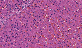
Which of the following is true about the cells pictured below?
They are liver hepatocytes
They store glucose in the form of glycogen
They have connections to bile canaliculi to secrete exocrine bile
All of the above
4
How are endocrine and exocrine products kept separate in liver hepatocytes?
By gap junctions around bile canaliculi
By having different cells secrete each product
By tight junctions around bile canaliculi
They are not kept separate and are encouraged to mix
3
In the image below, what metabolite is excessively accumulating within the hepatocytes to cause steatosis?

Carbohydrates
Lipids
Bile
Proteins
2
Which of the following is FALSE about the area of the pancreas highlighted below?

It is the Islet of Langerhans
It secretes insulin and glucagon
It has an extensive duct system
All of the above are true
3
What cell in the endocrine pancreas is paired with its correct secretion?
Beta cells- Insulin
Alpha cells- Glucagon
Delta cells- Somatostatin
All of the above
4
What organ is labeled correctly in the image below?

Pancreas
Thyroid
Liver
All of the above
2
What organ is pictured below?

a. Liver
b. Pituitary
c. Thyroid
d. Pancreas
d
What organ is pictured below?

a. Liver
b. Pituitary
c. Thyroid
d. Pancreas
c
Which of the following is part of the endocrine pancreas?
a. Acinar cells
b. Pancreatic ducts
c. Islet of Langerhans
d. All of the above
c
What organ is divided into follicles that contain colloid?
a. Pituitary
b. Pancreas
c. Thyroid
d. Liver
c
What statement is FALSE about the thyroid gland?
a. It is important in metabolism regulation
b. It helps in muscle control and bone maintenance
c. It requires a good supply of iodine to function
d. It secretes TSH
d
What organ contains the cell indicated by the arrow below?

A. Pancreas
B. Pituitary gland
C. Thyroid gland
D. Liver
D
Which of the following is true about the pancreas
It has both endocrine and exocrine functions
It secretes hormones like insulin and glucagon
It contains a duct system with acinar cells
All of the above
4
which of the following is true about endocrine glands
A. They secrete hormones into the bloodstream
B. They affect distant tissues
C. They have an extensive duct system
D. A and B
E. All of the above
D.
Which of the following is true about the structure of the thyroid gland?
a. It is made of follicles that contain colloid
b. Simple squamous epithelium lines the follicle
c. Parafollicular cells secrete thyroglobulin
d. It is avascular
a