knowt logo

Anatomy and Phys

Skeletal System

General

  • By birth most cartilage has been converted to bone, except for articular cartilage (end of long bones)

  • epiphyseal plates (where diaphysis and epiphysis)

  • Once you run out of cartilage, you stop growing

  • Bone marrow

    • Red marrow makes blood vessels

    • Yellow marrow stores fat

  • Bone remodeling

    • bones change overtime as they are used

    • if move arm a lot, scapula will gradually get larger

    • A fetus’s bones start as cartilage

    • By birth most cartilage has been converted to bone, except for articular cartilage (end of long bones)

  • Epiphyseal plate

    • site of bone growth, allow us to grow taller

    • this are has not ossified until late puberty

  • Epiphyseal line

    • appears where growth has stoped

    • no cartilage left

  • More muscle mass, bigger bones

  • Swimmers have thinner femurs than football players bc less gravity

  • Bone matrix

    • Passages of vessels and nerves

  • In an xray, bones look white, cartilage looks gray/black

  • Long bone anatomy (femur, radius, etc)

    • ends of bones called epiphysis

    • middle of long bones called diaphysis

    • hard bone surrounds everything

    • bone marrow in center

    • Spongy bone, inside of hard bone

    • Periosteum: connective tissue membrane on diaphysis

  • osteoblasts: cells that build up bones

    • paste calcium from blood on bones

  • osteoclasts: eat away at bone if not enough calcium

  • Scoliosis: exaggerated lateral curvature

    • should never be lateral curvature

  • Kyphosis: exaggerated outward rounding

    • hunchback

    • in infants, can mean some vertebrae fused together

    • as grow older, happens to some people

  • Lordosis: excessive curvature of the lumbar

    • visible when person lays on hard surface

  • C1-7

  • T1-12

  • L1-5

Fractures and the Healing Process

  • Youth and old age are when fractures are common

    • Youth because of poor choices and excess activity and sports

    • elderly because of bone thinning

  • Younger you are faster bones will grow

    • babies can take around two weeks to recover from fractures

  • Fracture: any type of break

  • Six main types

    • Comminuted: bone in multiple fragments

    • Compound fracture: bone is external

    • Compression: bone is crushed, usually from vertical pressure, in the vertebra and on elderly

    • Depressed: hollow bony cavity pushed in, usually the skull

    • Impacted: comminuted fracture (joining part shattered into many pieces) when bone pieces are driven into each other

    • Spiral: ragged bone break that occurs during twisting (pillsbury biscuits can opening)

    • Greenstick: bone breaks incompletely (like a young twig, or green stick)

Healing process

  1. hematoma - blood vessels are ruptured when pone breaks, a blood-filled swelling forms, bad bone cells are deprived of nutrition and die

  2. fibrocartilage callus, cartilage skeleton working inside hematoma to fix bone

  3. Osteoblasts and osteoclasts migrate to area and multiply to replace fibrocartilage with spongy bone, a bony callus

  4. Bone remodeling occurs, bony callous is removed and replaced with strong permanent bone

Negative Feedback Mechanism:
It occurs when the original effect of the stimulus is reduced by the output

Muscular System
Movement of joints

  • Hinge joint

    • elbow

  • pivot joint

    • Head of radius rotating against ulna

  • saddle joint

    • carpal/metacarpal joint in thumbs

  • condyloid joint

    • atlas to occipital

  • ball and socket joint

    • shoulder/hip

  • gliding joint

    • articular process between vertebrae

Muscles

  • Skeletal muscle

    • found throughout body

    • striated (striped/ribbed)

    • attached to bony skeleton and skin

    • contract quickly and strong but tire easily

    • controlled by conscious movement and reflexes

  • Smooth muscle

    • found in the walls of hollow, visceral (internal) organs

    • not striated

    • involuntary

    • arranged in sheets or layers

    • contracts slowly and steadily

    • pushes material in one direction in the body

  • Cardiac muscle

    • found only in the heart, thickest in the ventricles of the heart

    • striated

    • involuntary

    • arranged in spirals or “figure 8s”

    • functions through producing steady contractions but can be stimulated to move more rapidly when needed

Four types of muscles

  • Prime movers (agonists)

    • main responsibility

    • flexing elbow, agonists are the biceps

  • Antagonists

    • triceps are the antagonists to biceps, extends when biceps contract

  • Synergists

    • helps prim

  • Fixators

    • stablize vertebrae

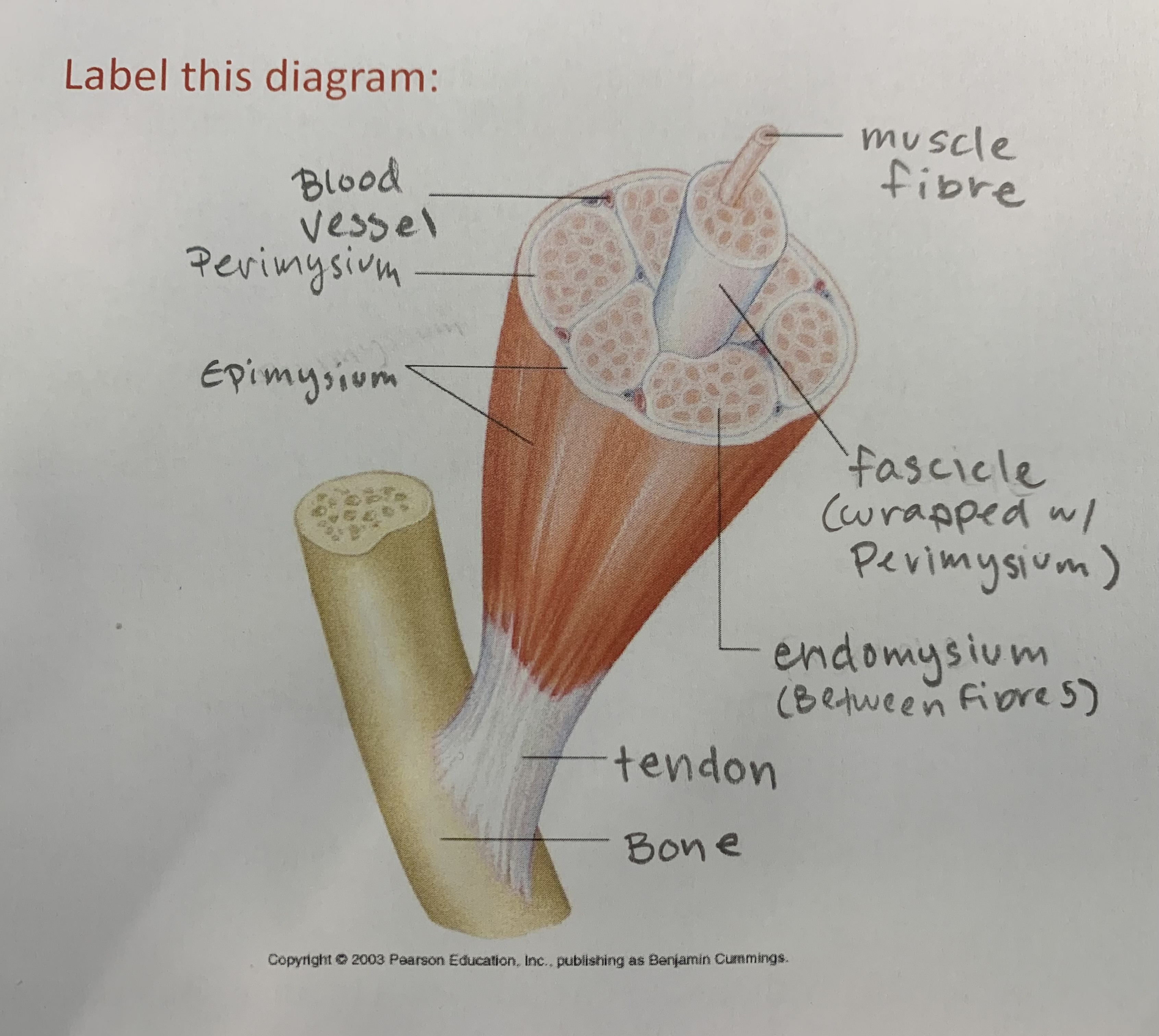
A tendon serves to move the bone or structure. A ligament is a fibrous connective tissue that attaches bone to bone, and usually serves to hold structures together and keep them stable

Muscle naming

  • direction of fibers (obliques = slanted, rectus = straight)

  • size (maximus vs minimus)

  • location (temporalis = near temporal bone)

  • origins (biceps = two origins)

  • location of attachment

  • shape (deltoid = triangle shape)

  • action (flexors = extend)

Muscle contraction

  • actin: thin filaments

  • myosin: thick filaments with myosin heads

Contracting

  1. brain signal sends neurotransmitter to muscle

  2. Acetylcholine (ACh) goes into muscle

  3. ACh binding opens gates and results in sodium rush

  4. triggers calcium to be released into muscle cell

  5. Calcium ions bond to actin and exposes myosin grabby sites

  6. the myosin heads can then attach to and pull on the actin

Not shortening myosin or actin, just sliding together to become more packed in

Releasing

  1. ATP molecules are brought to myosin heads to detach them from the actin (rigor mortis, no ATP when dead so muscles get rigid)

  2. Calcium gets removed by active transport

  • for reflexes, the nerve impulses come from the brainstem instead of brain

Nervous System

General
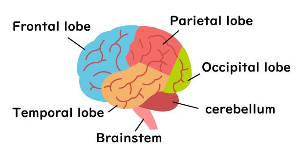
Cerebrum = cerebral hemisphere = lobes of brain

more “wrinkles” means smarter because more surface area

Cerebellum = “mini brain”, controls coordination, first affected by alcohol

Brain stem = lizard brain, because it preforms the basic functions all things need to do

  1. mid brain

    1. visual auditory

    2. reflexes

    3. maintains consciousness

  2. pons

    1. relay to send info to the thalamus and cerebellum

    2. does subconscious stuff like snoring

  3. Medulla

    1. relay to send info to the thalamus

    2. automatic behaviors are processed (breathing, digesting, etc)

Corpus Callosum = in between the two halves of the brain

  • responsible for connections where one side of the brain connects to the other side of the body

    • left occipital controls right eye

  • For some seizure disorders they cut the corpus callosum

    • weird results when patient can only draw half a drawing (thinking they’ve drawn a full one)

The Meninges = surrounds the brain = layers underneath the skin, periosteum, and cranium

  • Dura Mater (tough mother) = thick, leathery, protective

  • Arachnoid Mater (spidery layer) = holds blood vessels

  • Pia Mater (gentle mother) = super thin, keeps brain moist, membrane

  • Meningitis

    • meninges infected, put pressure on brain

    • often if caught early enough, drill into skull to skull to prevent damage

    • some parts of brain will die without oxygen for three minutes

    • comes from viruses and bacteria

Ventricles = fluid filled chambers with cerebrospinal fluid

  • in brain and spinal cord

  • “brain floating in fluid”, protects from impact

Brain Parts:

  • lateral ventricles (first and second ventricles) - produce and secret cerebralspinal fluid

  • corpus callosum - connects two sides of brain, insures communication

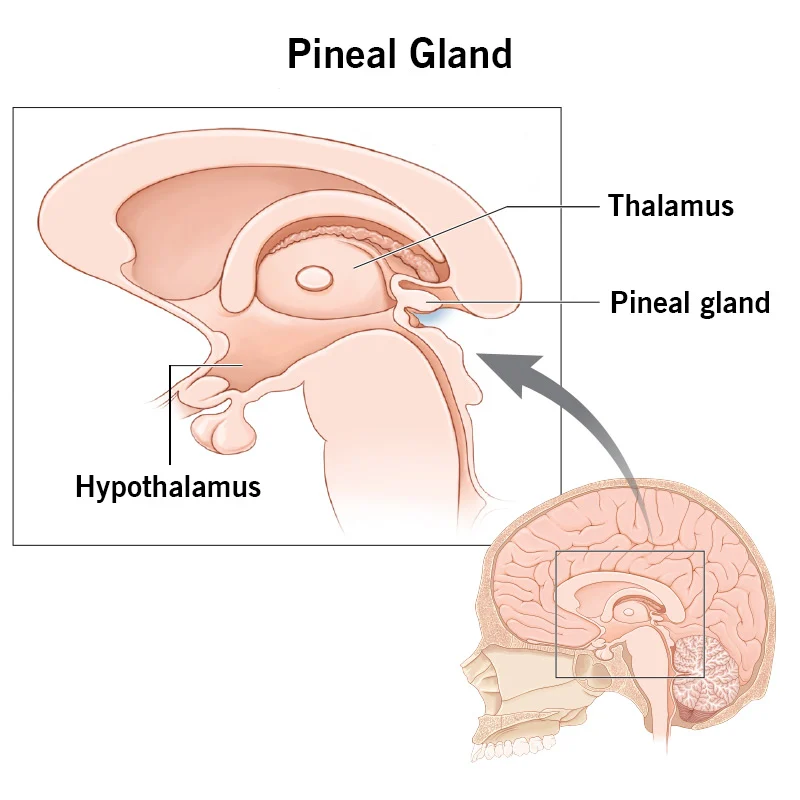
  • pineal gland - regulates circadian rhythm

  • hypothalamus - maintains homeostasis by influencing automatic nervous system and hormones

  • thalamus - all motor/sensory info (except smell) pass through

Reflex arcs: neural pathway that controls a reflex

  1. receptor

  2. sensory neuron

  3. integration center

  4. motor neuron

  5. effector

Cranial Nerves

  • 12 pairs

I. Olfactory: sensory (Olfaction, smell)

II. Optic: sensory (vision)

III. Oculomotor: motor and sensory (most eye movement)

IV. Trochlear: motor and sensory (moves eye)

V. Trigeminal: motor and sensory (face/mouth sensory, chewing muscles)

VI. Abducens: motor and sensory (Abducts the eye)

VII. Facial: motor and sensory (expression, taste)

VIII. Vestibulocochlear: sensory (equilibrium and cochlear)

IX. Glossopharyngeal: motor and sensory (taste, gag reflex)

X. Vagus: motor and sensory (gag reflex, parasmpathetic motor regulation of visceral organs)

XI. Accessory: motor and sensory (shoulder shrug)

XII. Hypoglossal: motor and sensory (swallowing, speech)

Vision

Eye

  • Extension of a brain

  • like a camera, takes the imagery, but can’t process it so it sends it to the brain

  • to focus it, it flips it through the lens, so we actually “see” things upside down, they are just processed right side up

  • At any given time, only a 1/6 of the eye is exposed

  • In most animals, it is round like a ball or a balloon

  • interior apparatus associated with the eye

  • nasolacrimal duct, nose side of the eye leak tears (inner eye)

  • lacrimal glands (outer above eye)

  • Tears contain antibodies and lysozymes (enzymes that kill bacteria)

Muscles

  • 4 rectus muscles (up, down, left, right)

  • 2 oblique muscles (kinda unnecessary?)

    • Inferior oblique (elevates, moves lateral)

    • Superior oblique (depress, moves lateral)

Tunic Layers of the Eye

  • (Conjunctiva) - covers the sclera, is a membrane

  • Sclera - the whites of the eyes, protective, thick, durable

  • Choroid - middle layer, blood rich tunic, dark pigment to prevent light from scattering in eye (so light doesn’t bounce around)

  • Retina - inner back of the eye, delicate tunic, contains millions of receptor cells (rods and cones) that receive and respond to light

Internal Structures of the Eye

  • Two hard lenses - bend light to focus on retina

    • Cornea - outer

    • Lens - inner

  • Iris - expands and constricts based on light

  • Pupil - hole in iris that enlarges and shrinks

  • Ciliary bodies - pull on the iris to dialate and constrict to regulate light imput

  • Aqueous Humor - watery just behind cornea, helps bathe cornea and give it oxygen and nutrients (because blood doesn’t flow here, and it still needs oxygen)

  • Vitreous Humor - thick, jelly like substance which makes up bulk of eyeball, give structure and maintains ocular pressure (where the lens resides)

Photo Receptors:

The back of the eye is the retina, contains thousands of photo receptors

  • Rods - slender, elongated neurons, allow us to see in black and white and shades of grey, also allow peripheral visions

  • Cone - fatter more pointy, come in blue, green, and red (primary colors of light), if you are missing a color of rod, could be color blind, allows us to see in color and focus clearly on objects in the center of our view

Vision

  1. light enters eye as wavelenght

  2. go through cornea, iris, pupil, lens

  3. go to the retina and hit the rods and cones

  4. they activate and send action potential (electrical impulses) to the brain through the optic nerve

  5. brain can process what the eye is seeing

  • left and right eyes are processed by the occipital lobes on the oposite side of the brain

The blind spot

  • produced by the fact that there are no photoreceptors in the part of the retina where the optic nerve connects (bc blood vessels)

Diseases:

myopia (nearsidedness)

  • when the distance between the cornea and the retina is too long

  • light has already focused and unfocused by the time it hits the retia

hyperopia (farsidedness)

  • when the distance between the cornea and the retina is too short

  • light not yet in focus when it hits the retina

astimatism

  • cornea curved unevenly

  • not properly focused on the retina

presbyopia

  • age related

  • lens can change shape to bring things in focus, when people age, lens gets less focused

  • why reading glasses exist

Reflexes

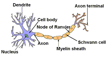
Connects dendrite to axon terminal to create a nerve fiber

  • Cannabis takes dendrites and slowly shrinks them down until the cell body looks more bulby, slowing the circuits

  • Alcohol will damage the myelin sheaths

Reflexes

  • rapid, predictable, involuntary responses to stimuli

  • babies have a grasp reflex (grab finger), step reflex (if you put their feet on flat ground they will step their feet)

  • Autonomic reflexes

    • regulate activity of smooth muscles, heart, and glands. Also regulate digestion, excretion, etc.

  • Somatic reflexes

    • reflexes that stimulate skeletal muscle

Five elements of a reflex arc

  1. sensory receptor (receptors in eye): receives stimulus

  2. Sensory neuron: brings message to central nervous system

  3. Integration center: in CNS, often in spinal cord. receives message and links to response

  4. motor neuron: sends message to muscle, gland, or organ

  5. Effector: muscle gland or organ that responds

Ear

  • allows us to hear

  • maintain balance and equilibrium

Sections

  • outer (heaing)

  • middle (hearing)

  • inner (balance and hearing)

    • balance - cemicircular canals

Parts

  • pinna (auricle) - outer/visible

  • External auditory canals - focuses sounds into middle ear

    • cereminous glands like under skin and excrete wax

  • Tympanic membrane (ear drum) - receives sound waves and sends them to bones in middle ear

    • only vibrates freely when pressure on both sides are equal (ear popping)

  • Otitis externa (swimmers ear) - infection of external auditory canal

  • otis media (inflamations of the middle ear)

    • due to bacterial infection as a result of fluid and pus in the middle ear cavity from the eustacian tube

Middle ear

  • ear drum vibrates the ossicles, the tiniest bones in your body (mallus, incus, stapes - mallet, anvil, stirrup)

  • stapes (last bone) vibrates against oval window

  • eustachian tube - connects ear with back of the throat

    • equalizes air pressure on both sides of eardrum

    • normally closed or flattened and opened when chewing

  • oval window - stapes presses on window, sends fluid of the inner ear in motion

  • round window - smaller inferior, pressure valve, dissipates pressure from vibrations

inner ear

  • consists of. a maze of bony chmabers called the bony labyrinth (osseus labrinth)

  • filled with perilymph, a plasma

  • plasma brushes against the organs of corti, “trapdoors”

  • organs of corti brush against hairs

  • semicircular canals

    • x,y,z chambers, filled with fluid

    • when fluid moves, tells body where you move

  • vestibules - helps with balance and determining up and down

    • uses otoliths, tiny stones made of calcium salts embedded in a gel-like membrane

  • information from the cochlea and semicircular canals will be sent to the brain by the vestibulocochlear nerve

Smell and taste

The type of receptors allow us to smell and taste are called chemoreceptors because the receptors respond to chemicals dissolved in solution\

Smell

  • thousands olfactory receptors in superior part of nasal cavity, extremely sensitive

  • olfactory hairs project from nasal epithelium, continuously bathed in mucus, boogers

  • chemicals in are dissolve in mucus, stimulates olfactory receptors, send signal to brain

  • the impulses brought to the olfactory cortes of the brain catalog the smell instantly, connect to memories too

  • olfactory cortex is tied to limbic system causing the above

  • sensors “adapt” quickly so we can be habituated to a smell and not notice it anymore

Taste

  • smell dictates more about taste than taste receptors

  • 90% of what we perceive we taste is from smell

  • 100,000 taste bugs in mouth

  • most on tongue, some on soft pallet and inner cheeks

  • saliva acts as the solvent in the mouth like mucus in the nose

  • Papillae are the small bumps on the tongue, contain taste buds

    1. sharp filiform papillae/foliate (white dots)

      1. back sides of tongue

    2. rounded fungiform papillae

      1. front part

    3. circumvallate papillae

      1. far back tongue

  • Gustatory cells in taste buds respond to chemicals dissolved in saliva

  • the cells contain gustatory hairs which receive chemical stimuli and send it to the brain

  • lower part of parietal lobe is the gustatory area

  • no area in the tongue do different taste, all are activated by any taste

Cardiovascular system

General

Heart

  • sits in the middle of the thoracic cavity (chest

  • Sits tilted, with apex pointing towards left lung

pericardium - dura mater of the heart, protective sac

  • decreases the friction of beating

    • if any of the vessels in the heart burst (possibly from friction) it is a heart attack

myocardium - the muscle that does the pumping, thick

endocardium - lining of the chambers, what the blood touches, thin

Structure of heart (chambers):

Right Atria Left atria

Right Ventricle Left ventricle

Four valves:

  • Atrioventricular valves (AV) (between atria and ventricles) - open to allow blood into ventricles, close when ventricles contract so blood doesn’t go back into atria

    • Tricuspid valve - right side

    • bicuspid (mitral) valve - left side

  • Semilunar valves - allow blood to leave heart

    • Pulmonary valve - lungs

    • Aortic valve - body

Blood:

Main function of the cardiovascular system is transportation. It transports:

  • oxygen

  • nutrients (glucose, salt, etc)

  • Waste products of cell metabolism (e.g. CO2, urea)

  • hormones

  • water

  • immune cells

  • heat (blood is approx. 100.4˚F)

a healthy adult has 5-6 quarts of blood which make up 8% of body weight

Blood is made of

  • plasma (55%): yellow, liquid, non living part

  • formed elements (45%): living cells or parts of cells

If you centrifuged blood, formed elements would go to the bottom and plasma would go to top

Plasma:

  • function: to maintain blood volume and to transport blood cells/molecules

  • 90-92% water

  • water is absorbed from large intestine and goes to plasma

Types of blood cells

  • RBC - red blood cells (erythrocytes) - 45%

    • carry oxygen to cells and carry away carbon dioxide

    • resemble donuts with indent instead of hole (biconcave disc)

    • strong affinity for oxygen due to the presence of hemoglobin

    • can carry up to four oxygen molecules

    • approx. 5 million per one ml of blood (5 mil - male, 4.5 mil - female)

  • WBC - white blood cells (leukocytes) - <1%

    • Protect body against pathogens (important part of immune system)

  • Platelets - <1%

    • allow for blood clotting when injured and scouting

Blood clotting

  1. platelets and damaged cells release prothrombin

  2. fibrin threads are produced and trap red blood cells inside

Homeostatic imbalance

  • anemia, decrease in body’s ability to carry oxygen

    • have a lower RBC count

    • Abnormal or deficient hemoglobin

    • people with it are more tired as they cannot produce enough energy (ATP) which takes oxygen

Blood vessels:

  • Arteries

  • Arterioles (smaller arteries)

  • Capillaries (smallest blood vessels)

  • Veins

  • Venules (smaller veins)

Arteries: A for away

  • take blood away from heart

  • have a pulse when palpated

  • do not have valves

  • located deeper in body/tissue

Carotid artery: major artery that feeds the brain on either side of neck from aorta

  • left carotid artery comes straight from the aorta

  • right carotid artery splits off the brachiocephalic artery, other side in split is right subclavian artery (left subclavian artery branches directly off aorta)

Veins:

  • carry blood to the heart

  • veins do not have a pulse, lost the pressure needed

  • veins have valves to prevent the back flow of blood, especially on long vertical paths up legs to heart

  • veins are located more superficially in the body/tissue, so they are what you can see through skin

Respiratory System

Works directly with the cardiovascular system to supply all cells with oxygen and disposing of CO2

Upper organs of respiratory system:

  • Nose: takes in air (purifies (hair and mucus) and humidifies (mucus) air)

  • Pharynx (back of throat): muscular passage way for air and food to pass through

  • Epiglottis: protects opening of larynx when swallowing so food doesn’t go into windpipe

  • Larynx (voice box): routes air and food into proper channels

Lower organs of respiratory system:

  • Trachea (windpipe): airway into lungs/bronchi, lined with cilia which moves mucus (& dirt, etc) up and out of lungs. Rigid due to walls reinforced with c-shaped rings of hyaline cartilage (also found in the ends of long bones).

  • Lungs: includes bronchi, bronchioles, and alveoli

  • Bronchi: branching tubes that lead to bronchioles (smaller tubes) that lead to the lungs.

  • Alveoli: tiny air sacs where gas exchange occurs. walls ar every thin to allow for diffusion of O2 and CO2 molecules

Circulation of blood throughout the body

two main circuits

Pulmonary circuit

  1. starts with un-oxygenated blood

  2. right atrium

  3. right ventricle

  4. pulmonary arteries

  5. lungs

  6. pulmonary veins

  7. left side heart

  8. ends with oxygenated blood

Systemic circuit

  1. starts with oxygenated blood (from pulmonary circuit)

  2. left atrium

  3. left ventricle

  4. aorta

  5. systemic arteries

  6. capillaries throughout body

  7. systemic veins

  8. right side of heart

  9. ends with un-oxygenated blood (which goes to the pulmonary circuit)

Heartbeat

Systole: ventricles contract, blood leaves heat

  • Blood leaves ventricles

  • Status of valves:

    • AV (atrio ventricular) valves: tricuspid/bicuspid closed

    • Semilunar valves open

    • wont let blood back to the ventricles

Diastole: ventricles relax, blood enters

  • AV valves open

  • Semilunar valves close

heartbeat described as lub-dup

  • lub is systole

  • dup is diastole

  • heartbeat what is heard is sound of valves closing

a heart murmur is due to the valves not folly closing, as a result some leakage of blood occurs and the sounds are a bit swishy instead of strong and distinct

Heart contractions:

  • heart muscle contractions do not rely on nerve impulses from the brane

  • this is because of “internal pacemakers” that initiate and coordinate

  • brain can still affect it

Heart rate is controlled by:

  • autonomic nervous system

    • accelerate or decelerate heart rate depending on situation

  • Intrinsic conduction system

    • Sinoatrial node (SA node) “pacemaker” located in upper right part of atrium, shoots electricity at AV node

    • AV node (atrial ventricular node) passes impulse to AV bundle

    • AV bundle transmits it to the Purkinje fibers which radiate out to contact both ventricles

    • This causes ventricles to contract in an upward motion

    • pushes blood with force into major arteries

    • SA node → AV node → AV bundle → Purkinje fibers

Homeostatic imbalance

  • Angina pectoris - crushing chest pain

    • myocardium deprived of oxygen

    • if too long, heart becomes ischemic and may die

    • if this occurs myocardial infarction (heart attack) is likely

    • heart attack can lead to state of fibrillation

      • rapid uncoordinated shuddering of heart muscle

    • Heart can no longer pump blood properly so major organs including brain are deprived of oxygen

Reproductive System

Male

Main function of male reproductive system

  • produce sperm

  • deposit sperm into female

  • produce testosterone (to develop secondary sex characteristics, eg. body hair, low voice, etc)

Scrotum

  • pouch that houses testes and epididymis

  • function: keeps testes at temperature lower than normal (3 degrees lower)

    • when it is cold will contract towards body, hot away from it

    • one testes is higher than the other to lessen chance of loss due to impact

Duct system (sperm flows):

  1. testes

    1. produce sperm and testosterone

    2. each approx. 1.5” long and 1” wide

    3. Covered by a white capsule known as the tunica albuginea “white coat”

    4. Inside are seminiferous tubules (800ft long) which produce sperm

  2. Epididymis

    1. first part of duct shape

    2. comma shape sits above testes (in scrotum)

    3. Long highly coiled tube (20ft)

    4. Stores immature sperm to allow them to mature (20 days)

  3. Vas deferens

    1. tube that runs upward through epididymis into pelvic cavity arching over bladder

    2. main job: propel live sperm from epididymis into urethra

    3. contracts during ejaculation

    4. where a vasectomy is preformed

  4. Ejaculatory duct

    1. near prostate gland

    2. opens to propel live sperm

    3. end of vas deferens, connects to urethra

  5. Urethra

    1. tube that carries sperm and urine out of body

Accessory Glands (create fluid for sperm to “swim in”):

Produce the bulk of the semen

Semina vesicles

  • two of them (behind prostate gland)

  • main function: produce 60% of semen

  • has a basic solution, sperm cannot survive in acidic conditions

Prostate gland:

  • located inferior to bladder

  • secretes a milky fluid that activates the sperm (when the sperm touches it the flagella starts wiggling)

  • inflammation is common in men, 3rd most common cancer in men

Bulbourethral glands (formerly called the Cowper’s glands)

  • tiny pea sized glands

  • produce a lubricant to help with copulation

  • lubricant also cleanses urethra of acidic urine

Spermatogenesis (sperm production)

  • begins at puberty and continues throughout lifetime

  • every days males produce millions of sperm

  • complete process of making sperm 64-72 days

Semen:

  • dilutes sperm to increase mobility

  • approx. 2-5 ml each ejaculation

  • approx. 50-130 mil sperm each ml

Fertility issues

  • first test is semen analysis

    • sperm count

    • motility of sperm

    • morphology of sperm (2 heads, 2 tails, abnormal head/cap, etc)

    • semen vol

    • semen pH

    • fructose content

  • Causes of male infertility

    • obstructions in duct system

    • hormonal imbalances

    • pesticides

    • excessive alcohol consumption

    • smoking

    • other factors

  • Factors that decrease sperm production

    • Antibiotics

    • Radiation

    • Pesticides

    • Marijuana

    • Tabacco

    • Excessive alcohol consumption

    • Laptop computers (used on laps)

    • Tight pants

Female

Main function of female reproductive system

  • produce (mature) eggs

  • Receive sperm

  • nurture and protect fetus

  • Produce and feed newborn baby milk

  • Produce estrogen and progesterone

ovaries

  • two, one on each side

  • size of an almond

  • inside are follicles

  • inside follicles are oocytes, immature eggs

fallopian tubes

  • first part of duct system

  • 4” long

  • fertilizations occurs here

  • tubes are not indirect contact with ovaries

  • end of fallopian tubes have finger-like projections called fimbriae

  • fimbriae create wave/fluid currents beckoning released eggs to the fallopian tubes

  • fallopian tubes are lined with cilia that move eggs down tube

  • journey takes 3-4 days

Ectopic pregnancy is when the fertilized egg gets stuck in fallopian tubes

Uterus:

  • located superior to vagina

  • function: receive/nourish fertilized egg

  • hollow, pear sized

  • bottom opening is the cervix, dilates during pregnancy

Vagina

  • thin walled tube

  • 3-4 in long

  • receives penis during copulation

  • birth canal for baby exit

  • menstrual flow passes through here

  • opening is covered with a thin fold of tissue (hymen) until this membrane wears away or is ruptured

Oogenesis

  • at birth infant girls born with about 2 million oocytes (immature eggs)

  • at puberty oogenesis begins (at this time girl has approx. 400,000 oocytes)

  • usually one egg per month ripens and is released from one ovary (ovulation)

  • on average, women will only release approx. 500 eggs in a lifetime

Making eggs

  • each month, one oocyte finishes meiosis

  • FSH-promotes follicle development

  • LH surge triggers ovulation

Menstrual Cycle

  • 28 day cycle controlled by ovaries

    • day 1-5 menses

      • menstruation

      • causes drop in estrogen

    • day 6-14 proliferative stage

      • thickening of endometrium

      • causes increase in estrogen and progesterone

      • ovulations occurs around day 14, caused by LH spike

    • Day 15-28 secretory stage

      • Corpus luteum (empty follicle) releases progesterone which continues to build endometrium

Fertilization

  • egg is viable for 12-24 hours

  • sperm survive for 3 days

  • fertilization occurs in fallopian tube

  • ~12 days later, implantation occurs

  • hormone (hCG) stimulates production of estrogen, and progesterone, maintaining endometrium

    • hCG is the hormone detected by pregnancy test

  • placenta develops

Contraception

Most important points:

  1. if mature enough to have sex, must be mature enough to be responsible with contraception (or a child)

  2. rhythm method and pull out are not affective

  3. use a method that one will actually use/follow through with

Contraception: preventing pregnancy (not STIs)

  • only physical barriers will prevent STDs

Main ways to prevent pregnancy

  • prevent fertilization (sperm no touch egg)

    • Physical:

      • condoms (Mr. Itow, 2023 “Condoms are cheap. Pay for a condom, not a kid.”)

        • put it on correctly

        • if accidentally put on the wrong way, throw away

        • Expiration dates

        • don’t use oil or silicone based lubricants, only water based, because others can dissolved condom

        • don’t double condom

        • size matters (if too small, burst, if too big, leak or slip off) (nasa large, jumbo, massive male pee cup size, google it)

      • female condom

        • don’t use along side with male condom

      • diaphragm

      • cervical cap

      • tubal ligation

    • Chemical:

      • Spermicide

      • Emergency contraceptive pill (95% failure rate)

        • blast of hormones, so is not good for you

  • preventing implantation (oopsies sperm is in egg, but egg cannot attach to uterine wall)

    • IUD

      • 3-6 years

      • local distribution of hormones, won’t go through whole body

    • Emergency contraceptive pill

      • blast of hormones, so is not good for you

      • take as soon as possible

  • Preventing gamete release (no sperm, no egg)

    • vasectomy (snip snip 🤭)

    • female: hormone control

      • Birth control pill

        • take at the exact same time every day

        • one dose missed, not effective birth control for 2-3 weeks

      • patch

        • like a nicotine patch, but leeches hormones

      • Depo-Provera shot: progestin only

        • blast of hormones

STIs

3 Important points:

  1. abstinence is the only 100% protection

  2. condom use reduces the risk of only some STIs

  3. Regular testing for STIs, especially if sexually active

Three types:

  • Parasites: things that live on your body

    • Scabies: skin mites

      • burrow under skin and deposit eggs and feces

      • spread by skin to skin contact

      • causes intense itching at night

      • small red bumps and rashes develop 4-6 week after contact

      • lotions and sprays for infected partners, and same treatment as lice

    • Crabs: pubic lice

      • members of head lice family

      • transfer between genital hair

      • small red bumps

      • killed by special soap

  • Bacteria (some curable with antibiotics, but becoming resistant)

    • Chlamydia

      • rate of infection in women is 3x rate of males (cause disease ridden *deposit* of fluids)

      • can lead to infertility

      • increases risk of HIV

      • pelvic inflammatory disease

      • often asymptomatic, should get tested often

    • Gonorrhea (“the clap”)

      • 2-21 days after contact

      • usually asymptomatic

      • can lead to PIV and infertility

      • pus

      • blindness in newborns if pregnant

      • infection of joints, heart valves, etc.

    • Syphilis (“the great imitator)

      • 7x higher in men (George O’Malley who?)

      • 9-90 days after contact

      • Early: small painless sores on genitals

      • Mid: rash on hands, feet, other parts of body

      • Late: mental illness, blindness, heart damage, death

      • can cross placenta, birth defects, stillbirth

  • Viruses (not curable, life long)

    • HPV

      • genital warts

      • skin to skin contact, sexual activity

      • associated with cervical cancer and tumors of the vulva, anus, and penis

      • get vaccine

      • most people will have genital HPV at some time in their life

      • warts can be removed but may reoccur

    • Hepatitis B (HBV)

      • inflamed liver

      • shared toothbrushes, needles, and razors or sexually

      • mother to child

      • jaundice, fatigue, abdominal pain, vomiting

      • liver failure/cancer, death

    • Genital Herpes

      • Type 1: cold sores and fever blisters in mouth (or genitals)

      • Type 2: blisters and ulcers on genitals

      • 1 in 5 people above 12 have had an infection of simplex 1 or 2

      • symptoms after 3-7 days

    • HIV/AIDS

      • transmitted sexually as well as needles an razors

      • mother to child

      • no cure, but there are meds that not only freeze progression but prevent spreading

  • youth make up a quarter of the sexually active population, but are responsible for 50% of diseases

  • male condoms are not good at preventing parasites

  • should get tested often