Ch. 7 Excitation of Skeletal Muscle
1/28
Earn XP
Description and Tags
Flashcards about Excitation of Skeletal Muscle, Neuromuscular Junction, Motor End Plate, ACH Secretion, Postsynaptic Muscle Fiber, Acetylcholinesterase, End Plate Potential, High Safety Factor, NM Junction Drugs and Myasthenia Gravis.
Name | Mastery | Learn | Test | Matching | Spaced |
|---|
No study sessions yet.
29 Terms
What innervates skeletal muscle?
Large myelinated nerve fibers originating in motoneurons in the anterior horns of the spinal cord
Describe the neuromuscular junction
skeletal muscle is innervated by nerve fibers in the anterior horn and each branch innervates individual muscle fibers
the nerve ends make a junction with the muscle fiber where action potential will travel
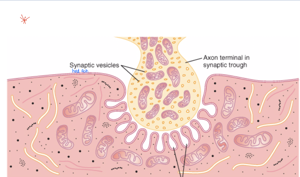
What is the motor end plate?
A complex of branching nerve terminals that invaginate into the surface of the muscle fiber but lie outside the muscle fiber plasma membrane.
the terminal nerve ending does not touch the plasma membrane
The axon terminal contains mitochondria for energy synthesis (in cytoplasm) of acetylcholine (absorbed and released by synaptic vesicles)
ACH will travel into the synaptic space meeting acetylcholinesterase (enzyme that degrades ACH)
What is the synaptic gutter or trough?
Invaginated membrane of the motor end plate.
What is the synaptic space or cleft?
Space between the nerve terminal and fiber membrane.
What is the role of the axon terminal in ACH synthesis?
Synthesizes acetylcholine (ACH) in the cytoplasm, which is then absorbed into synaptic vesicles.
What is the function of acetylcholinesterase?
Degrades acetylcholine into acetate and choline in the synaptic space.
What happens when a nerve impulse reaches the neuromuscular junction (nerve endings meet muscle fiber)?
Calcium channels open, allowing Ca into the nerve terminal, attracting vesicles to the neural membrane and dumping ACH into the synaptic space via exocytosis.

Where are acetylcholine receptor complexes located?
Located at the mouth of the subneural clefts; composed of 5 subunit proteins (2 alpha, 1 beta, delta, gamma).
Form a tubular channel as receptor sites lay side by side
What happens when the channels of the acetylcholine receptor complex open?
Sodium flows into the cell, making it more positive.
channel will remain closed till 2 ACH molecules attach to the 2 alpha subunit proteins causing a confirmational change and sodium will flow in
the potential change inside the muscle fiber membrane is the end plate potential which initiates an AP that spreads along the membrane causing a contraction
What is the end plate potential?
A potential change inside the muscle fiber membrane after ACH binds to the receptor (causing a conformational change) and Na slows into the cell
How is ACH removed rapidly from the synaptic space?
Acetylcholinesterase degradation and ACH diffusion out of the synaptic space.
What causes fatigue of the neuromuscular junction?
Results from diminished ACH vesicles due to rapid stimulation of the nerve fiber.
Give examples of drugs that enhance the signal at the NM junction. How does this occur?
Methacholine, carbachol, and nicotine.
The drugs are destroyed by acetylcholinesterase slowly and depolarization areas are localized. During repolarization, the areas initiate another action potential leading to muscle spasms (constant contraction)
Give examples of drugs that inactivate acetylcholinesterase. How does this occur?
Neostigmine, physostigmine, and diisopropyl fluorophosphate.
With each successive nerve impulse, ACH accumulates in synapse and stimulates muscle fiber repetitively (no acetylcholinesterase) = muscle spasms
What is Myasthenia Gravis?
An autoimmune disease where antibodies attack acetylcholine-gated sodium ion transport proteins, leading to weak end plate potentials.
causes paralysis because of neuromuscle junctions inability to transmit enough signals to muscle fibers
What are Transverse Tubules?
Internal extensions of the cell membrane that help get the AP deep within the fiber to all the myofibrils.
cause the release of calcium from the sarcoplasmic reticulum
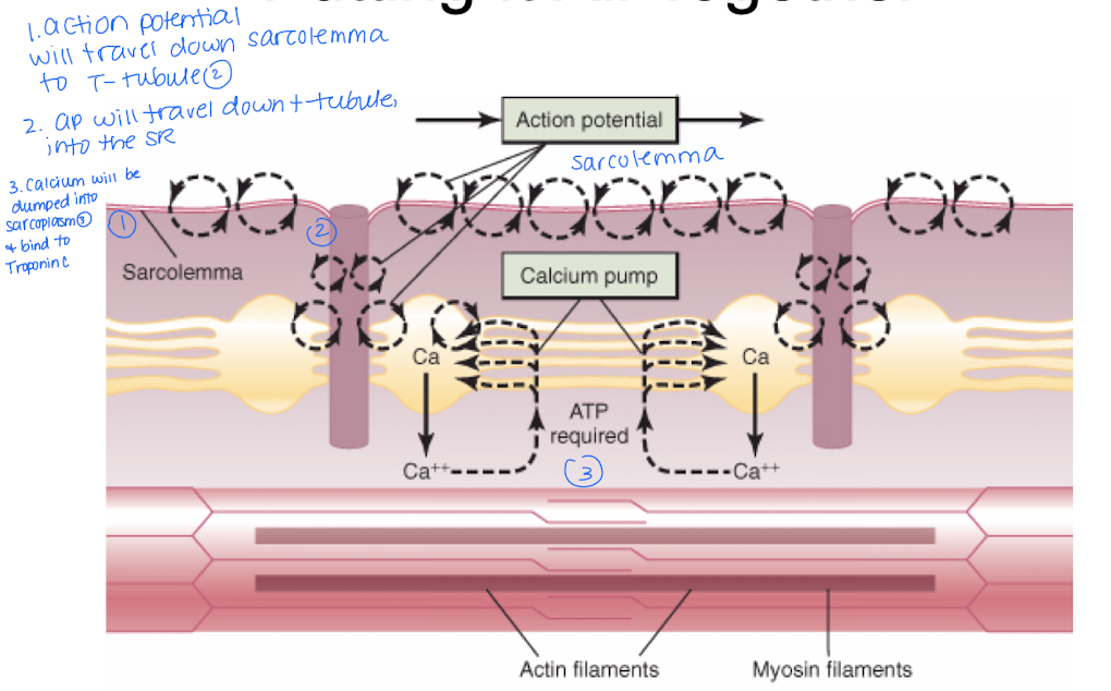
Explain this model.
Action potential occurs along the sarcolemma and travels down to the T-tuble into the sarcoplasmic reticulum
Calcium will be released from the SR and bind to Troponin C
Describe the events of the sliding filament model
Explains HOW a MUSCLE CELL contracts
Action potential is generated in a motor nerve and ACH moves to the synaptic space via exocytosis
Acetylcholine receptor acts as a gated ion channel
AP will occur down the sarcolemma to the T-tuble
T-tubules spread impulses
Sarcoplasmic reticulum releases calcium and allowing the binding of troponin C
Myosin heads protrude towards binding sites
ADP dissociates from myosin head.
Where is the action potential generated in a motor nerve?
Propagated along the axons to the terminal end, releasing acetylcholine into the synapse.
Putting it together Part 1
Describe the action potential generated in the motor nerve
impulse is propagated along the axons in the terminal end
axon releases neurotransmitter ACH into the synpase
ACH crosses the space and binds to receptors on sarcolemma
Putting it together Part 2
ACH Receptor acts as Gated Ion Channel
ACH binds to receptor creating a conformational change = charge becomes negative
Sodium will rush into the cell creating an AP
Putting it together Part 3
T-tubules spread impulses
Sarcolemma has T-tubules or invaginations that run perpendicular towards the fibers
The tubules spread the impuse deep into the cell and stimulate the SR
Putting it together Part 4
SR releases Calcium
Calcium binds to Troponin C
Troponin changes its conformation, moves tropomyosin from active site and reveals the crossbridge binding site on actin, allowing muscle contraction to occur.
Putting it together Part 5
Myosin heads protrude towards binding sites
ATP is hydrolyzed by ATPase (resets myosin head active site opening)
Myosin head binds to actin site
Pi dissociates from myosin
Myosin head pulls actin filament, causing contraction and Z disc shortens (concentric contraction)
Putting it together Part 6
New ATP will be loaded onto the myosin head and head will dissociate from binding site
Released energy used to return the head to “cocked” position
Describe how the sarcoplasmic reticulum releases calcium and its effect.
Calcium binds to Troponin-C, causing it to change conformation, move tropomyosin, and reveal the crossbridge binding site on the actin filament.
What is the role of ADP dissociation from the myosin head?
New ATP is loaded onto the myosin head, causing it to dissociate from the binding site using released energy to return to the cocked position.
When does the repeated process of AP stop?
Once calcium is pumped back into the SR = muscle relaxes