Humerus
1/14
There's no tags or description
Looks like no tags are added yet.
Name | Mastery | Learn | Test | Matching | Spaced | Call with Kai |
|---|
No analytics yet
Send a link to your students to track their progress
15 Terms
What is the humerus
A type of long bone that connects the upper limbs to the shoulder girdle.
Located between and articulates with the shoulder and elbow joints
diagram
how to tell which view
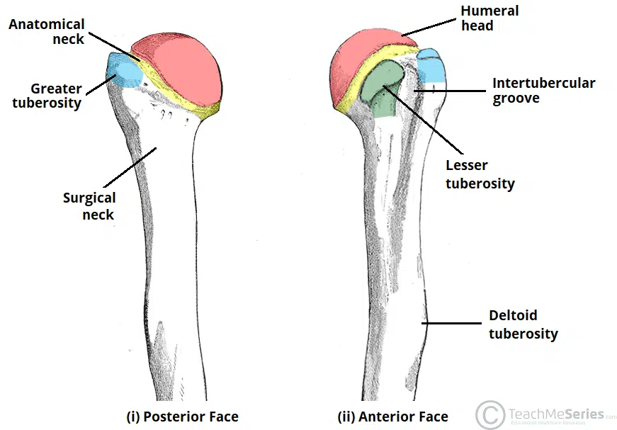
Anterior - bicipital groove, coronoid fossa, radial fossa and capitilum
Posterior - olcraneon fossa
Features of the proximal end
Head of humerus - rounded covered in cartilage
Anatomical neck - adjoining the head, distal to the articular surface and proximal to tuberosities. Location of growth plate.
Surgical neck - imaginary horizontal line across proximal shaft, distal to the tuberosity. Common fracture site
Greater tuberosity - posterolaterally the supraspinatus tendon is attached to the superior aspect
Lesser tuberosity - anteriorly, tendon of the subscapularis muscle is attached
Bicipital groove - between tuberosities
proximal extremity
articulates with glenoid cavity to form glenohumeral joint
synovial ball and socket joint
articulations
The proximal region of the humerus articulates with the glenoid fossa of the scapula to form the glenohumeral joint (shoulder joint).
Distally, at the elbow joint, the capitulum of the humerus articulates with the head of the radius and the trochlea of the humerus articulates with the trochlear notch of the ulna.
Features of shaft (diaphysis)
Cylindrical in cross section, flattened and wider at distal extremity. Has 3 key features;
deltoid tuberosity on antero lateral surface, attaches to deltoid muscle
spiral groove runs obliquely, forwards and downwards transmits radial nerve
nutrient foramen located on antero-medial surface, allows small blood vessels to pass through medullary cavity
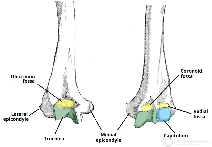
Features of distal end
The lateral and medial borders of the distal humerus form medial and lateral supraepicondylar ridges. The lateral supraepicondylar ridge is more roughened, providing the site of common origin of the forearm extensor muscles.
Immediately distal to the supraepicondylar ridges are extracapsular projections of bone, the lateral (superior to capitulum) and medial epicondyles (superior to trochlea). Both can be palpated at the elbow. The medial is the larger of the two and extends more distally.
Capitulum - lateral condyle. Rounded, articulates with head of radius
Trochlea - medial condyle. Lateral to the trochlea is the capitulum, which articulates with the radius
distal features continued
capitulum - smaller or 2 articular surfaces, articulates with radial head
trochlear - larger of distal articular surfaces, pulley shaped and articulates with trochlear notch of ulna
radial and coronoid fossae - 2 smaller depressions on anterior surface, located immediately above capitulum and trochlear
olecranon fossa - deep depression on posterior aspect of lower humerus, located above trochlear and accomodates olcranon process of ulna during extension of elbow
ulnar groove - marked groove between medial end of trochlear and medial epicondyle, transmits ulnar nerve
distal - depressions
located on the distal portion of the humerus are three depressions, known as the coronoid, radial and olecranon fossae. They accommodate the forearm bones during flexion or extension at the elbow.
Ossification
Shaft - one centre, 8th/9th week intraterine
Upper end;
one centre in head - 6 months
one centre in greater tuberosity - 2 years
one centre in lesser tuberosity - 5th year
all centres unite to form one epiphysis at year 6 and fuse with shaft at 20 years
Lower end;
one centre in capitulum - 2 years
one centre in medial epicondyle - 5th year
Trauma and pathology of humerus
fracture of surgical neck
fracture of humeral shaft
supracondylar fracture
condylar fracture
bone metastases
malignant bone tumours
Surgical neck fracture
Cause is FOOSH, usually in elderly
Treatment usually is a sling
Shaft fracture
Middle third; spiral, oblique or comminuted
Cause - FOOSH, direct blow or high velocity injuries
Sling or cast
risk damage to radial nerve, leading to unopposed flexion of the wrist, known as wrist drop
Supracondylar fracture
Fracture of the distal humerus just above the elbow joint. The fracture is typically transverse or oblique, and the most common mechanism of injury is FOOSH. It is more common in children than adults.
The brachial artery can be damaged. The resulting ischaemia can cause uncontrolled flexion of the hand – as flexor muscles become fibrotic and short.
The Gartland classification is used for these fractures:
Type 1 is minimally displaced
Type 2 is displaced with but with an intact posterior cortex
Type 3 is completely off-ended.
Type 1 can usually be managed conservatively with an above elbow cast whereas types 2 and 3 typically require surgical fixation with crossed, bi-cortical k-wires.