ligand gated ion channels and G-protein coupled receptors
1/22
There's no tags or description
Looks like no tags are added yet.
Name | Mastery | Learn | Test | Matching | Spaced | Call with Kai |
|---|
No study sessions yet.
23 Terms
receptor
a molecule that recognises specifically small molecules whose binding brings about the regulation of a cellular process
Ligand-gated ion channels (ionotropic receptors)
They form a channel between the extracellular and intracellular environment of the cell/organelle. The channel is made up of subunits which can vary
Cellular change occurs in milliseconds
How do ligand-gated ion channels alter cellular function?
Ligand has to bind to the channel causing conformational change so then the ion can pass through via the electrochemical gradient. This occurs in milliseconds.
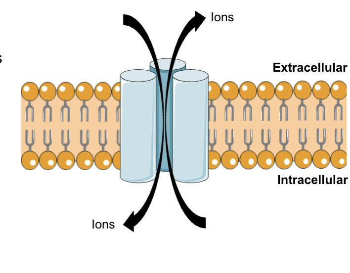
How does this alter cellular function?
Activate signalling pathways and alter the excitability of a cell
Role of ligand gated ion channels in depolarisation
They are responsible for that initial depolarisation event which causes the release of neurotransmitter
What is depolarisation and how does it work?
This occurs in excitatory ligand-gated ion channel e.g. Acetylcholine receptor
A ligand binds to its ligand-gated ion channel - causes a flow of positive ions to go inside the cell.
The inside of cell becomes more positive compared to outside (depolarisation) which results in a voltage change and triggers the voltage-gated ion channels to open. (Na+ moves into the cell)
So the action potential moves along.
What is hyperpolarisation and how does it work?
This occurs in inhibitory ligand-gated ion channels e.g. GABA receptor
A ligand binds to its ligand-gated ion channel - causes a flow of negative ions to go inside the cell.
The inside of cell becomes more negative compared to outside (hyperpolarisation) - this means no activation of voltage-gated sodium channels so no action potential is generated.
Examples of LGICs
Nicotinic acetylcholine and GABA A receptors
What is the neuromuscular junction?
Allows for communication between nervous system and skeletal muscle to initiate contraction
Motor nerves from spinal cord synapse with skeletal muscle fibres
Each branch form an end plate region on a single muscle fibre as the motor end plate
neurotransmitter = acetylcholine (ACh) receptor = nAChR (nicotinic acetylcholine receptor)
How does the neuromuscular junction work?
Acetylcholine binds to nicotinic acetylcholine receptor - causes Na+ to move inside the cell which results in depolarisation of the skeletal muscle cell
This drives a voltage change - triggers influx of Na+ via voltage gate channels into the skeletal muscle cell which results in an action potential
This triggers an increase second messenger which is Ca2+ in this case
This causes muscle contraction
What is a neuromuscular blocking agent (NMB)?
Acts as an antagonist ( has affinity for a receptor but no intrinsic activity, which prevents the receptor from being activated by the endogenous agonist)
Prevents acetylcholine from binding to receptors so muscle contraction cannot occur - paralysed the muscle
Use of NMBs
Tracheal intubation - ventilation of lungs allows delivery of O2 and removal of CO2 by protection against aspiration of gastric contents.
The larynx is a muscle (what tracheal intubation passes through) - NMBs can be used to relax the larynx and allow easy passage of the intubation tube into the trachea
Can also be used for surgical procedures specifically to cause relaxation of abdominal muscles as they can prevent tearing
Also can be used for therapeutic hypothermia - reduce body temperature to 32-34 degrees as it can improve neurological recovery if the patient has suffered from a stroke - the NMBs prevent shivering and should only be taken once patient is anesthetized.
What are G protein-coupled receptors compromised of?
7 transmembrane domains, extracellular N terminus, intracellular C terminus
They are coupled to a G protein which has a trimeric structure - Gα, beta and gamma subunits
Cellular change occurs in seconds
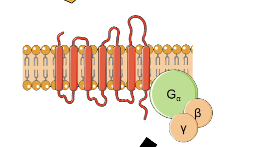
How do G protein-coupled receptors work?
Agonist binds to one of the extracellular N-terminus which causes it to be activated. This stimulates the g-protein and the Gα separates from the beta and gamma subunits. It moves along the membrane, altering the effector molecule's basal activity, activating or inhibiting the effector molecule. This triggers a cellular response.
What are the Gα subunits and their role?
Gαs, Gαi, Gaq, Gα12 - this determines where the receptor inhibits or activates
Binds GDP/GTP and has GTPase activity
Effector molecule for Gαs?
adenylyl cyclase - when increased stimulation it increases cAMP
Effector molecule for Gαi?
adenylyl cyclase - when increased stimulation it decreases cAMP
Effector molecule for Gαq?
phospholipase C - when stimulated it increases DAG and IP3
What are the G beta and gamma subunit
There are 5 beta and 12 gamma which can form unique beta and gamma combinations - the effectors are usually ion channels, enzymes, and kinases
Smooth muscle and α1-adrenoreceptors
Arteries and arterioles contain a circular layer of smooth muscle - constriction of SM reduces the blood vessel diameter whilst relaxation of SM increases the diameter
Change in diameter of blood vessel has a large impact on blood pressure
Tone of SM controlled by: circulating humoral factors, sympathetic and parasympathetic nervous system, paracrine factors
α1-adrenoreceptors antagonists
antagonist binds to α1-adrenoreceptors on vascular smooth muscle cell
this blocks adrenaline (released from adrenal medulla due to stress) from binding to the receptors
This inhibits the G alpha Q subunit from activating phospholipase C which means IP3/DAG is not activated so Ca2+ will not be released
This results in relaxation of smooth muscle
Uses of α1-adrenoreceptors antagonists
Pheochromocytoma - tumor of the adrenal gland, secretes huge amount of adrenaline/noradrenaline, doxazosin (antagonist) reduces BP before and during surgery
Benign prostatic hyperplasia - urinary retention due to urethral obstruction, SM relaxtion of urethra, reduces urethral resistance, improves urine flow
Examples of G-protein coupled receptors
Dopamine and muscarinic acetylcholine receptors