lab ass. respiratory system (exam 3)
1/129
There's no tags or description
Looks like no tags are added yet.
Name | Mastery | Learn | Test | Matching | Spaced |
|---|
No study sessions yet.
130 Terms
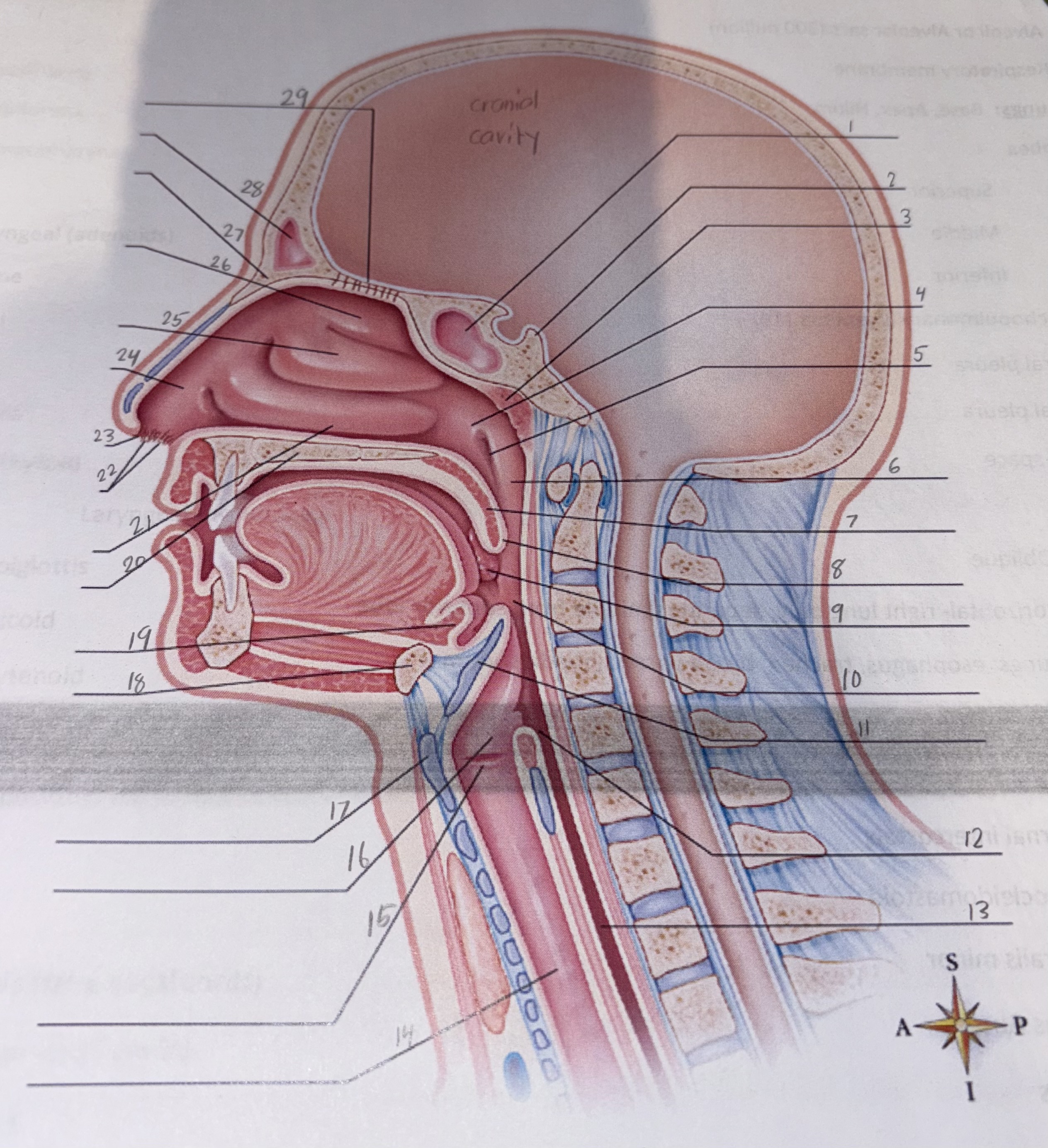
what view is this
midsagittal section through upper respiratory tract. the nasal septum has been removed to reveal the turbinates’ (nasal conchae) of the lateral wall and the nasal cavity.

1
sphenoid sinus

2
sella turcica

3
pharyngeal tonsil

4
posterior naris

5
opening of auditory (eustachian) tube

6
nasopharynx

7
soft palate

8
uvula

9
palatine tonsil

10
oropharynx

11
epiglottis (part of larynx)

12
laryngopharynx

13
esophagus

14
trachea

15
vocal folds (part of larynx)

16
larynx

17
thyroid cartilage (part of larynx)

18
hyoid bone

19
lingual tonsil

20
hard palate

21
inferior nasal concha

22
vibrissae

23
anterior naris

24
vestibule

25
middle nasal concha of ethmoid

26
superior nasal concha of ethmoid

27
nasal bone

28
frontal sinus

29
cribriform plate of ethmoid bone

what view is this showing
nasal septum consists of the perpendicular plate of ethmoid bone, vomer, and the septal and vomeronasal cartilages

1
frontal sinus

2
cribriform plate of ethmoid bone

3
sphenoid sinus

4
pharyngeal tonsil

5
palatine bone

6
maxilla

7
incisive foramen

8
vomeronasal cartilage

9
vomer
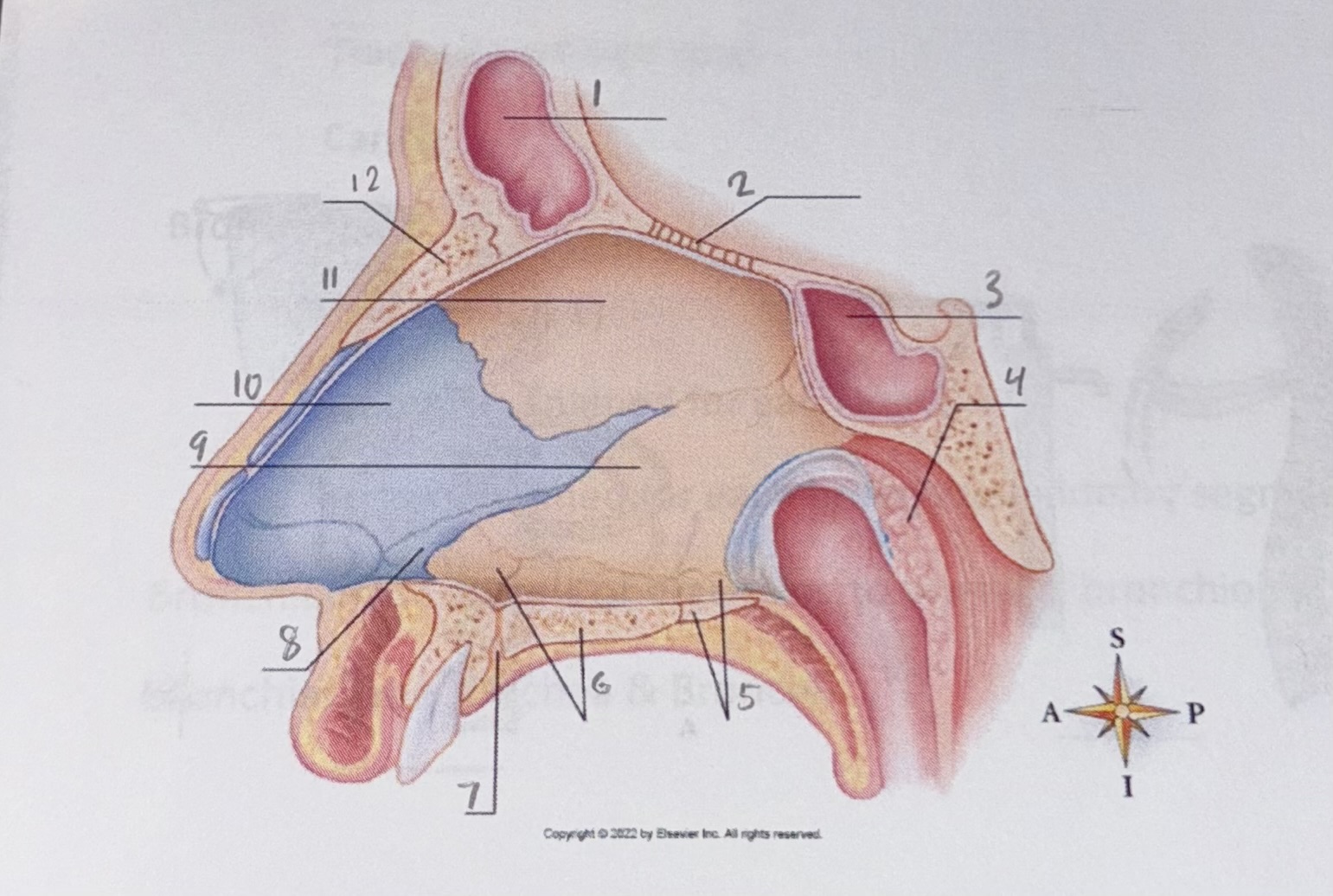
10
septal cartilage

11
perpendicular plate of ethmoid bone

12
nasal bone

what is this view showing
anterior view of the paranasal sinuses
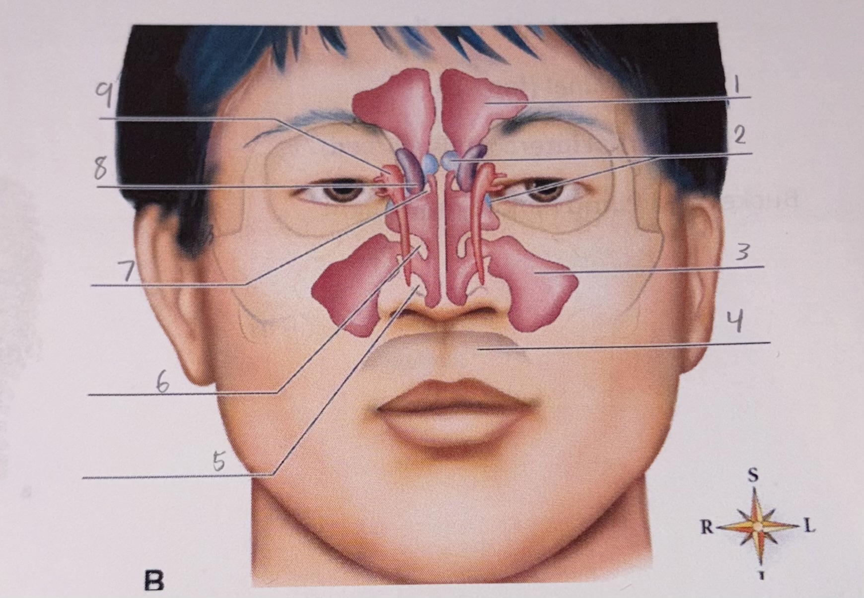
1
frontal sinus

2
sphenoid sinus

3
maxillary sinus

4
oral cavity

5
inferior concha

6
middle nasal concha of ethmoid

7
superior nasal concha of ethmoid

8
ethmoid air cells

9
lacrimal sac

what view is this showing
A: anterior view B: posterior view of larynx. some softer tissues of the larynx and surrounding structures have been removed to make it possible to see the cartilages of the larynx

1
epiglottis

2
superior thyroid notch

3
laryngeal prominence (Adams apple)
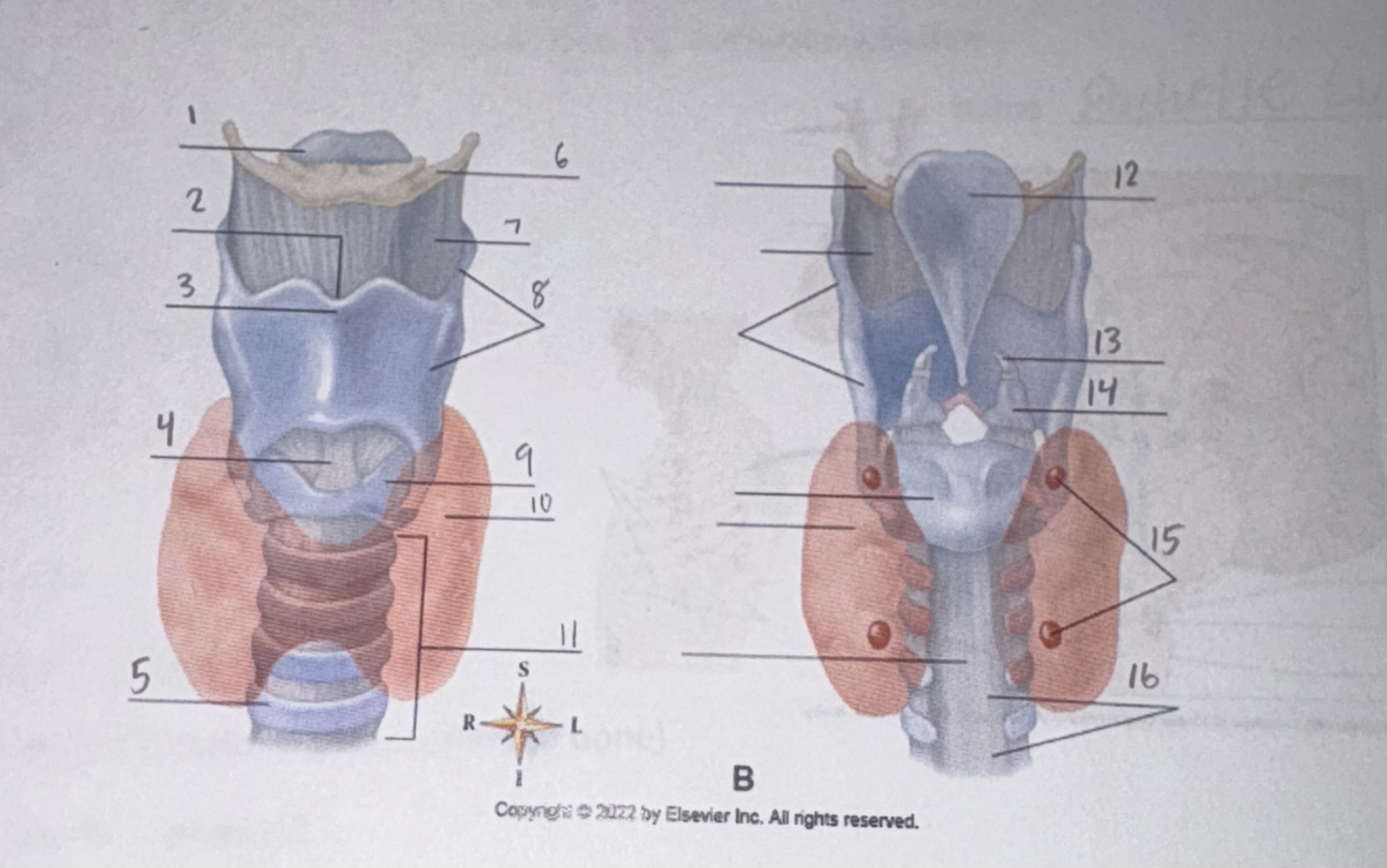
4
cricothyroid ligament

5
tracheal cartilage

6
hyoid bone

7
thyrohyoid ligament

8
thyroid cartilage

9
cricoid cartilage

10
thyroid gland

11
trachea

12
epiglottis

13
corniculate cartilage

14
arytenoid cartilage

15
parathyroid glands

16
membranous part of trachea

what view is this showing
sagittal section of the mucosal lining of the larynx, with its folds and underlying muscles and ligaments visible

1
hyoid bone

2
epiglottis

3
vestibular fold (false vocal cord)
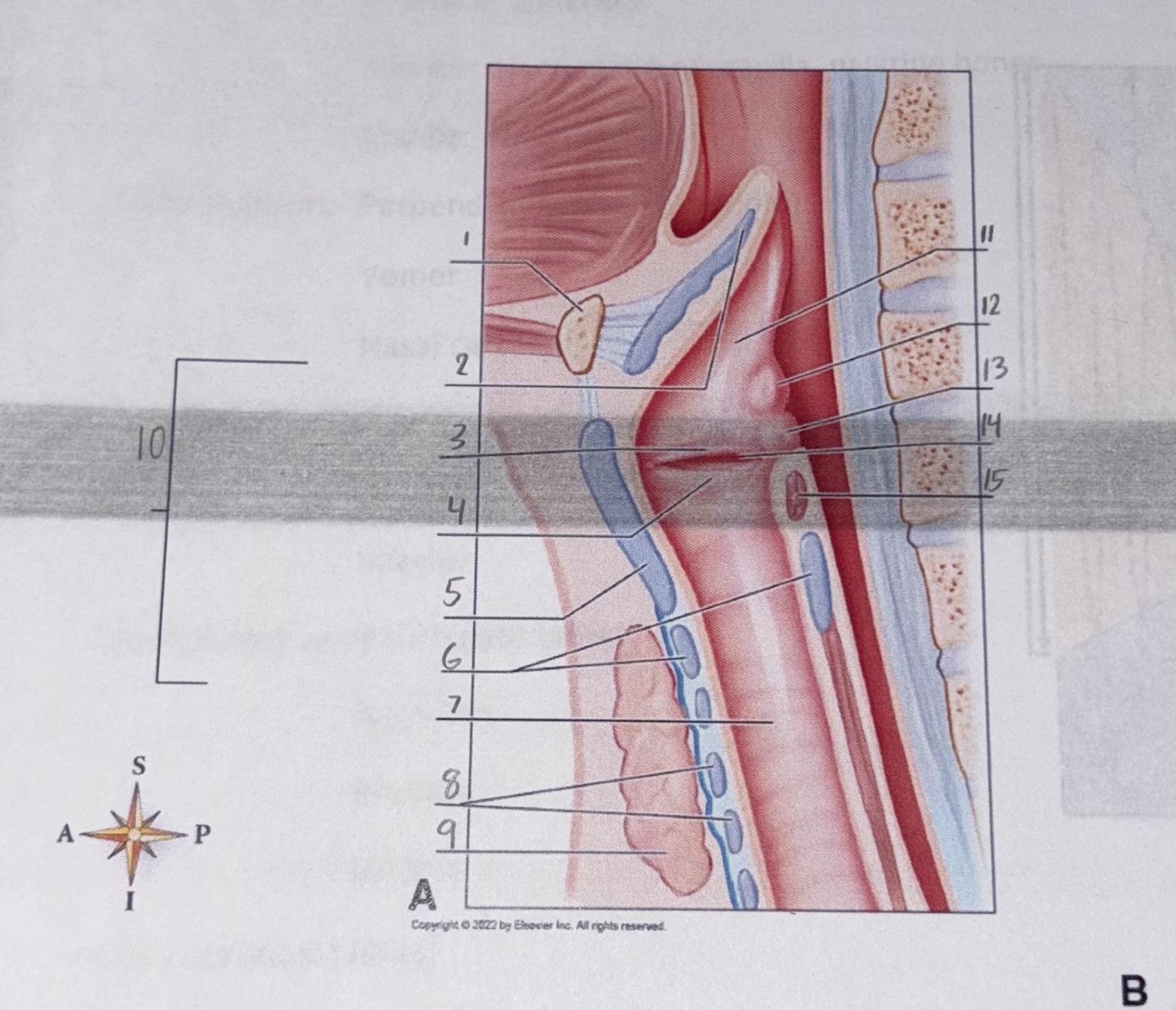
4
vocal fold (true vocal fold)

5
thyroid cartilage (Adams apple)

6
cricoid cartilage

7
lumen of trachea

8
cartilages of trachea

9
thyroid gland

10
larynx

11
vestibule

12
cuneiform cartilage

13
corniculate cartilage

14
vestibule

15
arytenoid muscle
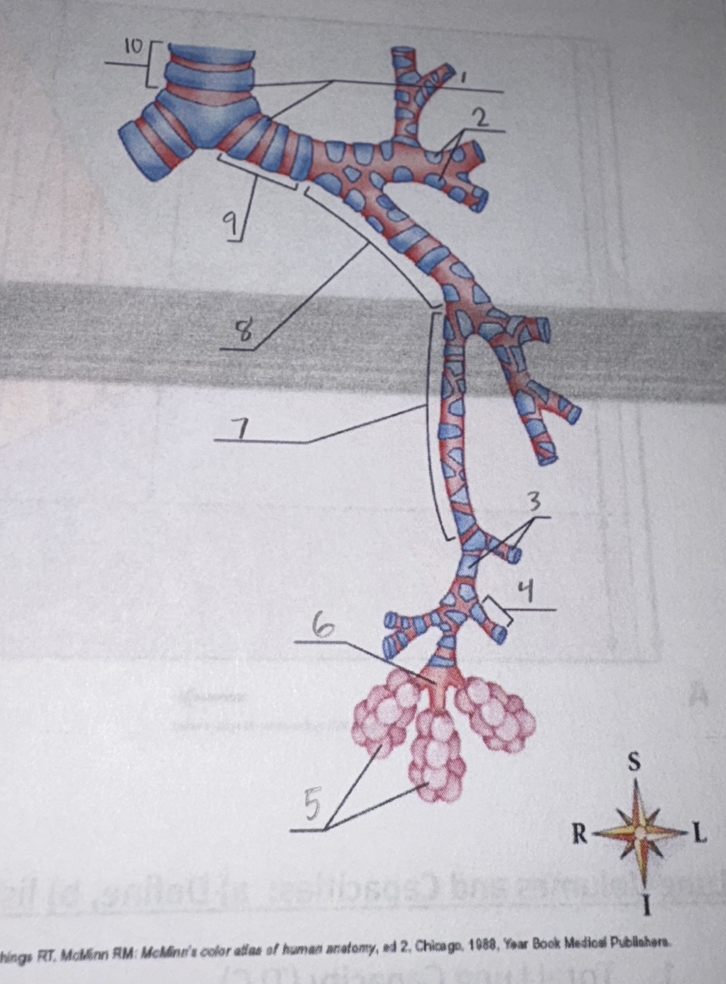
what view is this showing
various levels of branching from the primary bronchi (first level of branching) to the alveolar sacs (20th-23rd levels of branching)

1
cartilage rings

2
cartilage plates

3
bronchioles

4
terminal bronchiole

5
alveolar ducts and alveolar sacs

6
respiratory bronchiole

7
tertiary bronchi

8
secondary bronchus

9
primary bronchus

10
trachea

what view is this showing
medial view of the right lung; note the hilum

1
right pulmonary bronchus

2
pulmonary artery