KIN 1Y03 Lecture 33: Skeletal Muscle Gross Anatomy Pt1
1/35
There's no tags or description
Looks like no tags are added yet.
Name | Mastery | Learn | Test | Matching | Spaced |
|---|
No study sessions yet.
36 Terms
origin/head vs insertion vs belly
Origin/Head: muscle end attached to more stationary of 2 bones – ū more proximal
Insertion: muscle end attached to bone with greatest movement – ū distal
Belly: largest portion of hte muscles between origin and insertion – able ot have multiple bellies (eg. bellies)
tendons vs ligaments
ligaments = connect bone to bone
tendons = connect muscle to bone
aponeurosis
very broad tendon (u sheet-like)
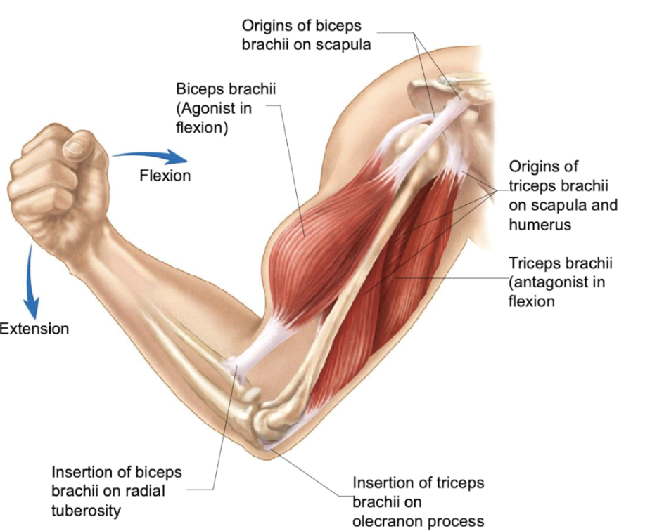
agonist vs antagonist
[Agonist: muscle that, when contracted, will cause an action
[Antagonist: a muscle working in opposition to agonist
⇒ both will vary based on movement:
Bicep contracting ⇒ cause flexion @ elbow ⇒ agonist
⤷ triceps = antagonist
Elbow going from flexion ⇒ extension
⤷ bicep = antagonist
⤷ tricep = agonist
synergists
muscles that work together to cause a movement
Prime mover: plays a major role in accomplishing movement
Fixators: stabilize joints crossed by the primer mover; prevents movement of the origin of the prime mover
⤷ eg.scapula ⇒ ū just “floating” but fixed in place that allows other joints to move
examples of muscles named according to… location
Location: pectoralis (chest), gluteus (butt), brachial (arm)
examples of muscles named according to… size
Size – ū group of muscles: maximus (largest in group), minimus (smallest), longus (longest), brevis (shortest), vastus (huge), major (larger-causing more action), minor (smaller)
examples of muscles named according to… shape
Shape: deltoid (triangle), quadratus (shape), teres (rounded from cross-section), trapezius (trapezoid)
examples of muscles named according to… action/function
Action/function: abductor, adductor, masseter (mastication/chewing), extensors
examples of muscles named according to… orientation or direction of fibers
Orientation or direction of fibers: rectus (fibers in erect fashion, parallel to midline), transverse (perpendicular to midline), oblique (@ angle)
examples of muscles named according to… origins/insertions
Origin and insertion: sternocleidomastoid (attaches to sternum, clavicle, and mastoid process on temporal bone), brachioradialis (attach on radius + brachial region)
examples of muscles named according to… number of heads
Number of heads: biceps (2), triceps (3)
examples of muscles named according to… number of muscles
Number of muscles: quadriceps (4 diff muscles)
What muscle groups allow for flexion, extension and rotation + lateral flexion for neck ?? (idk relationship between question)
Flexion: are ū muscles deep within the neck along and originate from anterior margins of the vertebral bodies ⇒ then extend up to occipital bone
⤷ anterior muscles that allow for flexion
Extension: posterior neck muscles attached to occipital bone and mastoid process
Rotation and lateral flexion (tile of neck to shoulder): lateral and posterior groups
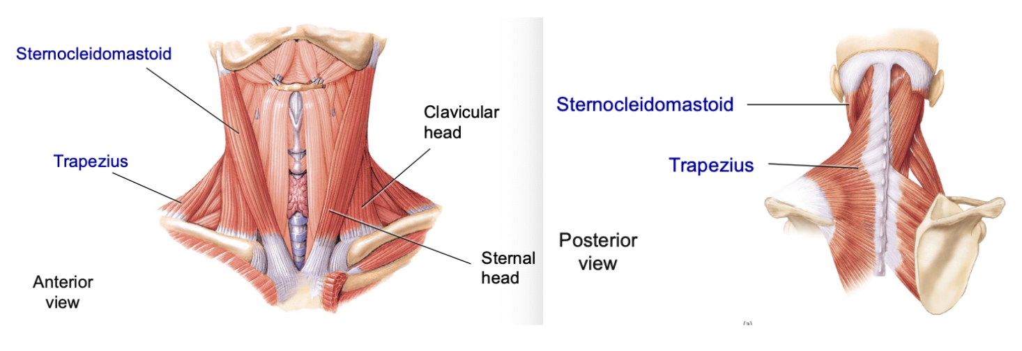
sternocleidomastoid
prime movers that originate on manubrium of sternum and clavicle
⤷ will extend up and insert on mastoid process
⤷ lateral muscle ⇒ will cause forward flexion of head if all muscles are contracting OR tilting ear to sternum (working both muscle heads)
⤷ has 2 heads – the sternal head – originates from the manubrium of the sternum & clavicular head – originates from the medial third of the clavicle

Trapezius
prime mover posterior muscle that is trapezoid shaped
⤷ has fibers in many diff DIR (which dictate the way muscles contract)
⤷ movement for neck → extension + lateral flexion
muscles that move the vertebral column is divided into deep and superficial groups
Deep group: short muscles that go from vertebra to vertebra
Superficial group: extends from vertebrae to ribs
⤷ eg. erector spinae: prime mover of the back extension with 3 subgroups
Subgroups – spinalis thoracis (ū medial), longissimus muscle (ū intermediate), iliocostalis muscle (ū lateral) [DONT NEED TO KNOW, JUST KNOW IT HAS 3 SUBGROUPS]
erector spinae
prime mover of the back extension with 3 subgroups
scalene
thoracic muscles associated w rib cage
scalene: u elevates the first 2 ribs during inspiration (bc it doesn’t have anything above them)
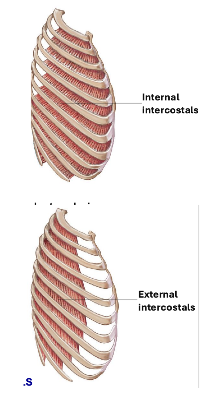
external intercostal
thoracic muscles associated w rib cage
External intercostal: elevate the ribs
⤷ muscles are found in between the ribs → goes superficial to deep as they go from inferior margin of 1 rib to superior border of the rib below
⤷ during contraction, ribs will expand up and down
Muscle DIR = “hands in pocket” oblique angle, pointing towards the midline
internal intercostal
thoracic muscles associated w rib cage
Internal intercostal: depress ribs during expiration
⤷ job: pull inferior ribs back into place
⤷ deeper muscles than external intercostals
⤷ DIR of muscles = perpendicular to “hands in pocket”
transversus thoracis
thoracic muscles associated w rib cage
Transversus thoracis: depresses ribs during expiration
⤷ work w internal intercostals to depress ribs during expiration
⤷ run from sternum to costal cartilages
diaphragm
dome-shaped muscle that runs upward-medially/inferior-superior DIR
⤷ separation between thoracic and abdominal cavity
inspiration vs expiration + what happens to the ribs and diaphragm
Inspiration: “breathing in”
Ribs – will move upward and outward as the external intercostal muscles contract
Diaphragm – contracts and moves downward, increasing the volume of the chest cavity
⤷ will create a vacuum & cause the lungs to expand and draw air in from the outside (↑ volume of the thoracic cavity)
Expiration: “breathing out”
Ribs – move downwards and inwards
Diaphragm – relaxes and moves upwards
⤷ will decreases the volume of the thoracic cavity – increasing the pressure inside the lungs and forces air out.
abdominal wall
Will flex and rotate vertebral column, decrease volume of abdominal and thoracic cavities ⇒ “compression”
Compression movement will help in forced expiration, vomiting, defecation, urination, childbirth
Muscle fibers has a criss-cross pattern of muscles, adding to the strength of the abdominal wall to support organs – enforced with muscles bc no bone

rectus abdominis
most medial (only medial muscles)
⤷ fibers run up/down in rectus formation
⤷ gives the “six-pack” appearance
⤷ muscles separated in intervals by CT, involved in flexion of vertebral column + some compression

external abdominal oblique
superficial muscles, where fibers run @ oblique DIR (“hands in pocket”)
⤷ flexion and rotation of vertebral column + compression

Internal abdominal oblique
deep muscles, where fibers run perpendicular to “hands in pocket”
⤷ flexion, rotation, and compression

transversus abdominis
fibers that run in transverse (horizontal) DIR, responsible for compression of the abdominal cavity
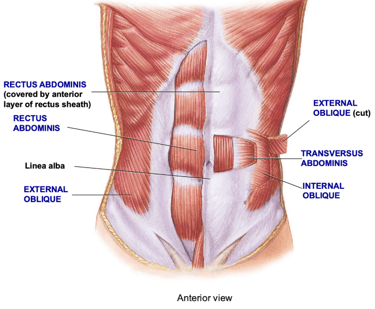
linea alba
CT @ center line, where all the muscles will attach to the linea alba

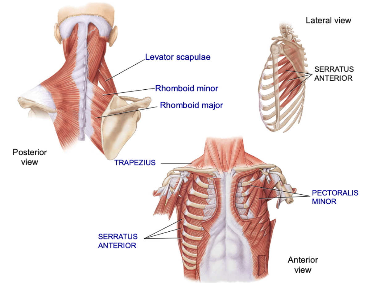
scapular movement
muscles that attach the upper limbs to the body and move/stabilize the scapula and clavicle
⤷ will originate on the axial skeleton – limbs are connected to the scapula
incl. trapezius, levator scapulae, rhomboideus/rhomboid (minor and major), serratus anterior, pectoralis minor

trapezius
trapezoid looking muscle on the posterior side, responsible for elevation and depression of the scapula + rotation

levator scapulae
deep to trapezius, responsible for elevation/rotation of scapula
⤷ goes up to the cervical vertebrae and inserts @ top point of scapula

rhomboideus/rhomboid + 2 components
runs from vertebrae to medial border of scapula, will pull scapula towards vertebrae that “square” shoulders
Composed of 2 muscles:
Rhomboid minor: lower, smaller
Rhomboid major: higher, bigger

serratus anterior
holds scapula in place on the thoracic cage
⤷ anterior to teh scapula
⤷ runs from teh lateral edge of series of ribs and runs underneath scapula to medial border of scapula @ midline
⤷ responsible for movements of arms held anterior (“boxers muscles”)
⤷ looks like serrated knife

pectoralis minor
Pectoralis minor: goes from the 3rd-5th ribs, extending up and inserts onto the coracoid process of scapula ⇒ wrapping around shoulder joints
⤷ job: depresses scapula and pulls on top of scapula