Lecture 4 - Synaptic Transmission I (Electrical & Chemical Synapses)
1/19
There's no tags or description
Looks like no tags are added yet.
Name | Mastery | Learn | Test | Matching | Spaced | Call with Kai |
|---|
No analytics yet
Send a link to your students to track their progress
20 Terms
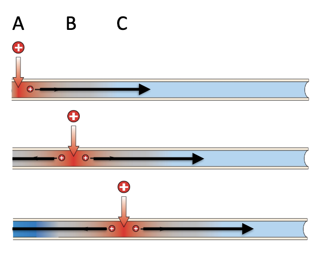
Active vs Passive Action Potential propagation
Unmyelinated → only active, slower
How to increase the speed of AP propagation?
By increasing the efficiency of passive conduction (charge travels further without decay)
Cable theory
model for studying the passive conduction of electrical signals along a fiber
Rm → insulation of the membrane
Ri → resistance of the cytoplasm to allowing the passing of AP
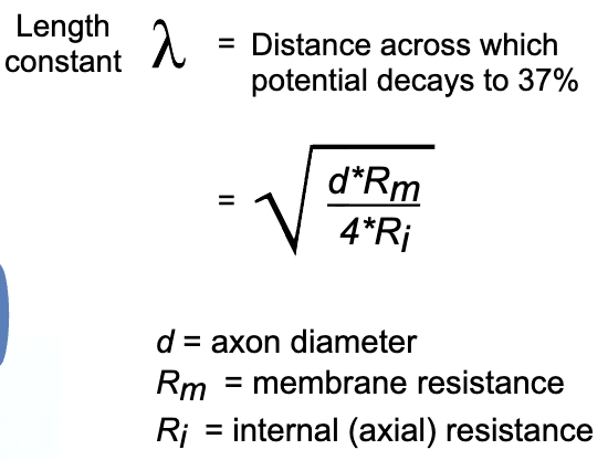
How to increase the speed of passive conduction along axons?
Increasing axon diameter (ex. giant squid axon)
Increasing Rm (ex. myelination)
Decreasing Ri (already low and constrained by cytoplasm composition)
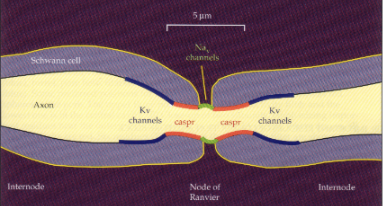
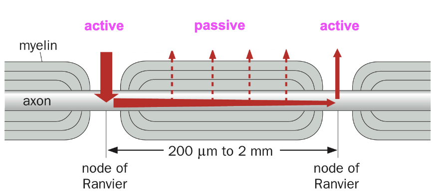
Nodes of Ranvier
Intermittent regeneration of signal (active conduction)
caspr: prevents sodium channels from migrating away
After NaV depolarizes, the Kv channels repolarize (under myelin to not interfere)
Saltatory conduction
In myelinated cell, AP jumps from one node to the next
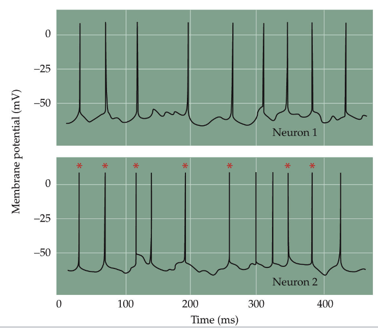
Electrical synapses
More common in invertebrates, not just neurons
Fast, synchronous, high threshold (many inputs must integrate → low false positives)
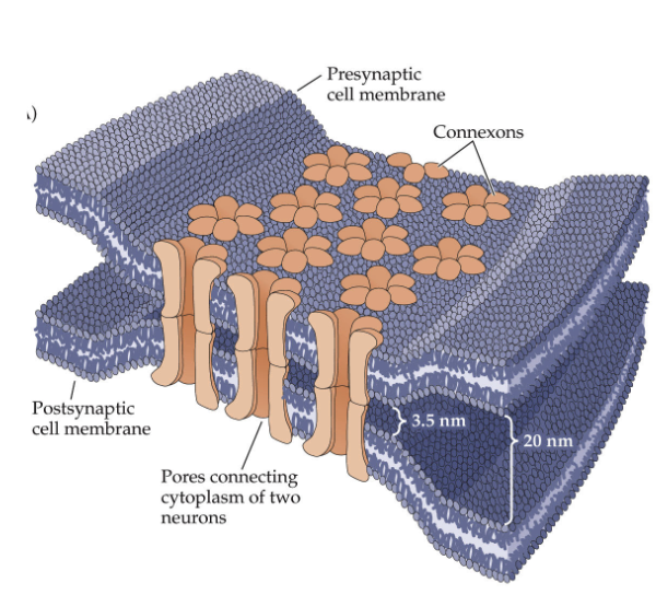
~3 nm gap spanned by gap junctions (tunnels)
Connections: axon-axon, axon-dendrite, dendrite-dendrite
Gap Junctions
6 connexins → 1 connexon (nonspecific, can be ex or in, bidirectional, reliable)
Electrical coupling: pass ions & molecules directly
PSP lower in 2nd cell because not all ions pass through (leaky, Rm)
Common in early development (“fire together, wire together”)
Synchronizes local astrocyte networks → distributed regulatory networks
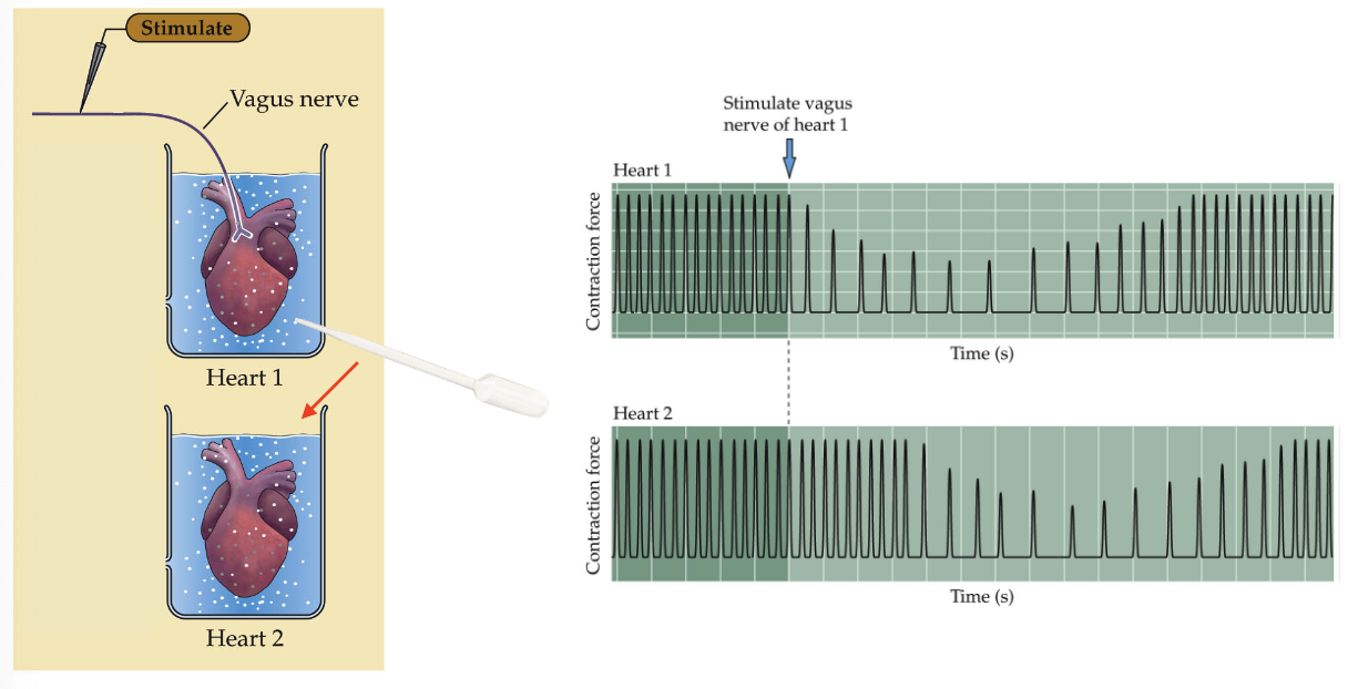
Otto Loewi - Evidence for chemical neurotransmission
Stimulated vagus nerve of frog heart → slowed
Transferred fluid to second heart → slowed
Some chemical substance transmitted the nerve signal → later found to be ACh
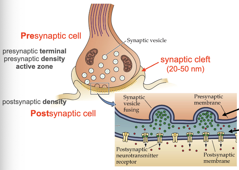
Presynaptic density active zone
protein-dense region on the presynaptic membrane where synaptic vesicles dock, fuse, and release neurotransmitters
Types of neurotransmitters
Amino acids
Amines
Peptides
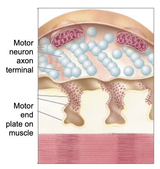
Neuromuscular junction (NMJ)
Large, accessible, reliable
Motor neuron axon (vesicles packed with ACh) connects to motor end plate (folded membrane with ACh receptors)
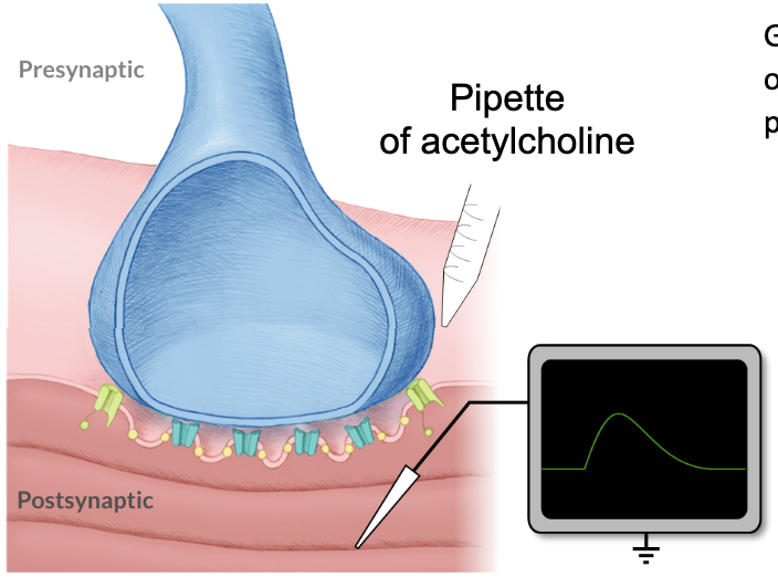
Katz’ Hypothesis
Applied ACh to NMJ & observed EPPs
Saw EPPs that were similar to that obtained from electrical stimulation
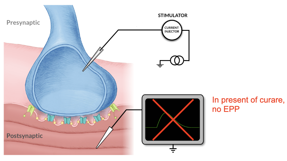
Applied curare to NMJ (blocks ACh receptors to cause paralysis)
No EPP in the presence of curare
Stimulation of presynaptic neuron → generates postsynaptic potential (PSP) → EPP
Nerve impulse triggers ACh release
ACh binds to ionotrophic receptors on end-plate membrane
Sodium channels open → Na+ enter muscle fiber
Muscle membrane depolarizes → EEP
If strong enough to reach threshold → AP → muscle contraction
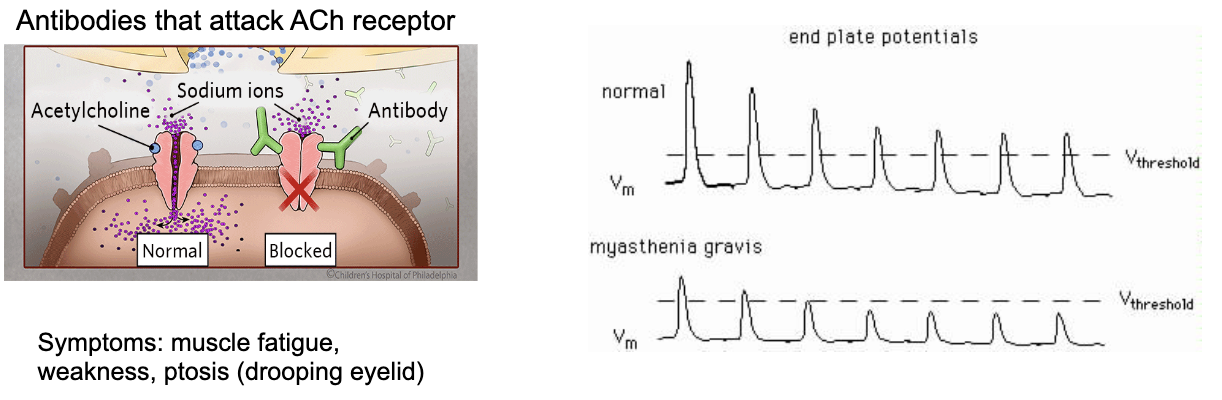
Myastenia gravis (MG)
NMJ autoimmune disease - antibodies that attack ACh receptor
ACh blocked, EPPs can’t reach threshold
Symptoms: muscle fatigue, weakness, ptosis (drooping eyelid)
Solution: 1. Rescue AChR, 2. Increase ACh release, 3. Reduce ACh degradation
Neostigmine: slows degradation of ACh by inhibiting acetylcholinesterase
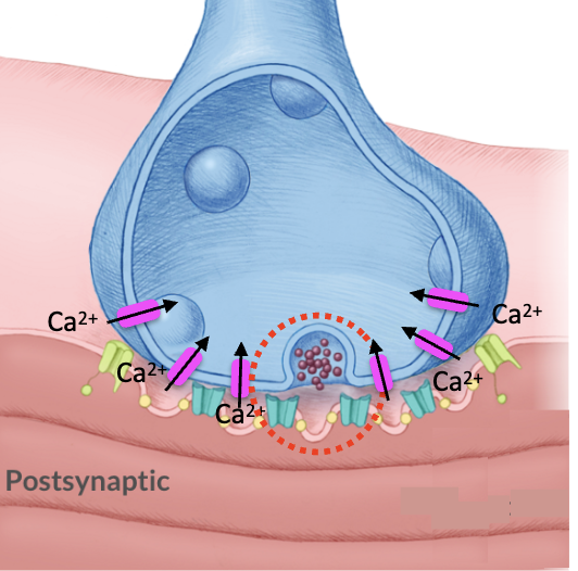
How does AP in motor neuron cause ACh release? Hypothesis: Ca++ influx
Evidence:
1. Increasing extracellular Ca++ at NMJ leads to larger EPPs
Adding Ca++ buffer abolishes EPPs
Injection of Ca++ into presynaptic terminal drives EPPs
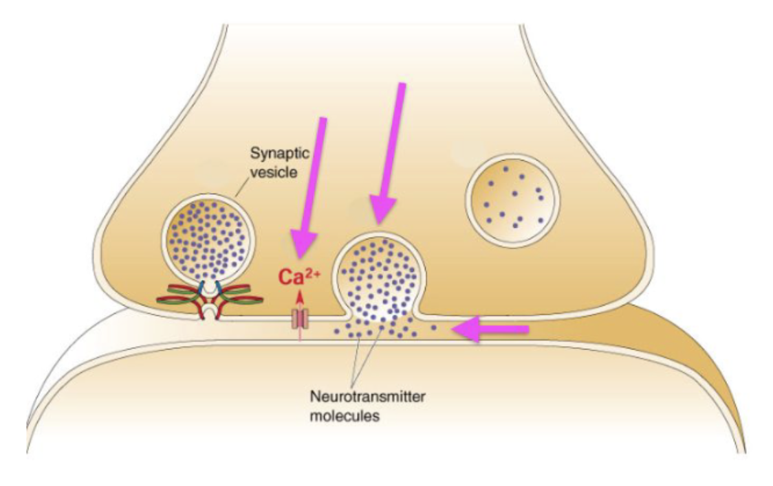
How are neurotransmitters (like ACh) released?
Action potential reaches presynaptic terminal
Depolarization causes voltage-gated Ca++ channels to open → influx of Ca++
Ca++ binds to synaptotagmin → SNARE complex fuses docked vesicles
Exocytosis: neurotransmitters released into synaptic cleft
Receptor binding → enzymatic breakdown (AChE) → vesicle recycling
Difference between EPSP, EPP, mEPP, and AP?
EPSP: Excitatory Postsynaptic Potential, small local depolarization that is graded (size depends on how much NT is released), caused by release of glutamate, decremental, many ESPS must summate to reach AP threshold at Hillock/AIS
EPP: End-Plate Potential, large depolarization at NMJ that is normally enough to reliably exceed the threshold, caused by release of ACh
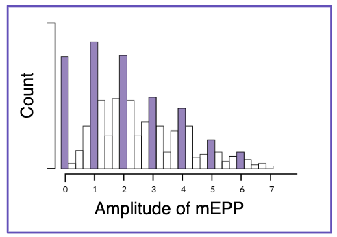
mEPP: Miniature End-Plate Potential, small spontaneous depolarization at NMJ, random fusion of single vesicles of ACh, used to come up with Quantal Hypothesis
AP: Action Potential, all-or-none, non-decremental
Quantal hypothesis
Neurotransmitters released in discrete packets of uniform size → predicted synaptic vesicles
Confirmed by electron microscopy
mEPPs occurred spontaneously
mEPPs got larger after neostigmine treatment (suggesting they are triggered by ACh)
Katz reduced extracellular Ca++ → EPPs became very small
Discrete amplitudes, multiples of the smallest spontaneous mEPP
Clostridium botulinum
Cleaves SNARES to block vesicle release, flaccid paralysis
Used to treat muscle spasms (cerebral palsy) and cosmetic (Botox)