3 Major Groups of Microbes
Prokaryotes
Eukaryotes
Acellular Infectious Agents
Scientific Names
Carl Linnaeus (1700s)
Genus Species
Ex. Homo sapien
Microbes
Essential component of a healthy human
Human Microbiome
Group of microorganisms living in/on your body & don’t usually cause disease
Prevent disease by competing with pathogens
Aid with digestion
Promote immune system development
Pathogens
Disease-causing microbes
Robert Hooke (1665)
Used crude microscope to view individual cells
Bread mold (“Microscopial Mushroom”)
Beginning of Cell Theory
Cell Theory
All living organisms are composed of cells
Antoine Van Leeuwenhoek (1674)
Amateur lens grinder
Built microscopes that could view living microorganisms
Called microorganisms “little animacules”
Spontaneous Generation vs Biogenesis (1668 - 1800s)
Spontaneous Generation: Forms of life can arise from non-living matter.
Biogenesis: Living matter only arises from pre-existing life forms.
Louis Pasteur (1861)
Used swan neck flasks to disprove spontaneous generation.
Showed that microscopic yeast (fungi) converts sugar to alcohol using fermentation.
Absence of oxygen - anaerobic
Souring occurs when bacteria turns the alcohol into vinegar.
Presence of oxygen - aerobic
Solution: Heat beer or wine after fermentation to kill bacteria and prevent spoilage - pasteurization.
Germ Theory of Disease
Microorganism cause certain diseases.
Joseph Lister (1860s)
Used Phenol to clean surgical instruments and treat surgical wounds.
Drastically reduces incidents surrounding surgical wound infections.
Led to development of disinfectants and antiseptics.
Robert Koch (1876)
first proof that bacteria cause disease.
Investigated cause of anthrax.
Establish a sequence of experiment steps that could be used to find the causative agent of other diseases
Koch’s Postulates
Isolated bacteria
Showed that particular bacterium was present in all cases of the disease
Injected bacterium into healthy specimens
Specimen contracted anthrax and died
Re-isolated bacteria from injected specimens and showed they were identical to him first sample
Showed that specific microbe was the cause of particular disease
Edward Jenner (1796)
Developed smallpox vaccine
Observed that people previously sick from cowpox did not contract smallpox.
Purposefully inoculated a young boy with cowpox, fell ill, recovered and became immune to smallpox.
Paul Ehrlich (1909)
Noticed that certain dyes stain bacteria differently than they stained a animal cells
Proposed that a chemical might be found that would harm disease causing microbes without harming the host.
Discovered Salvarsan, an arsenic derivative that could be used to treat syphilis.
Selective Toxicity
Ability to drug target sites relative to the microorganism responsible fro infection. Some sites are unique to microorganism or more essential to the survival of the microorganism than the host.
Alexander Fleming (1928)
Noticed mold inhibited bacterial growth on contaminated plates.
Produced first natural compound, penicillin.
First antibiotic
Compound naturally produced by molds or bacteria, inhibiting he growth of or kills other microorganisms.
Light Microscope
Light Microscope that uses visible light to observe a specimen
Compound Light Microscope: Uses two lenses to magnify image
Objective Lens: Lens closest to specimen & can magnify between 4x - 100x
Ocular Lens: The eyepiece & magnifies by 10x
Calculating magnification:
Total Magnification = Objective lens x Ocular Lens
Resolution
Ability to distinguish fine detail & structure
Ability to distinguish 2 points a certain distance apart
Light must pass between 2 objects for them to be seen as two separate things.
Need light of a short enough wavelength to fit between them, otherwise will appear as one object.
Resolution General Principle: the shorter the wavelength, the better the resolution.
Electron Microscope
Electrons instead of light, electrons travel in much shorter waves.
Resolving power is greater
Allows for greater magnification
Allows us to view viruses and internal cell structures
Electron Microscope - Transmission (TEM)
See internal structures
Very thin slices can be cut from sample
Samples generally stained with a metal to make structure opaque to electrons
Electron Microscope - Scanning (SEM)
See surfaces, less powerful
Atomic Force (AFM)
See’s molecules
Uses thin metal probe to scan a specimen revealing bumps and depressions
Stains
Microorganisms are almost colourless when seen though a microscope, stains make them easier to see.
Stains are composed of positively and negatively charges ions, one of which is coloured (chromophore).
Stain - Simple
One dye is used to highlight the entire microorganism
Steps:
Smear sample on slide
Fix with heat
Add stain
Was, dry, and view
How Stains Work
Bacteria have a net negative charge on their outer surface, this charge attracts stains with positively charged chromophores and repels stains with negatively charged chromophores.
Positive Stains - Simple
Stain binds to bacterium
Bacterium appears coloured
Background appears clear
Negative Stains - Simple
Will not bind to bacterium
Bacterium appears clear
Background is coloured
Stains - Differential
React different with bacteria, thus can be used to distinguish between them.
Eg. Gram Stain
Differentiates bacteria based on the structure of the cell wall
Gram Stain - Differential
Gram Positive: Bacteria with a thick cell wall retain the primary stain crystal violet and appear purple.
Gram Negative: Bacteria with a thin cell wall lose crystal violet during destaining, take on the colour of counterstain safranin and appear pink.
Endospore Stain - Differential
Stains internal structures of some bacteria
Primary stain colours endospores green
Counterstain colours the rest of cell red
Flagella Stain - Differential
Stains external structures
Mordant (dye fixative) thickens flagella so they can be observed under light microscope
Acid-Fast Stain - Differential
Detects presence of waxy compound in cell wall
Used to identify the genus Mycobacterium
Mycobacterium cell wall retains dye carbol fuschin
Counterstain with methylene blue stains non acid-fast bacteria and tissues blue
Capsule Stain - Differential
Detects thick layer of polysaccharide outside the cell.
Negative stain colours background
Positive stain colours the cell
Does not take up most dyes and remains colourless.
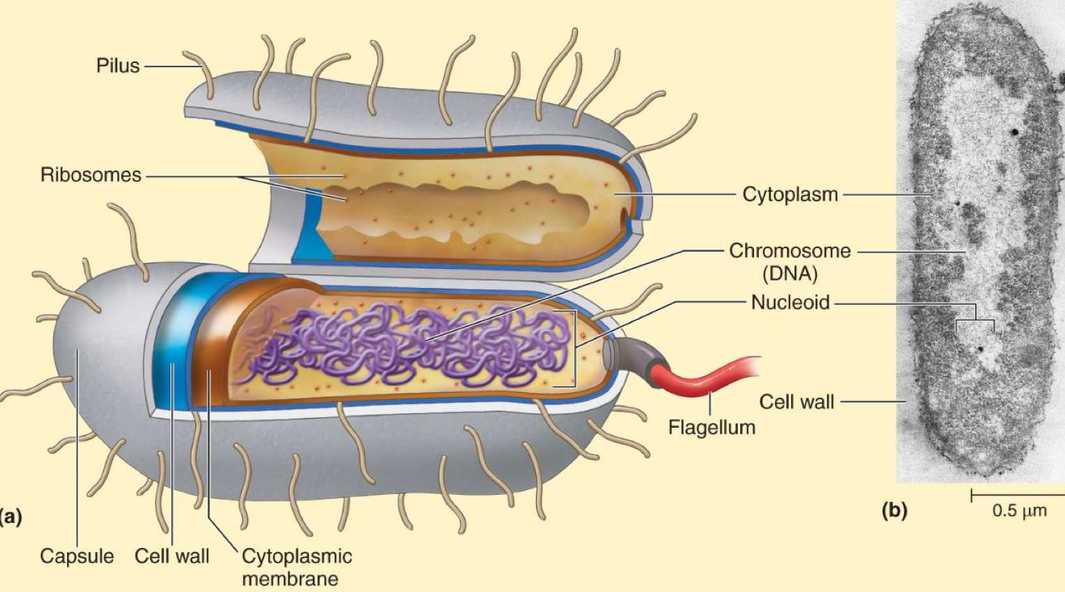
Prokaryote
DNA not enclosed within a nucleus
Usually arranged as one circular chromosome
Lack membrane bound organelles
Single celled organisms: Bacteria, Archaea
Eukaryotes
DNA found in nucleus, surrounded by nuclear membrane
DNA arranged as multiple chromosomes
Membrane bound organelles
Single celled or multicellular organisms
Algae, Protozoa, Fungi, Plants, Animals
Bacteria
Morphology
Coccus - Spherical
Bacillus - Rod
Vibrio - Curved
Spirillum - Spiral Shaped
Spirochete - Corkscrew Shaped
Bacteria Cell Structure
External Structures - Bacteria Capsule
Sticky, gelatinous layer external to the cell
Composed of polysaccharides, protein, or both
If layer is organized and firmly attached to cell wall, capsule
In some bacteria capsules a play a role in virulence
Protection against phagocytosis
Streptococcus pneumoniae
With capsule: causes disease
Without capsule: no disease
External Structures - Bacteria Slime Layer
Sticky, gelatinous layer external to the cell
Composed of polysaccharides, protein, or both
If layer is unorganized and loosely attached to the cell wall, slime layer
Often allow bacteria to attach to surfaces
Medical Implants, water pipes, teeth
Streptococcus mutans
Makes polysacccharide slime from sucrose
Attaches to teeth, leading to cavities
Bacteria - Flagella
Long protein appendages
Used in motility
Semi rigid helical, turns like a propeller
Flagella - Arrangement
Monotrichous: Single polar flagellum
Lophotrichous: Two or more flagella originating from one pole
Amphitrichous: Tufts of flagella originating from opposite poles
Peritrichous: Flagella distributed all over the cell
Flagellar Motility
Flagella turn causing cell to move in one direction - “run”
Periodically flagella reverse direction causing a random change in direction - “tumble”
Flagella allow chemotaxis
Movement toward or away from a stimulant
Toward nutrients (attractant)
Away from toxins (repellent)
Flagella protein can be used to distinguish among strains of species
Bacteria - Fimbriae
Short, hair like appendages
Hollow
Allow cell to adhere to surfaces
Contribute to pathogenicity
Bacteria - Pili
Short, hair like appendages
Hollow
Allows attachment of two bacteria to each other
Involved in transfer of genetic material between bacteria
Bacterial Conjugation
Pilus Formation
The donor cells (F+ cells) form a sex pilus and begin contact with an F- recipient cell.
Physical Contact between Donor and Recipient Cell
The pilus forms a conjugation tube and enables direct contact between the donor and the recipient cells.
Transfer of F-Plasmid
The F-factor opens at the origin of replication. One strand is cut at the origin of replication, and the 5’ end enters the recipient cell.
Synthesis of Complementary Strand
The donor and the recipient strand both contain a single strand of the F-plasmid. Thus, a complementary strand is synthesized in both the recipient and the donor. The recipient cell now contains a copy of F plasmid and becomes a donor cell.
Bacterial Cell Wall
Semi Rigid structure giving shape to the cell
Major function is to prevent rupture of the cell - protects against environmental changes
Useful in identification of bacteria - ie. Gram Stain
Composed of peptidoglycan
Mesh-like structure composed of polysaccharide and amino acids
Polysaccharide portion composed of 2 alternating monosaccharide covalently joined
N-acetyl glucosamine (NAG)
N-acetyl muramic acid (NAM)
Peptide portion composed of short changing of amino acid
Peptidoglycan
Polysaccharide chains run parallel
Peptide chains link polysaccharides together
Forms a mesh-like net surrounding cell
Completely different from anything found in animal cells
Many antibiotics have been discovered that act against peptidoglycan
Penicillin: inhibits production of peptidoglycan
Lysozyme: Degrades and is found in tears, saliva, mucous
Gram Positive Cell Wall
Made of thick layers of peptidoglycan outside of plasma membrane
Contains teichoic acids
Wall teichoic acids: Attached to peptidoglycan
Lipoteichoic acids: Attached to plasma membrane and extend through peptidoglycan
Have only one membrane - cytoplasmic membrane
Gram Negative Cell Wall
Thin peptidoglycan layer sandwiched between two membranes
Outer membrane made of phospholipid, proteins, and lipopolysaccharide (LPS)
Polysaccharide portion of LPS is composed of O-sugars
Useful for distinguishing negative bacteria
Lipid portion of LPS is toxic
Referred to as endotoxin
How Gram Stain Works - Positive Cells
Thick peptidoglycan traps crystal violet - stain purple
How Gram Stain Works - Negative Cells
Thin peptidoglycan does not trap crystal violet, and outer membrane gets disrupted by alcohol
Crystal violet is washed away
Safranin counterstain stains cells pink
Prokaryote - Cytoplasmic Membrane
Composed of phospholipid bilayer
Separates interior from outside environment
Serves as a semi-permeable barrier
Selectively allowed inflow and outflow of materials
Exists in a semi-fluid state
Antimicrobial Agents
Alcohols disrupt the membrane
Can be used as disinfectant
Bacterial Internal Components - Cytoplasm
Substance inside plasma membrane
80% water
Contains most of the stuff needed for life
Sugars, amino acids, nucleotide, etc.
Enzymes
Some functional structures
Bacterial Internal Components - Nucleoid
Contains bacterial chromosome (DNA)
All genetic information required for cell’s structures and functions
Not surrounded by nuclear membrane
May contain plasmids
Smaller double stranded DNA molecules
Contain non-essential genes - eg. Genes for antibiotic resistance
Bacterial Internal Components - Ribosomes
Site of protein synthesis (translation)
Made of protein and ribosomal RNA (rRNA)
Two Parts:
30s Subunit
50s Subunit
Together form the complete 70s ribosome
Ribosomes of bacteria differ from eukaryotic ribosomes
Eukaryotes have 80s ribosomes
Several antibiotics target bacterial ribosomes
Prevent bacteria from making new proteins
Bacterial Internal Components - Storage Granules (Inclusion Bodies)
Usually deposits or granules of nutrients, stored for late use
Examples:
Sulfur granules
Polysaccharide (Glycogen)
Lipid inclusions
Enzymes
Magnetite
Variety of inclusion bodies occur in different bacterial species - can serve as a basis for identification
Bacterial Internal Component - Endospores
Formed only by some Gram-positive bacteria
Special resisting structure - allows bacteria to enter dormant state
Extremely durable
Resist heat, desiccation chemicals, radiation
Some endospores can survive in boiling water for hours
Remains dormant until good growth conditions occur
Can for new population
Sporulation
Cell replicates its DNA
Septum forms, dividing cell into unequal compartments
Larger compartment engulfs the smaller
Peptidoglycan and other protective material forms around the foreshore - spore coat
Finished spore is freed from the mother cell as the mother cell dies
Eukaryotic Cell Structure
Includes microorganisms algae, fungi, protozoa and higher organisms, plants, animals.
Larger and more complex than prokaryotes
Genetic material housed in nucleus
Membrane bound organelles
Eukaryotic - Cytoplasmic Membrane
Composed of phospholipid bilayer
Separates interior from outside environment
Serves as a semi-permeable barrier
Selectively allowed inflow and outflow of materials
Exists in a semi-fluid state
Contains phospholipids, proteins, and sterols
Sterols make membrane rigid compared to bacteria
Eukaryotic - Cell Wall
Not all eukaryotes have one
Allows endocytosis
Simple structure compared to bacteria
Made of:
Cellulose
Chitin
Eukaryotic - Cytoplasm
Substance inside plasma membrane but outside nuclear membrane
Has complex internal structure - Cytoskeleton
Protein filaments on the inside of plasma membrane
Provides support and shape
Transports substance through the cell
Eukaryotic - Ribosomes
Larger and heavier than bacterial ribosomes
80s
Several antibiotics target bacterial ribosomes
Prevent bacteria from making new proteins
Eukaryotic - Membrane Bound Organelles
Structures with specialized functions
Not present in bacteria
Example:
Mitochondria: Site of energy production
Chloroplast: Site of photosynthesis in algae and plant cells
Eukaryotic - External Appendages
Flagellum & Cillia
Long flexible projections that contain protein and cytoplasm
Move in whip-like fashion
Can be used for:
Motility
Sweeping material past stationary cells
Has 9 + 2 array (9 pairs of microtubules with 2 in enter of ring)
Microtubules: Long, hollow tubes made of protein called tubulin
Bacterial Growth
Refers to increase in bacterial cell numbers
Not an increase in size of individual cells
Most bacteria reproduce by binary fission
Bacterial cell:
Elongates and makes copy of its DNA
Divides into two identical cells
Exponential Growth
Binary fission → Population of cells double every generation
Time required for population to double = generation time
Varies greatly between different bacteria
Bacteria Growth in Lab
Inoculation: Introducing microbes into a medium to start culture
Culture: Microbes Growing in a medium
Batch Culture
Closed System
Once started, no other nutrients added
When nutrients are used up - bacteria stomp growing
Continuous Culture
Open system
Nutrients are continuously added, wasted are continuously removed
Supports indefinite growth
Growth Curve in Batch Culture - Lag Phase
Period of adaptation
Cells adjust to new media and prepare to grow
Growth Curve in Batch Culture - Exponential Phase (Log Phase)
Period of maximal reproduction - cell numbers increase exponentially
Used to calculate generation time
Growth Curve in Batch Culture - Stationary Phase
Cells have reached maximum population density
Nutrients have been used in, or wasted have accumulated
No increase in cell number
Growth Curve in Batch Culture - Death Phase
Toxic waste products have accumulated
Cells die at uniform rate
Growth Curve in Batch Culture - Phase of Prolonged Death Phase
Sometimes a small fraction of population survived the death phase
May consume nutrients release from dying cells
Selects for the strongest cells in population
Environmental factors that influence bacterial growth
Temperature
Oxygen
pH
Osmotic Pressure
Nutritional Factors
Nutritional Diversity
Temperature Requirements
Each microbe species has its own temperature range
Usually spans about 30°C
Maximum: Lowest temperature supporting growth
Optimum: Temperature supporting best growth
Maximum: Highest temperature supporting growth
Psychrophiles
Cold loving
Grows between 5°C - 15°C
Killed at 20°C
Psychrotrophs
Very broad temperature range
Minimum: ~5°C
Maximum: ~30°C - 45°C
Optimum: 15°C - 30°C
The microbes that cause food to spoil in fridge
Mesophiles
Moderate temperature loving
Minimum: ~10°C
Maximum: ~45°C
Optimum: 25°C - 45°C
Most bacteria
Most pathogens have temperature optimum of 37°C
Thermophiles
Heat loving
Minimum: ~40°C
Maximum: ~80°C
Optimum: 65°C
Hyperthermophiles
Minimum: ~75°C
Maximum: ~121°
Restricted to very few places on earth where water reaches these temperatures
E.g. Deep ocean vents
Food Safety
Involves use of both hot and cold temperatures
Heat is used to kill mesophilic and psychrotrophic microbes
Cold temperature is used to slow growth
Only psychrotophs will grow in a refrigerator - and slowly
Obligate Aerobes
Require O2 for respiration (energy generation)
Facultative Anaerobes
Can use O2 for respiration but also grow in it its absence
Obligate Anaerobes
Cannot use O2 and are killed by it
Microaerophiles
Require O2 in low amounts, but killed in high concentrations
Aerotolerant Anaerobes
Cannot use O2, but are not killed by it
pH
Measurement of acidity or alkalinity
pH < 7 = Acidic
pH > 7 = Alkaline
pH of 7 = Neutral
Most bacteria grow at or near neutral
Bacteria that grow at low pH are Acidophiles
Bacteria that grow at high pH are Alkaliphiles
Osmosis
Movement of solvent molecules across a semi-permeable barrier
E.g. Movement of water through cytoplasmic membrane
H2O will move from area of high concentration to low
Hypertonic Solution
High solute concentration
Ex. Salt or sugar
Water flows out of cell
Cell dries up - Plasmolysis
Hypotonic Solution
Low solute concentration
Water flows into cell
Cell bursts - Osmotic Lysis
Isotonic Solution
Condition where solute concentration on outside of cell is equal to that inside the cell
Osmotic Pressure & Food Preservation
Some bacteria have adapted to life in high salt concentrations - requiring up to 30% NaCl
Extreme Halophiles
Blood has a salt concentration of about 0.9%
Does not inhibit the growth of most microorganisms
Nutritional Factors Influencing Growth - Carbon
required for all organic molecules - backbone of living matter
Heterotrophs - Take carbon from organic matter
Autotrophs - Use inorganic carbon
Nutritional Factors Influencing Growth - Nitrogen, Sulfur, & Phosphorus
Required in smaller amounts for synthesis of cellular material
E.g. Protein, nucleic acids, phospholipids, ATP
Nutritional Factors Influencing Growth - Trace Elements
Required in very small amounts
E.g. Iron, zinc, molybdenum
Essential to functions of certain enzymes