Cardiovascular and respiratory systems
1/18
Earn XP
Description and Tags
Melissa coy
Name | Mastery | Learn | Test | Matching | Spaced |
|---|
No study sessions yet.
19 Terms
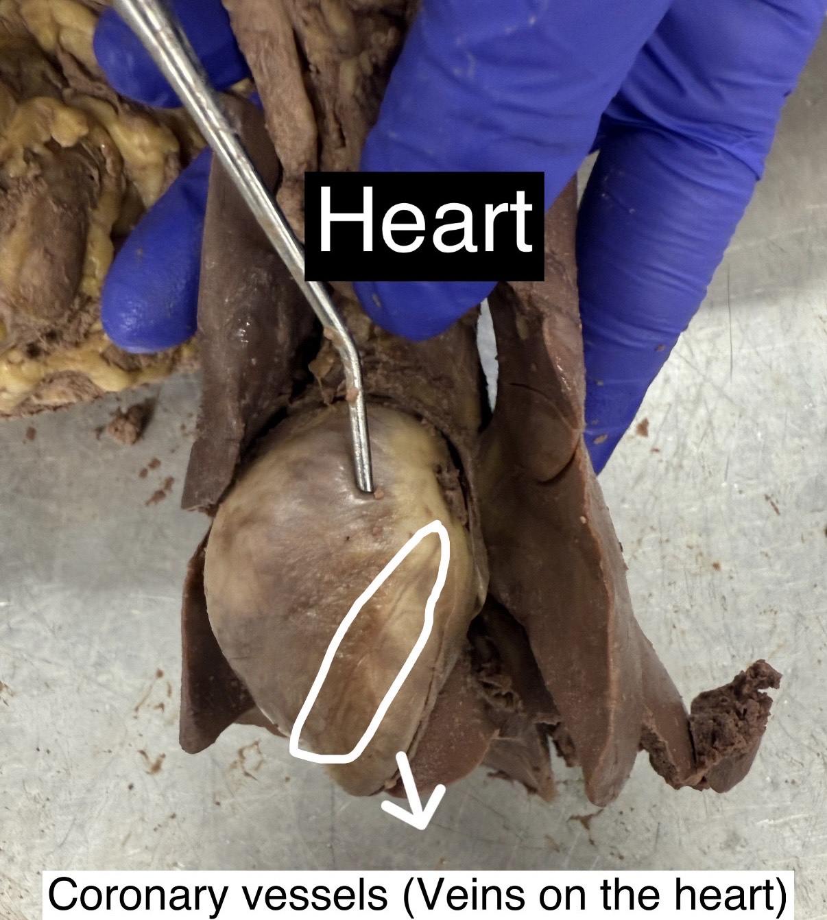
Heart and coronary vessels
The middle part that can open but point for the heart on the outside, and the coronary vessels is the veins on the heart
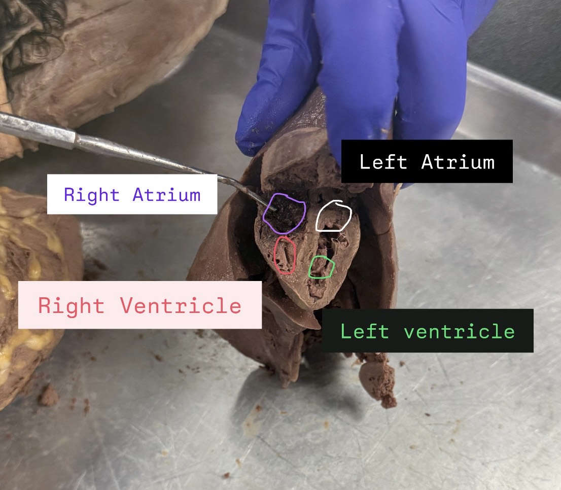
Right atrium, Left atrium, right ventricle, left ventricle
When looking at it, it will be opposite directions and atrium is on top and ventricle on bottom; in the inside of heart
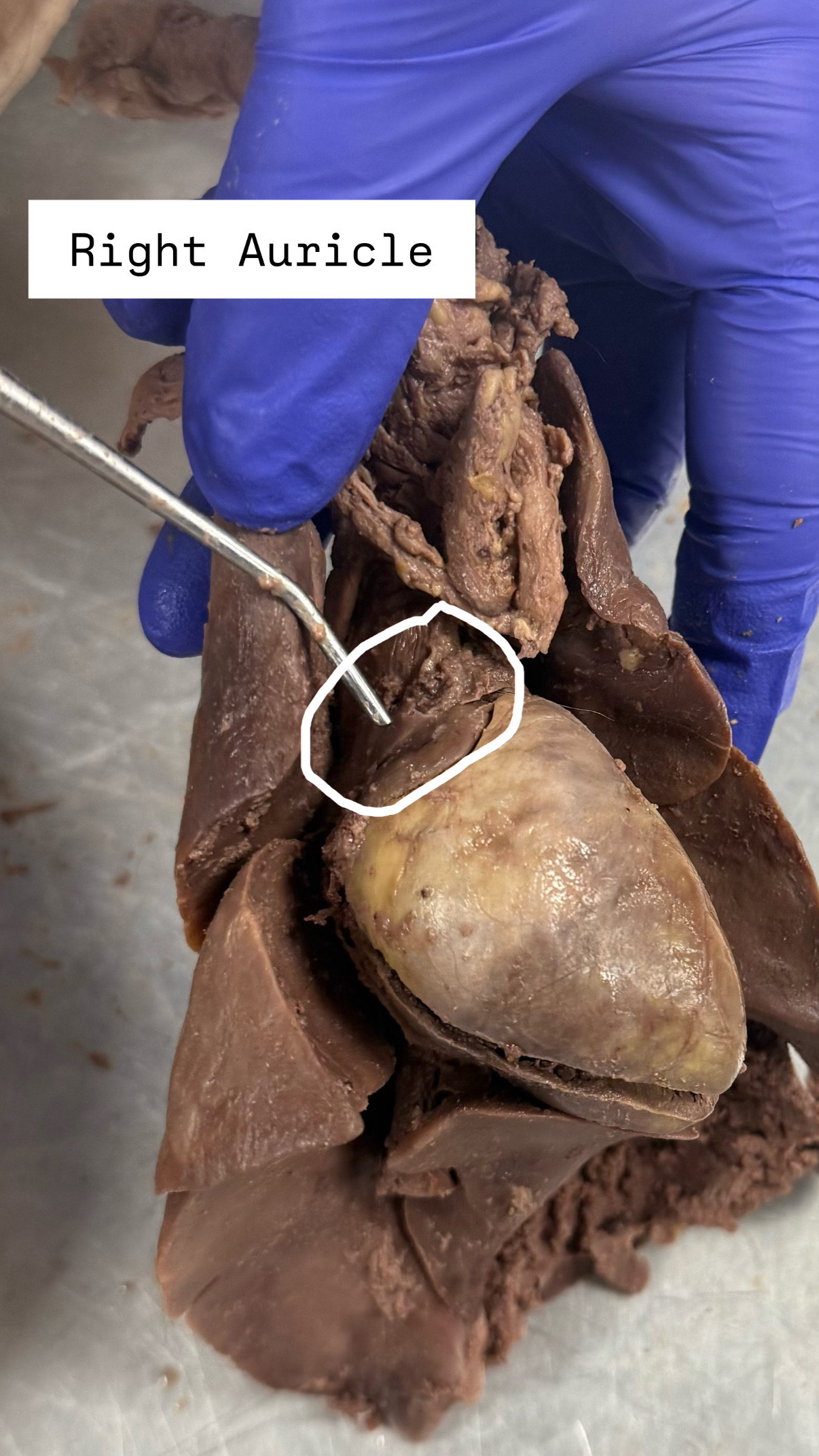
Right Auricle ("AW-ri-kuhl)
When looking at it, it will be opposite directions, they look like fans on the side
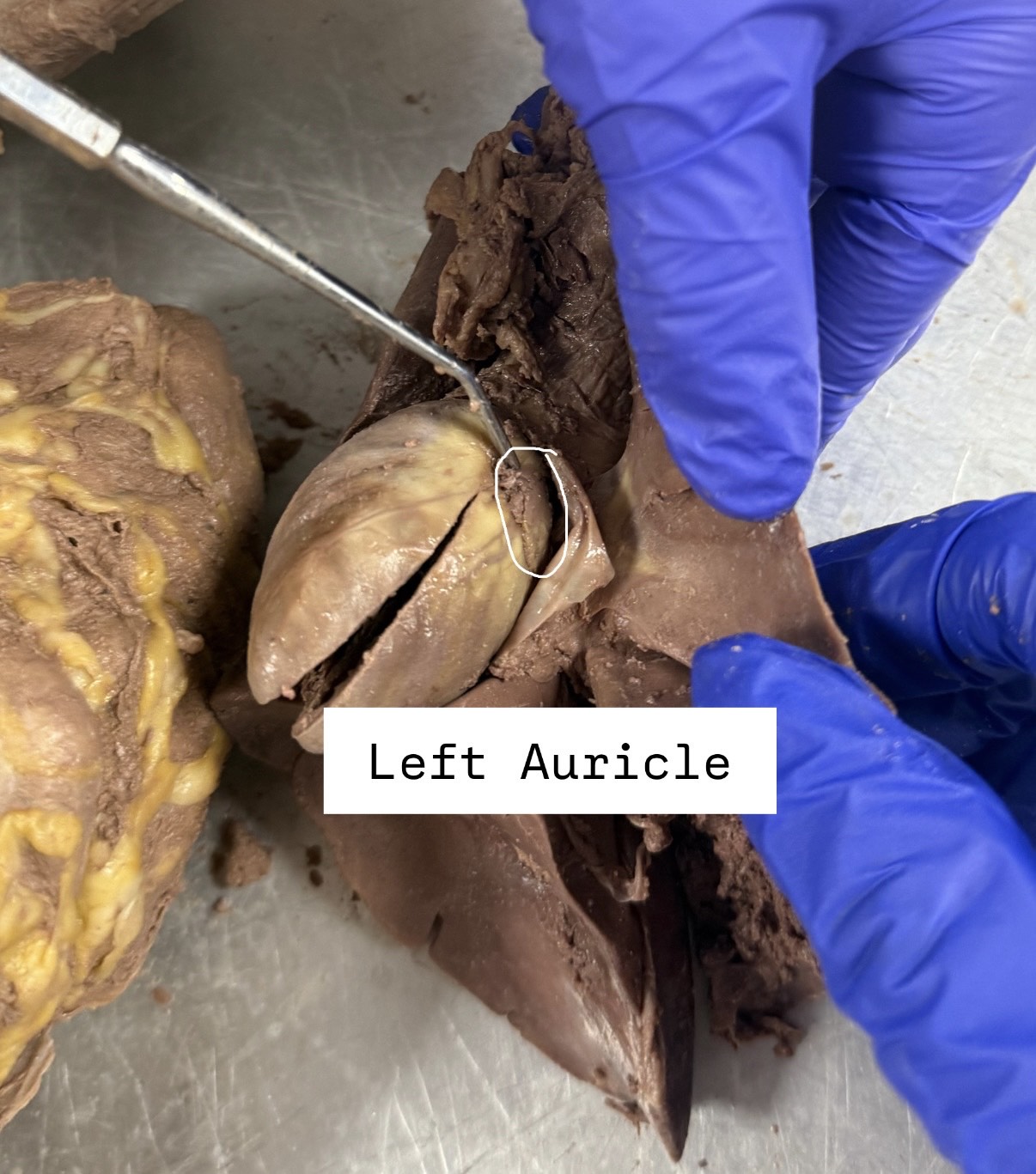
Left Auricle
When looking at it, it will be opposite directions, they look like fans on the side
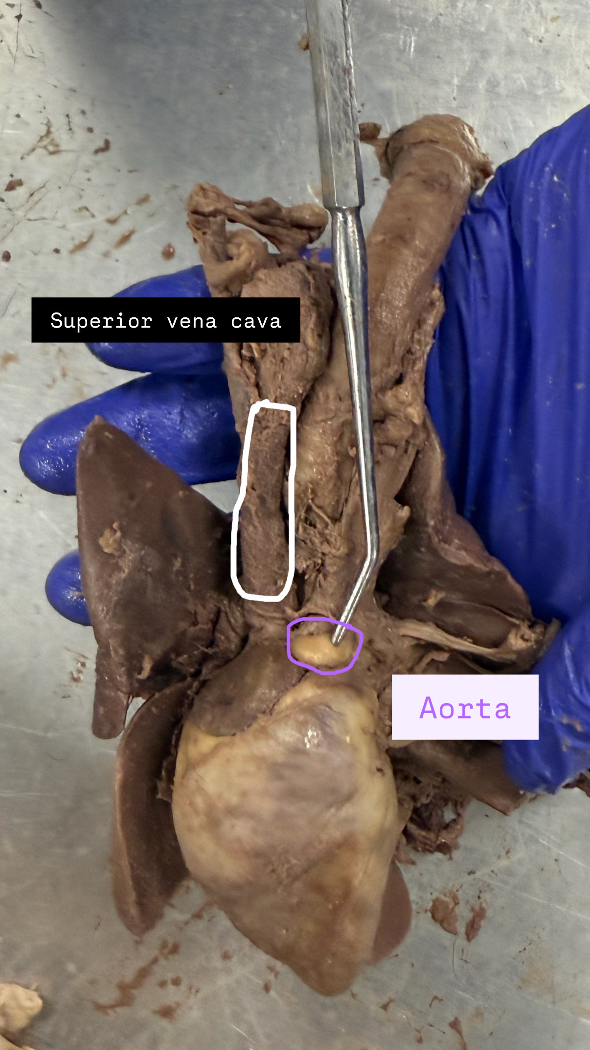
superior Vena Cava, Aorta
The left most thing sticking out for superior vena cava, the white ball for aorta
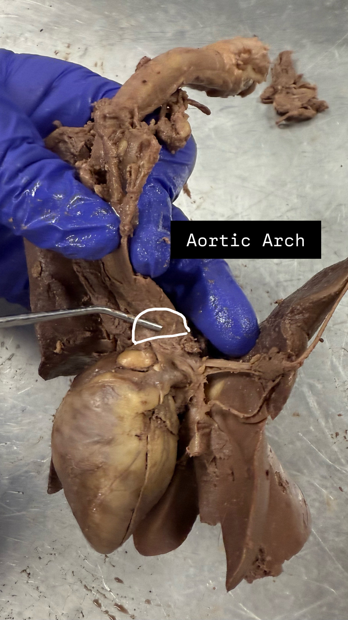
Aorta Arch
The arch by aorta, move it to the side, point to the middle
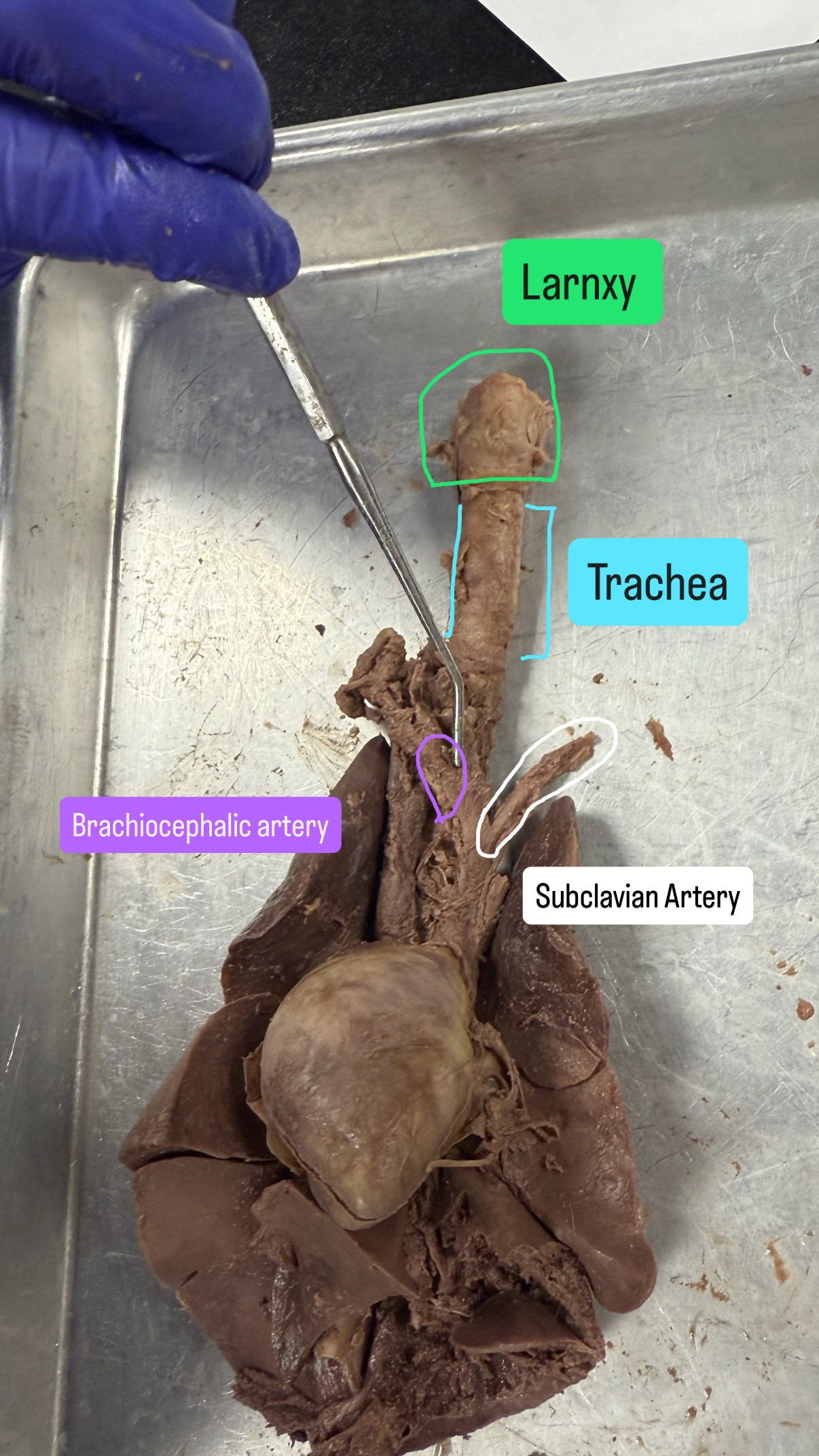
Subclavian artery, Brachiocephalic artery (BRA-key-oh-suh-FAL-ik), Larynx (LARR-inks), Trachea
Larynx will be the top, the trachea will be the middle, the artery will have two parts going away
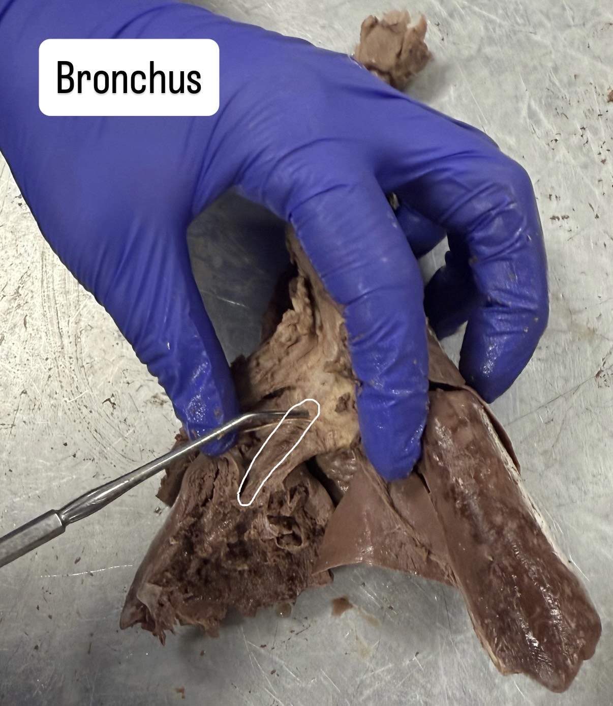
Bronchus
flip the heart over and search for the brown line
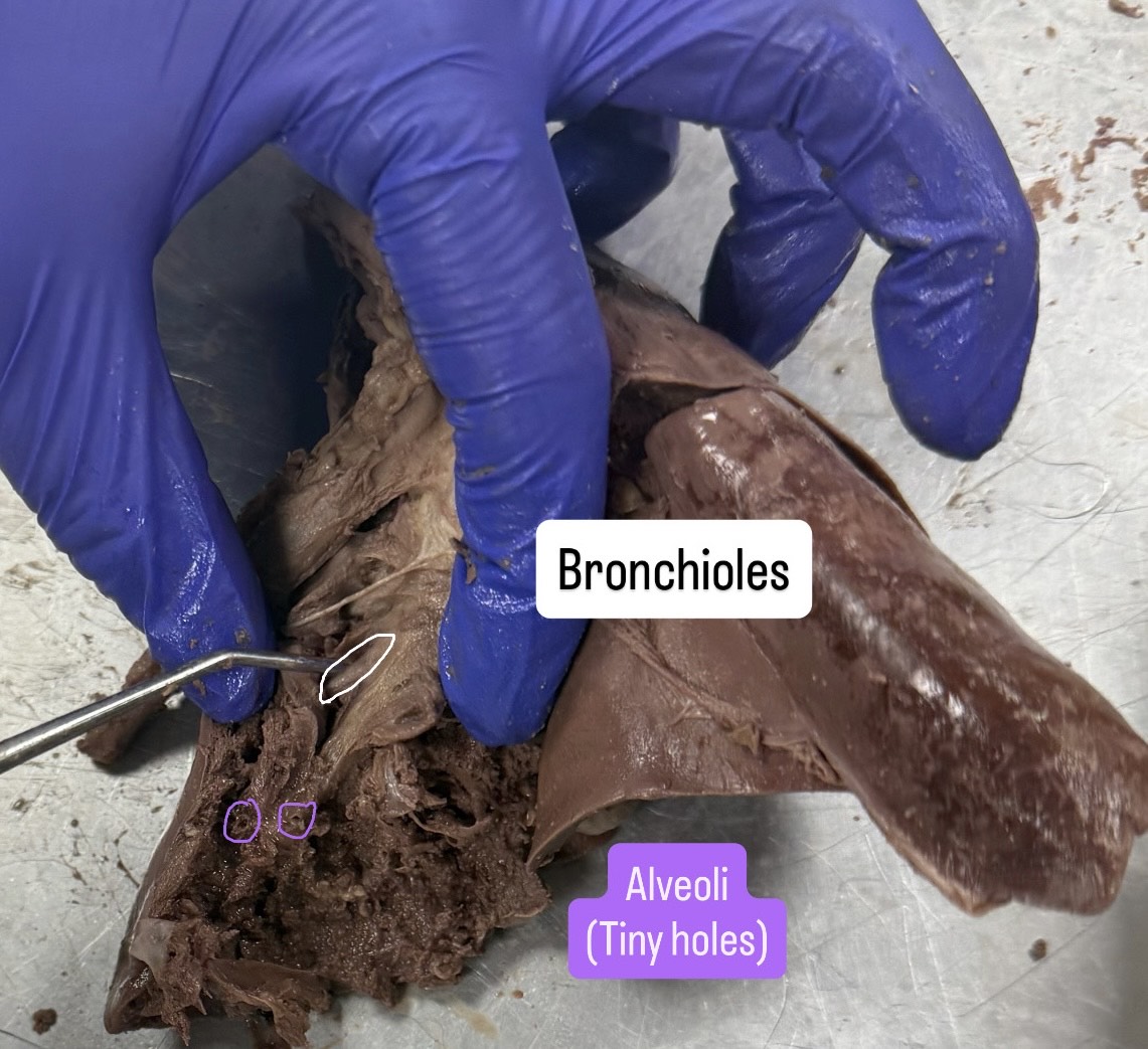
Bronchioles and Aveoli (al-VEE-oh-ly)
On the back of the heart, Bronchioles is the white horizontal line, Aveoli is the holes
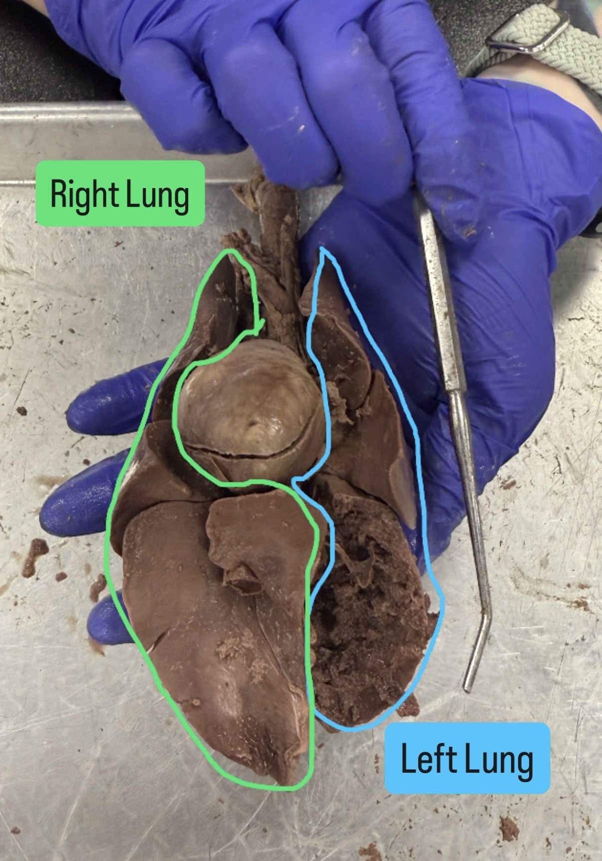
Right lung and left lung
when looking at it, make sure trachea is at top, it will be opposite that way in the right to left
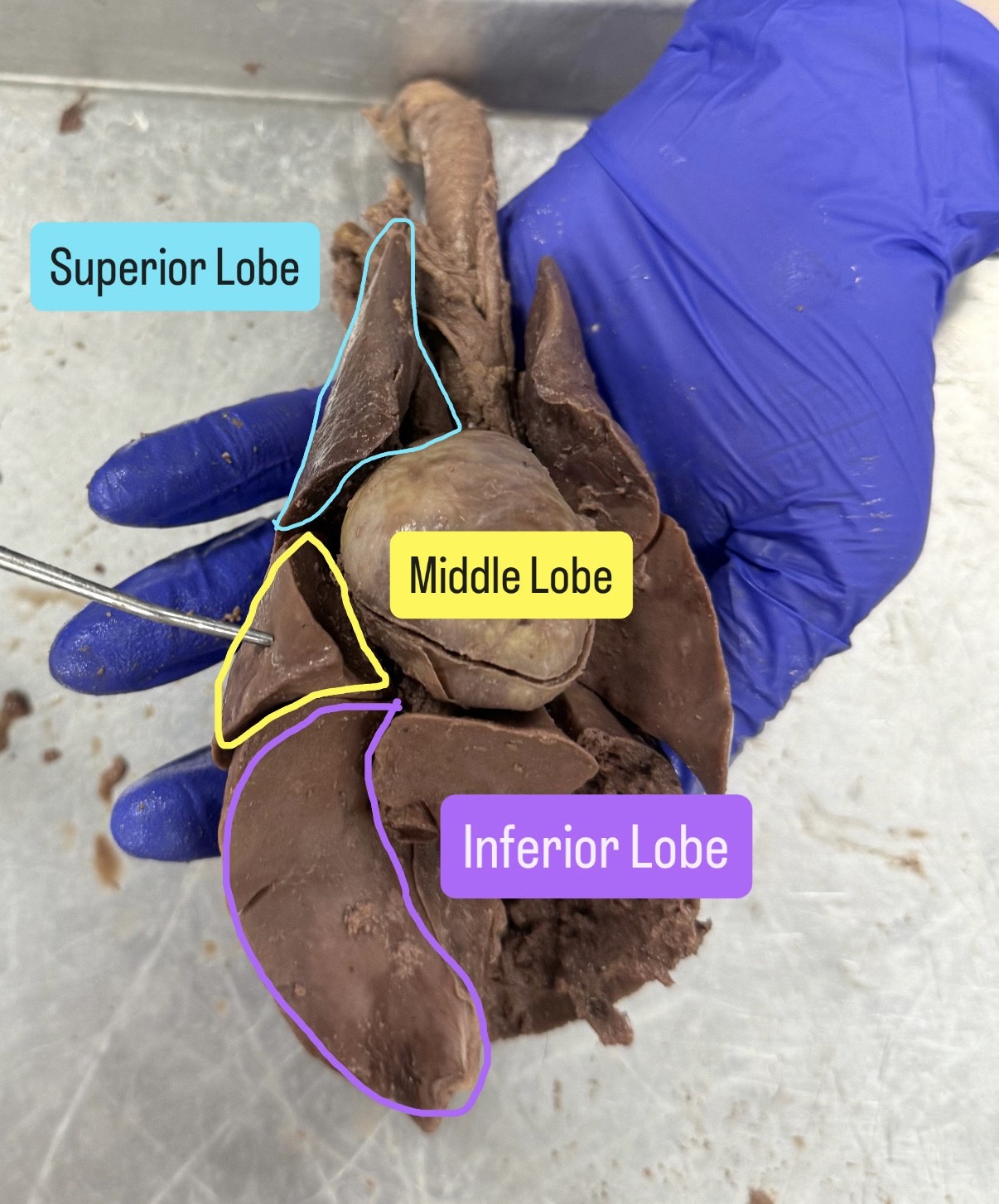
Superior lobe, inferior lobe, middle lobe
top, middle, bottom think of the names on the lungs with the trachea at the top
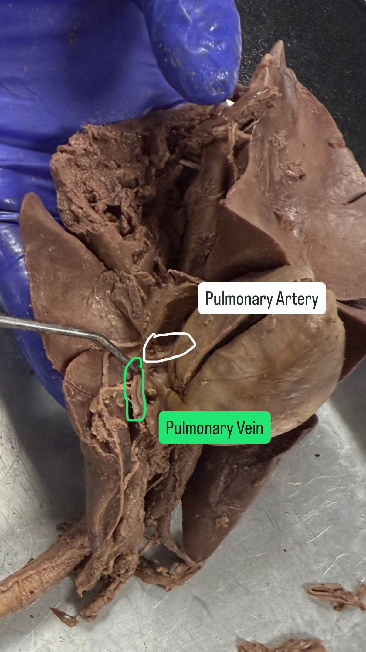
Pulmonary artery and vein
Turn it upside down with the trachea downwards and lean it towards the right side so the trachea is on the left, the artery is the white sliver of horizontal bar, and the vein is the top of the two looking bars but point on the top of the left one
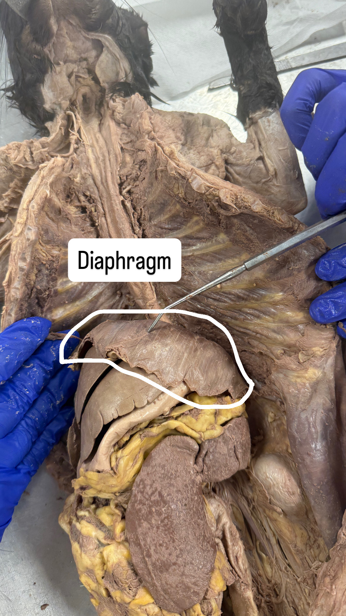
Diaphragm
Look inside the cat, it is the jellyfish looking part
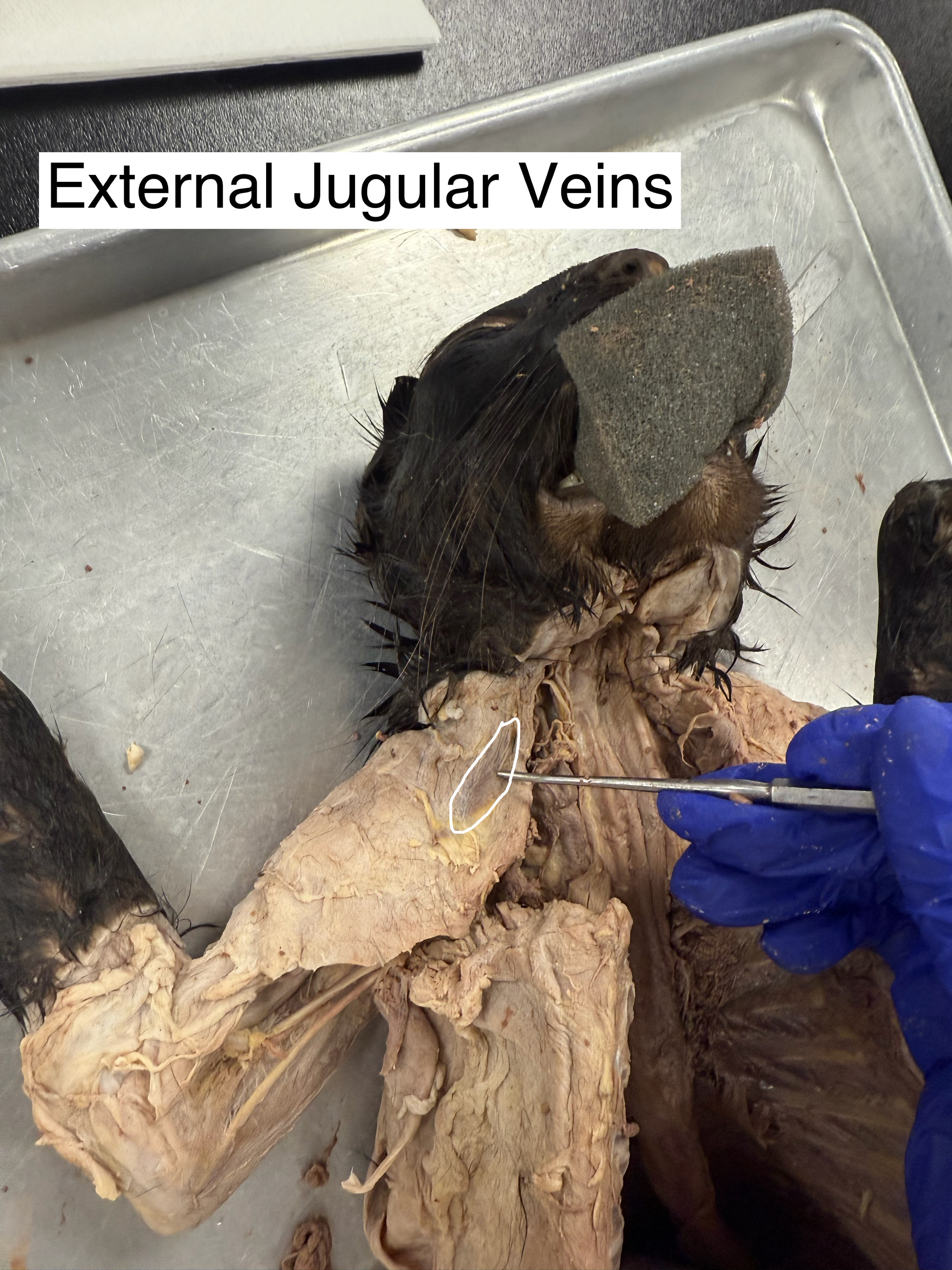
external jugular veins
left shoulder when looking at it, the black line
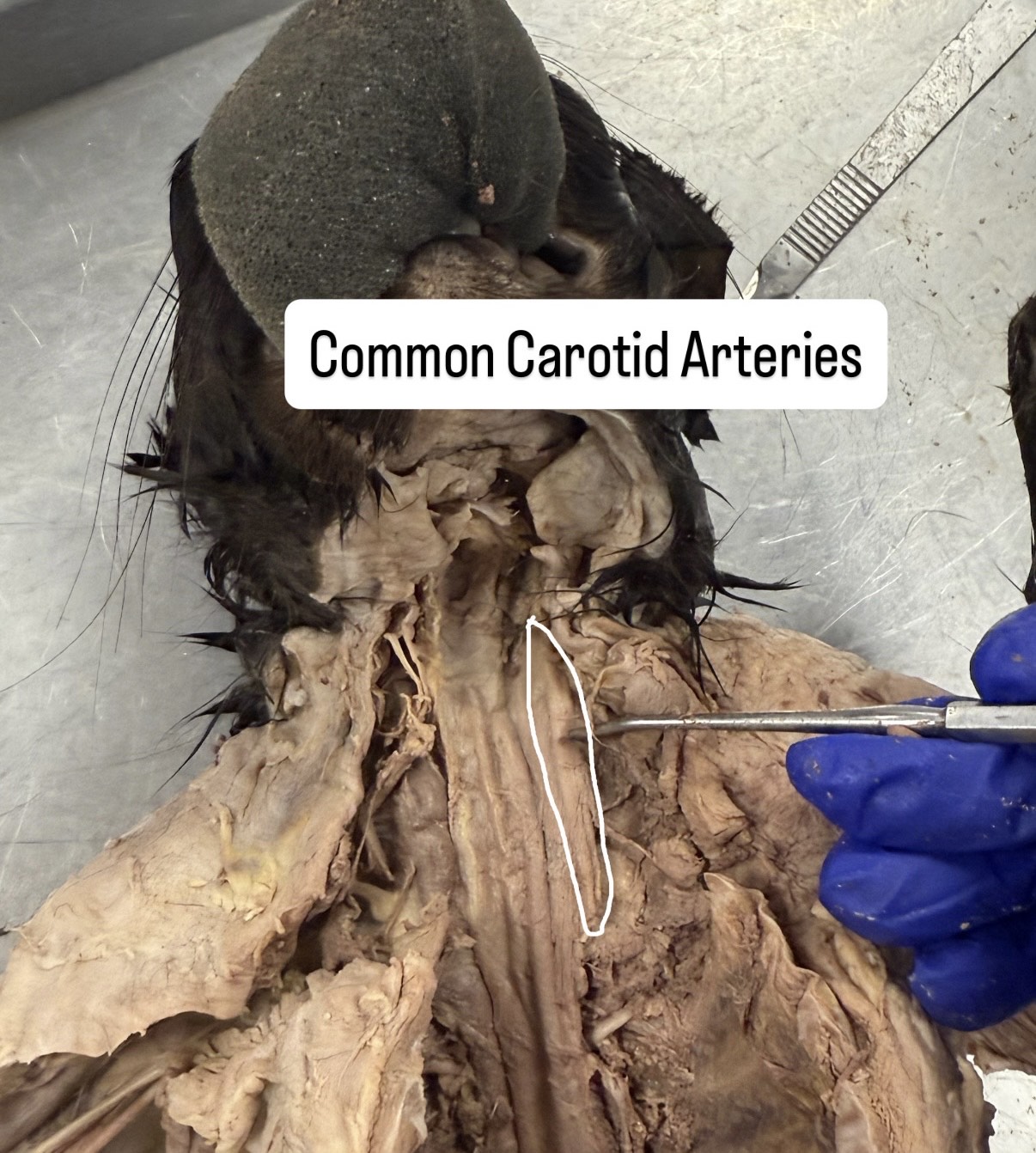
common carotid arteries
the neck, the right most popped out artery
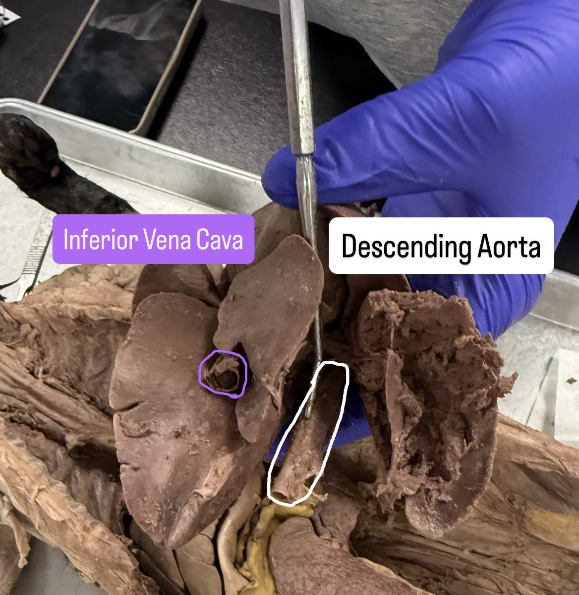
Descending aorta, inferior vena cava
have the heart up and the descending aorta will be by the left lung with a hole on the bottom. The inferior vena cava will be between the right lung with the hole sticking out of it
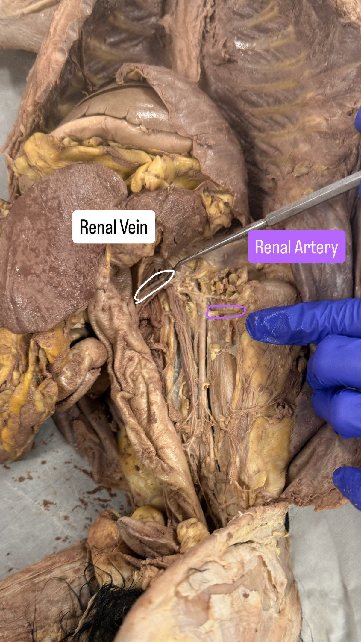
Renal vein and renal artery
Open stomach area (on the left most side push the stomach contents, it will be the top sliver of white bar for renal vein) (for renal artery it will be the second second most visible sliver of white bar for renal artery)
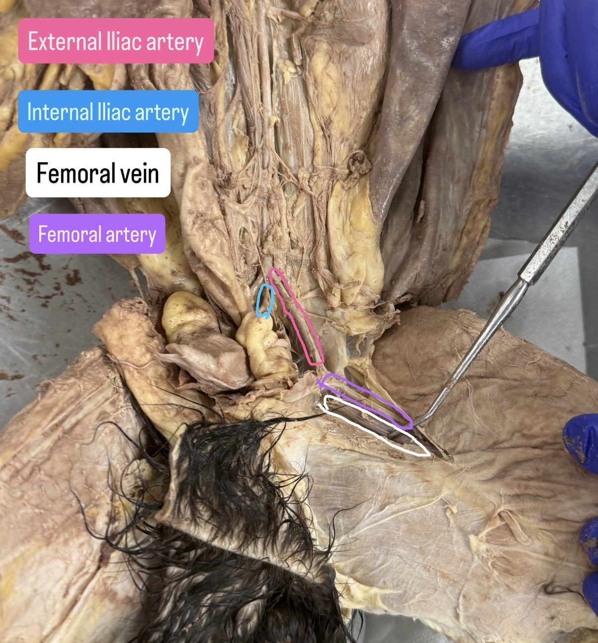
External iliac artery, internal Iliac artery, femoral vein, femoral artery
External Iliac artery is the long bar connected to the femoral artery. The internal iliac artery is the sliver of bar on the left side that is close to the external iliac artery (the lower bar one), the femoral vein is the one vein under the femoral artery, and the femoral artery is the one above the femoral vein and the one that connects with the external iliac artery
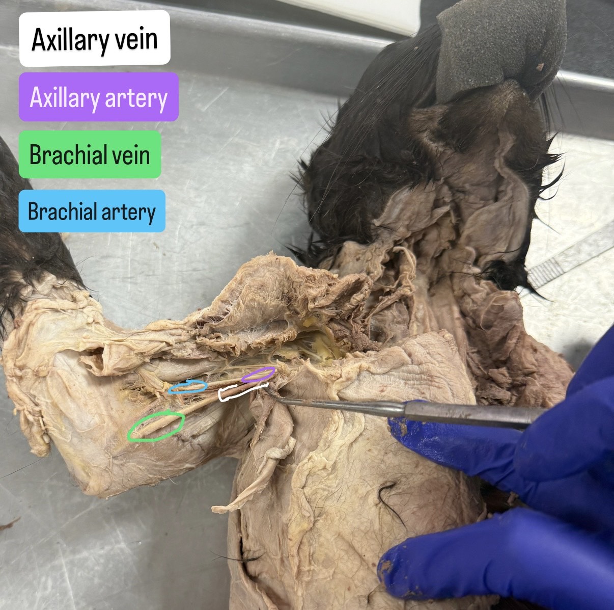
Axillary vein, axillary artery, brachial vein, brachial artery
Axillary vein is the bottom right side of the vein, the axillary artery is the top one on the right side of the artery, the brachial vein is the bottom left side of the vein, the brachial artery is the top left side of the artery. (Artery on top, Vein on bottom)