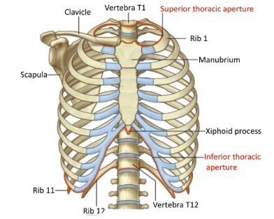
What is the superior border of the thorax defined by?
The first rib
Where does the trachea enter the thorax through?
The superior opening of the thorax
Name some bony structures of the chest wall.
Manubrium
Sternum
Xiphoid process
Clavicle
Ribs
Costal margin
What is the superior thoracic aperture made up by?
The anterior of the body of the T1 vertebra.
The medial borders of the Rib 1 (left and right)
The superior border of the manubrium
What is the inferior thoracic aperture made up by?
Anterior of the body of the T12 vertebra
Ribs 11 and 12
Costal margin
Xiphoid process
What do the bony structures of the chest wall look like?
What structures pass through the superior thoracic aperture?
Trachea
Oesophagus
Major blood vessels and nerves
What structures attach or pass through the inferior thoracic aperture?
Attachment for diaphragm
Descending oesophagus
Descending aorta
Ascending inferior vena cava
Give some characteristics of the structure of the thorax when breathing.
When we breathe, the whole structure of the thorax must be moved, we rely on a change in pressure between air inside and outside the body.
The structure must be rigid to not be damaged by changes in pressure, but also mobile, to permit movement to change pressure.
What is the main function of the chest wall? Are there any other functions?
The chest wall is well designed for breathing. It also has a level of protection, however there are lots of holes in between ribs and the gut is unprotected too.
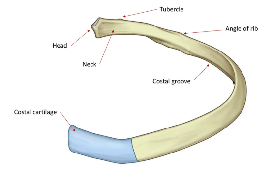
What are the structures of a typical rib, from posterior to anterior?
Head
Neck
Tubercle (articulates with vertebral column)
Angle of rib
Costal cartilage
Running along inferior border of ribs is the costal groove, where the intercostal artery, intercostal vein and intercostal nerve run.
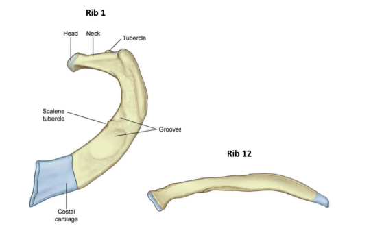
What does a typical rib look like?
What do the atypical ribs look like?
What are the numbered ribs which are atypical?
Ribs 1, 11 and 12
How can ribs be classified?
Ribs 1-7 - True ribs
Ribs 8-10 - False ribs
Ribs 11-12 - Floating ribs
How are ribs classified as true, false or floating?
This is based on the articulation of their costal cartilages
True ribs directly articulate with the sternum via costal cartilages.
False ribs indirectly articulate with the sternum via costal cartilage of rib 7. This forms the costal margin.
Floating ribs do not articulate with the sternum.
Generally, how do the ribs curve?
Downwards and outwards
What are the rib movements during inspiration?
During inspiration, there is a pump handle movement, increasing the volume of the thorax. There is a superior and anterior movement of the ribs and sternum.
Another movement occurs medially to laterally, where the ribs are elevated, also increasing thoracic volume. This is a bucket handle movement.
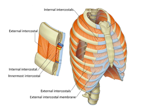
What are the three layers of intercostal muscles?
External intercostals (elevates the rib below while breathing in)
Internal intercostals (depresses the rib above while breathing out)
Innermost intercostals (depresses the rib above while breathing out)
What do the intercostal muscles look like?
How does each intercostal muscle fibre run?
External intercostal muscle fibres run from above to below, run anteriorly and inferiorly.
The internal and innermost intercostal fibres run anteriorly and superiorly.
What do the external intercostal muscles do?
External intercostal muscles will elevate and lift up the rib below.
What do the internal and innermost intercostal muscles do?
Internal and innermost intercostal muscles will pull the superior rib down and inwards.
Give some characteristics of the structure and the function of the diaphragm itself.
The diaphragm is the most important muscle for respiration.
It is dome shaped in its relaxed position, when it contracts it flattens out and moves inferiorly.
It inserts along the costal margin. It also has a central tendon.
Its movement will create a vacuum and air rushes in to fill this space.
Upon relaxation, it returns to its original position, forcing air out. The internal intercostal muscles also play a role in this.
What important structures pass through the diaphragm? At what vertebral levels do they do this?
The IVC (inferior vena cava) passes through the central tendon at vertebral level T8
The oesophagus passes through the muscular region posteriorly at T10
The aorta passes between the inferior and posterior most region at T12
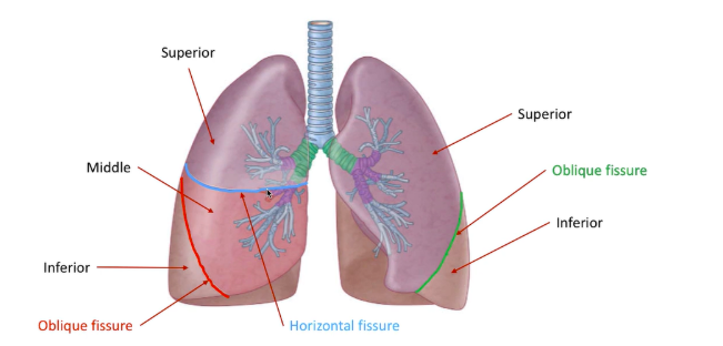
How many lobes does the left lung have? What are these lobes separated by?
The left lung has 2 lobes, the superior lobe and inferior lobe, separated by the oblique fissure.
How many lobes does the right lung have? What are these lobes separated by?
The right lung has 3 lobes, the superior, middle and inferior lobe, separated by the horizontal fissure superiorly and oblique fissure inferiorly.
What do the apices of the lungs extend above?
They extend above the superior border of the first rib.
What is the carina? What vertebral level is it at?
The carina is the bifurcation of the trachea at the vertebral level T4-T5, same as sternal angle.
What does the trachea bifurcate into?
The trachea bifurcates into the right main bronchus and left main bronchus.
What are the differences between the left and right main bronchi? Why is this clinically significant?
The right main bronchus is steeper than the left, it is also slightly wider.
This means if something is aspirated and is lodged in the lung, its likely in the right one.
What does the main bronchi split into?
The main bronchi then split into the lobar bronchi.
What would a diagram of the lobes of the lungs look like?
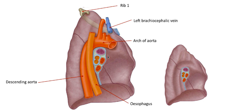
Which structures have caused the indentations on the left lung?
First rib
Left brachiocephalic vein
Arch of aorta
Oesophagus
Descending aorta
What do the associated structures of the left lung look like?
What structures have caused the indentations on the right lung?
First rib
Right brachiocephalic vein
Oesophagus
Superior and inferior vena cava
Azygos vein
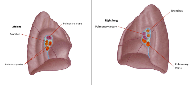
What is the hilum?
The hilum is the region on the medial surface of the lung where important structure pass in and out the lung.
What structures make up the hilum, from superior to inferior?
Pulmonary artery
Bronchus
Pulmonary veins
What does the hilum of the lungs look like?
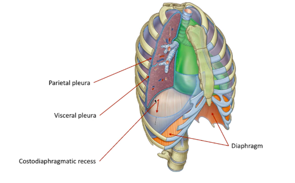
What is the pleura?
The pleura is a membrane around the lungs and the inside of the thoracic wall.
What is the pleura divided into?
It is divided into:
The parietal pleura, which lines the inside of the thoracic wall
The visceral pleura, which lines the outside of the lungs. It extends into fissures
The two layers are continuous with one another. They are continuous at the hilum.
What is between the two layers of pleura and what is its function?
Between the two layers is a small volume of serous fluid this is the parietal fluid, prevents friction between layers by lubricating them.
It also helps keep the layers against one another, as well as negative pressure.
This is important as the layers move over each other in respiration.
What is the costodiaphragmatic recess? How can it appear differently in medical imaging?
The costodiaphragmatic recess is a small recess where the ribs meet the diaphragm, it is a pocket in the plural cavity.
In medical imaging, it can appear blunted if there is a build-up of fluid.
What would a diagram showing the pleura of the lungs look like?
What is the most common clinical consideration of the lower respiratory tract?
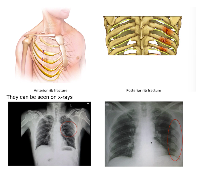
Rib fracture
What is the difference between an anterior rib fracture and a posterior rib fracture?
Anterior rib fracture is one on the anterior side of the body.
A posterior rib fracture is closer to the vertebra.
What is the difference between an anterior and posterior rib fracture on images?
Give some characteristics of pneumothorax.
A rib fracture can damage the surface of the lung, leading to air entering the pleural cavity.
This would prevent the adhesive affect that couples the thoracic wall and lung.
This can cause the lung to collapse as it moves away from the chest wall.
Give some characteristics of haemothorax.
If the rib fracture is severe, the cut edges of bone can damage neurovascular bundle in the costal groove, leading to blood building up in the pleural cavity.
This again prevents the adhering of the pleura to one another, leading to the lung collapsing.
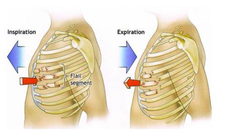
Give some characteristics of paradoxical chest wall movement.
This is where an isolated section of the chest wall does something different to the rest of the wall.
In the case of a flail segment, where a section is isolated, the segment will move in during inspiration and out during expiration due to pressure changes.
What does paradoxical chest wall movement look like in a diagram?
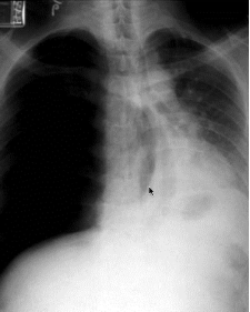
What is tension pneumothorax?
This is where air is drawn into the thorax but cannot escape.
This leads to an increase in pressure on the mediastinal structures, and can result in a change in their position.
What does tension pneumothorax look like in medical imaging?