Chapter 12 MCQ + Essay
1/23
There's no tags or description
Looks like no tags are added yet.
Name | Mastery | Learn | Test | Matching | Spaced | Call with Kai |
|---|
No analytics yet
Send a link to your students to track their progress
24 Terms
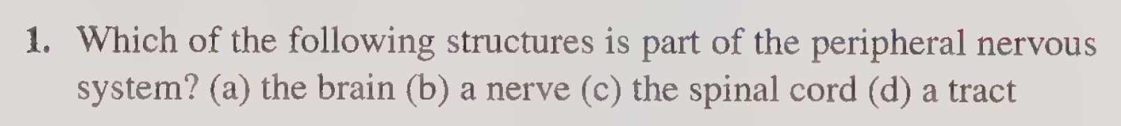
(b) a nerve.
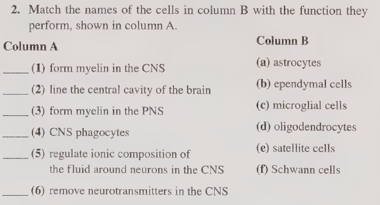
(1) d
(2) b
(3) f
(4) c
(5) a
(6) a
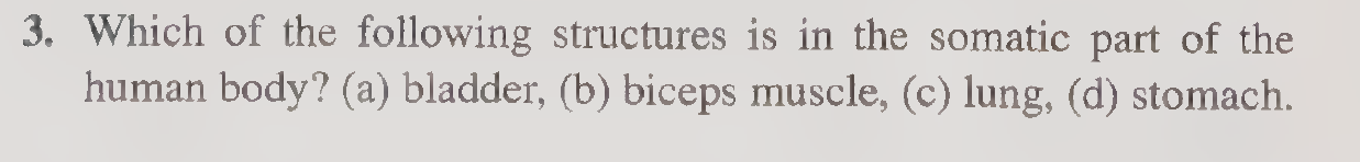
(b) biceps muscle.
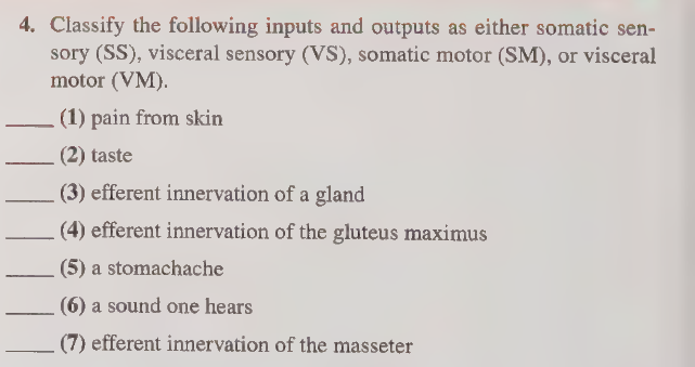
(1) SS
(2) VS
(3) VM
(4) SM
(5) VS
(6) SS
(7) SM
(b) a gland.
(b) motor neuron cell bodies.
(a) neuron cell bodies.
(c) axosomatic.
(d) the plasma membrane.
(b) neuron cell bodies in the gray matter.
(c) serial processing.
(d) axons of afferent and efferent neurons.
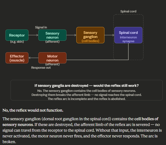
2, 1, 3.

Proprioception is unconscious body position sense carried ipsilaterally in the dorsal columns, lost in dorsal column lesions, and tested clinically with the Romberg sign.
Your body has sensors hidden in your muscles, tendons, and joints that are constantly tracking where every body part is — even without you looking.
This is why you can close your eyes and still touch your nose. Your brain already knows where your hand is.

Interneurons are CNS-only neurons that integrate and relay signals between sensory and motor neurons, forming the basis of complex processing, reflex arcs, and spinal cord circuitry.
Input | Interneuron | Output |
|---|---|---|
Sensory neuron | Processes & integrates | Motor neuron |

Sure! Here's the composition broken down the same way:
Composition:
🧠 Gray Matter:
Neuronal cell bodies → the "headquarters" where decisions are made.
Dendrites → receiving incoming signals.
Synapses → where neurons actually talk to each other.
Little to no myelin → that's why it looks gray.
White Matter:
Myelinated axons → long cables carrying signals from A to B.
Myelin sheath → the fatty wrapping that speeds up transmission and gives white matter its color.
No cell bodies, no synapses → no processing happens here, just transmission.
Location 🧠 In the Brain:
Gray matter → on the OUTSIDE (cortex — the wrinkly surface).
White matter → on the INSIDE (beneath the cortex).
Exception: deep gray matter nuclei sit inside the white matter (e.g., basal ganglia, thalamus).
🧠 In the Spinal Cord:
Gray matter → on the INSIDE (butterfly/H-shaped core).
White matter → on the OUTSIDE (surrounding the gray).
High yield: Brain and spinal cord are opposite to each other in terms of gray/white matter arrangement!

Neuron:
Large cell body (soma) with multiple processes
Nissl bodies (rough ER) — for protein synthesis
Cannot divide (post-mitotic)
Nucleus:
Large, round, pale (euchromatic)
Prominent nucleolus (large dark dot)
Pale = actively transcribing (high metabolic activity)

Neurons vs. Neuroglia
Neurons | Neuroglia | |
|---|---|---|
Structure | Large cell body, axon, dendrites, Nissl bodies | Smaller, vary by type, no axons |
Function | Generate & transmit electrical signals | Support, protect, and nourish neurons |
Location | Gray matter (cell bodies) | Throughout CNS & PNS |
Division | Cannot divide | Can divide |
Number | Less numerous | ~10x more numerous than neurons |
Neurons are the signal generators; neuroglia are the support crew — more numerous, able to divide, and found everywhere neurons are.

erve vs. Nerve Fiber vs. Neuron
Neuron | Nerve Fiber | Nerve | |
|---|---|---|---|
What is it | The entire cell | Single axon + its myelin sheath | Bundle of many nerve fibers |
Includes | Cell body, dendrites, axon | Axon only (+ covering) | Multiple fibers + connective tissue |
Analogy | The whole wire factory | One wire | A cable containing many wires |
A neuron is the whole cell; a nerve fiber is just its axon; a nerve is a bundle of many nerve fibers bound together by connective tissue.

PNS vs. CNS Nerve Damage PNS — Reversible:
Schwann cells guide and support regrowth of damaged axons
Schwann cells produce nerve growth factors
Connective tissue sheaths (endoneurium) act as a scaffold for regeneration
Axon can regrow at ~1mm/day'
CNS — Irreversible:
Oligodendrocytes cannot guide regrowth the way Schwann cells can
Astrocytes form a glial scar — physically blocks regrowth
CNS produces inhibitory factors that actively stop regeneration
No connective tissue scaffold present
PNS regenerates because Schwann cells guide regrowth along intact sheaths; CNS cannot because oligodendrocytes lack this ability and astrocytes form a glial scar that blocks regeneration.

Axon:
Single process that carries signals away from the cell body (efferent)
Can be myelinated or unmyelinated
Ends in axon terminals (synaptic knobs) that release neurotransmitters
Dendrite:
Multiple branching processes that carry signals toward the cell body (afferent)
Usually short and unmyelinated
Greatly increase the surface area for receiving signals
Dendrites bring signals in; axons send signals out.

They sit in the middle one end touching the periphery, one end reaching into the spinal cord so the signal goes straight through with no detours.
![<p></p><p class="font-claude-response-body break-words whitespace-normal leading-[1.7]">They sit in the middle one end touching the periphery, one end reaching into the spinal cord so the signal goes straight through with no detours.</p>](https://assets.knowt.com/user-attachments/9b97e3b9-87bb-4984-b694-70d92fb85abf.png)

Myelinated
One Schwann cell wraps around one axon — many times — forming a thick myelin sheath.
Creates nodes of Ranvier (gaps between Schwann cells) where the signal jumps.
Conduction is saltatory (fast) — signal leaps node to node.
Found in fibers requiring speed (motor, proprioception, touch).
Nonmyelinated
One Schwann cell loosely envelops multiple axons simultaneously — just tucking them into its surface, no wrapping.
No nodes of Ranvier.
Conduction is slow and continuous.
Found in fibers carrying pain and temperature (C fibers).
In myelinated fibers, one Schwann cell wraps one axon many times for fast saltatory conduction; in nonmyelinated fibers, one Schwann cell loosely cradles many axons with no wrapping, resulting in slow continuous conduction