Dissection 5 - The Orbit
1/40
There's no tags or description
Looks like no tags are added yet.
Name | Mastery | Learn | Test | Matching | Spaced | Call with Kai |
|---|
No analytics yet
Send a link to your students to track their progress
41 Terms
Abducent n.
What CN pierces the medial side of the lateral rectus m.?
Annular ring
Tendinous ring around optic n. where all 4 rectus muscles originate from
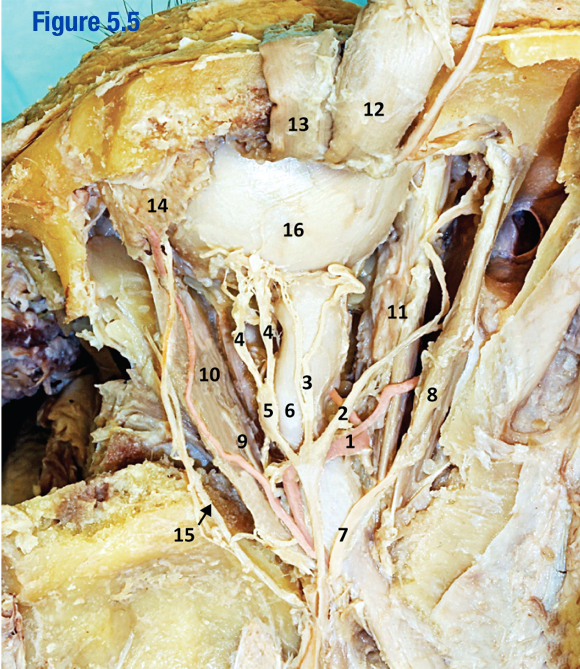
Ciliary ganglion; 5
Where para/pre from CN III synapse before going to ciliary m. and/or sphinctor pupillae (include number in attached image)

Conjunctiva
Transparent membrane covering the inner portion of the eyelid and sclera
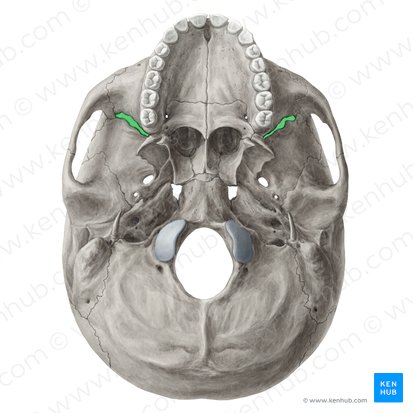
Cribiform plate
Part of ethmoid plate on the medial side of the orbit
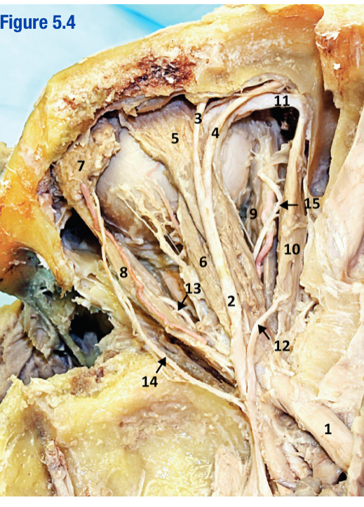
Frontal n.; 2
Branch of CN V1 that branches into supraorbital and supratrochelar n. (include number in attached image)

Infraorbital groove
Path for infraorbital n. (CN V2) to travel through on the floor of the orbit before exiting the infraorbital foramen
Infraorbital foramen
What hole does the infraorbital VAN pass through?
Inferior orbital fissure
What opening does the zygomatic n. and infraorbital VAN pass through to enter the orbit?
Inferior oblique m.
Eye muscle innervated by CN III; Elevation and ABduction
Inferior rectus m.
Eye muscle innervated by CN III; Depression and ADduction
Iris
Colored part around pupil
Lacrimal caruncle
On the medial corner of the eye (can be seen anteriorly)
Lacrimal gland
On the lateral upper corner of the eye; SS from CN V1 and Para from CN VII
Lacrimal n.
What n. is labeled 15?
Lateral rectus m.
Eye muscle innervated by CN VI; ABduction
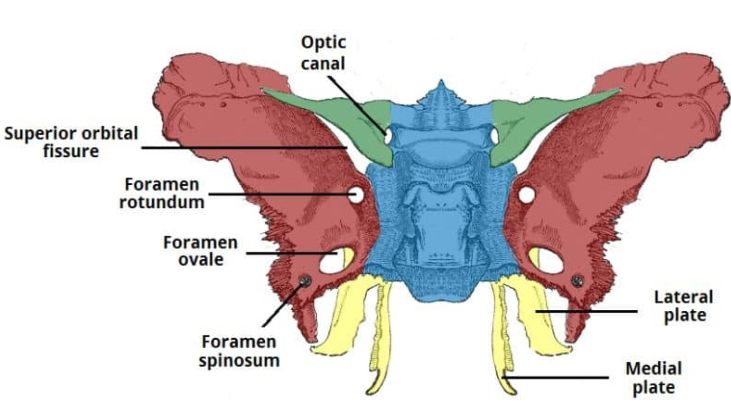
Lesser wing of sphenoid
What is the green colored bone?
Levator palpebrae superioris; 5
Innervated by CN III involved in raising eyelid; Located just below the frontal n. (Include number in diagram)

Long ciliary n.
Branch of nasociliary n. (3 in photo)

Maxilla
What bone contributes to the floor of the orbit
Medial rectus m.
Eye muscle innervated by CN III; ADDuction
Nasociliary n.; 2
Most medial branch of CN V1 (include number from diagram)

Nasolacrimal canal
What connects the orbit to the nasal cavity?
Occulomotor n. (CN III)
What nerve innervates 3 of the 4 rectus m. and carries SM and Para/pre
Ophthalmic a.
Branch of ICA that enters orbit with CN II (1 in image)

Optic n.
Travels through optic canal and carries SSA vision
Orbicularis oculi
M. of facial expression that allows for forceful blinking
Orbital plate of the frontal
What contributes to the roof of the orbit?
Periorbital membrane
What lays just below the bony roof of the orbit
Sclera
What is the whites of the eye?
Short ciliary n.
What n. branch from the ciliary ganglion? (4 in image)

Superior oblique m.
Goes through trochela before attaching tho the eye; Depression and ABduction; Innervated by CN IV
Superior ophthalmic v.
______ → Cavernous sinus
Superior orbital fissure
CN III, IV, V1, and VI enter orbit while superior orbital v. exits orbit
Superior rectus m.; 12
Eye muscle innervated by CN III; Elevation and ADduction (include number in image)

Supraorbital n.
What nerve is 3?

Supraorbital foramen/notch
Where does the supraorbital VAN exit?
Supratrochlear n.
What n. is 4?
Trochlea
Fibrocartilagenous sling that the superior oblique m. passes before attaching to the eye
Trochlear n.
What n. innervates superior oblique m.?
Zygomatic
What makes up the lateral wall of the orbit?