Gallstones and biliary colic
1/34
There's no tags or description
Looks like no tags are added yet.
Name | Mastery | Learn | Test | Matching | Spaced | Call with Kai |
|---|
No analytics yet
Send a link to your students to track their progress
35 Terms
What are gallstones?
small stones that form within the gallbladder, can take years to form
What are consequences of gallstones?
they can be asymptomatic , or they can present with acute disease
about 60% of these presentations are biliary colic, and the rest are acute cholecystitis, acute cholangitis, and pancreatitis
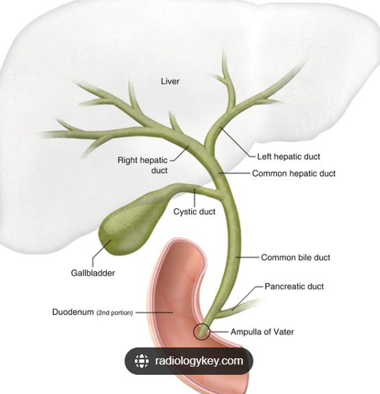
What is the anatomy of the biliary system?
the right and left hepatic ducts leave the liver and join to form the common hepatic duct. The cystic duct leaves the gallbladder, and joins the common hepatic duct, to become the common bile duct. The pancreatic duct joins the common bile duct to become the ampulla of vater, which opens to the duodenum through the sphincter of oddi.
What is cholestasis?
blockage to the flow of bile
What is cholelithiasis?
gallstones
What is choledocholithiasis?
gallstones in the bileduct
What is biliary colic?
intermittent right upper quadrant pain caused by gallstones irritating bile ducts
What is cholecystitis?
inflammation of the gallbladder
What is cholangitis?
inflammation of the bile ducts
What is gallbladder empyema?
pus in the gallbladder
What is cholecytsectomy?
surgical removal of the gallbladder
What is cholecystostomy?
inserting a drain into the gallbladder
What are types of gallbladder stones?
cholesterol stones- usually solitary and large made of cholesterol and calcium
pigment stones- black hard stones
mixed- multiple and irregular shaped
What is the epidemiology of gallstones?
more common in women
incidence increases with age
At age 30 – 5% of women and 2% of men have / have had gallstones. Aged 55 – 20% / 10%, age 70, 30% / 20%.
What are risk factors for gallstones?
fat
female
forty
fair
family history
fertile
diabetes, crohn’s, rapid weight loss
How are cholesterol stones formed?
cholesterol stones form when the concentration of micelles is not great enough to hold all the cholesterol in the micelles. For cholesterol stones to form there need to be three factors:
High concentration of cholesterol in the gallbladder
Gallbladder stasis
Products that promote the crystallisation of cholesterol
initially, cholesterols will form crystals which go on to form stones
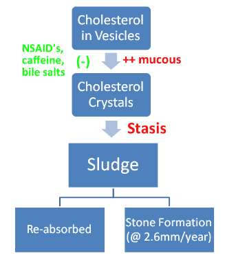
What inhibits stone formation?
caffeine
NSAIDs
bile salts
What exacerbates stone formation?
mucin, rapid weight loss, pregnancy, increased serum cholesterol, oral contraceptive, clofibrate
How are pigment stones formed?
increase in bilirubin load as a result of haemolytic anaemia
cirrhosis
pigments become less water soluble in the bile if the gallbladder is colonised by bacteria
What are symptoms of biliary colic?
severe, colicky, epigastric/RUQ pain, that can radiate to shoulder/interscapular region
often triggered by meals (high fat)
last between 3-8 hours
nausea and vomiting
no fever/raised inflammatory markers
Why can fatty foods trigger biliary colic?
Fat entering the digestive system causes cholecystokinin (CCK) secretion from the duodenum. CCK triggers contraction of the gallbladder, which leads to biliary colic. Patients with gallstones and biliary colic are advised to avoid fatty foods to prevent CCK release and gallbladder contraction
What are differential diagnosis for biliary colic?
GORD
peptic ulcer
IBS
pancreatitis
What investigations are done for biliary colic?
LFTs
ultrasound- gold standard diagnostic test
What could LFTs show in biliary colic?
bilirubin- normally drains through the bile ducts, raised bilirubin (with pale stools and dark urine) show biliary obstruction
ALP- non-specific marker made in the liver, biliary system, and bone. Raise in this with consistent symptoms could show biliary obstruction
ALT/AST- normally indicative of hepatocellular injury, but a slight raise, less than ALP, could show an obstructive picture
What is the first line investigation for gallstone disease?
US
What findings can be found on US for biliary colic?
gallstones in gallbladder or duct
bile duct dilation
acute cholecystitis (thickened gallbladder wall, fluid around gallbladder)
What is MRCP?
magnetic resonance cholangio-pancreatography
MRI scan that produces detailed biliary images. Normally used to investigate further if found bile duct dilation but no gallstones on US
What is endoscopic retrograde cholangio-pancreatogrophy?
ERCP
involves inserting endoscope down the oesophagus to the opening of the common bile duct in the duodenum.
the main indications is to clear stones in the bile duct
What can ERCP do?
Inject contrast and take x-rays to visualise the biliary system and diagnose pathology (e.g., stones or strictures)
Perform a sphincterotomy on the sphincter of Oddi if it is dysfunctional (blocking flow)
Clear stones from the ducts
Insert stents to improve bile duct drainage (e.g., with strictures or tumours)
Take biopsies of tumours
What are complications of ERCP?
bleeding
cholangitis
pancreatitis
What is management of gallstones?
is asymptomatic- conservative treatment
symptoms or complications- elective laparoscopic cholecystectomy
What is courvoisier’s sign?
palpable gallbladder- this is usually a sign of acute pathology, eg: malignancy/obstruction of the pancreas
gallstones result in shrunken, fibrotic gallbladder
What is cholecystectomy?
involves surgical removal of the gallbladder. It is indicated where patients are symptomatic of gallstones, or the gallstones are leading to complications
can be open or laparoscopic (preferred)
What are complications of cholecystectomy?
bleeding, infection- normally given prophylaxis >40
scars
damage to bile duct
stones left in duct
damage to other organs
anaesthetic risk
VTE- given prophylaxis
post-cholecystectomy syndrome
What is post-cholecystectomy syndrome?
non-specific symptoms attributed to changes in bile flow:
diarrhoea
indigestion
epigastric/RUQ discomfort
nausea
flatulence
intolerance of fatty foods