StemUp: OCR A A level Biology 2.1.1 Cell structure
1/85
There's no tags or description
Looks like no tags are added yet.
Name | Mastery | Learn | Test | Matching | Spaced | Call with Kai |
|---|
No analytics yet
Send a link to your students to track their progress
86 Terms
What is the basic function of a microscope? (3)
- Instrument which allows you you to magnify an object many times
- In order to observe structures / organisms
- Which are impossible to see with a naked eye
What are the 3 examples of microscopes? (3)
- Optical (light) microscope
- Transmission electron microscope (TEM)
- Scanning electron microscope (SEM)
What are the benefits of using a stain on your sample? (3)
- Increases the contrast of the image
- More internal structures are visible
- Image obtained is much clearer
What is differential staining? (3)
- When multiple stains are used
- Each stain binds to a specific cell structures or cells
- Common feature of electron microscopes
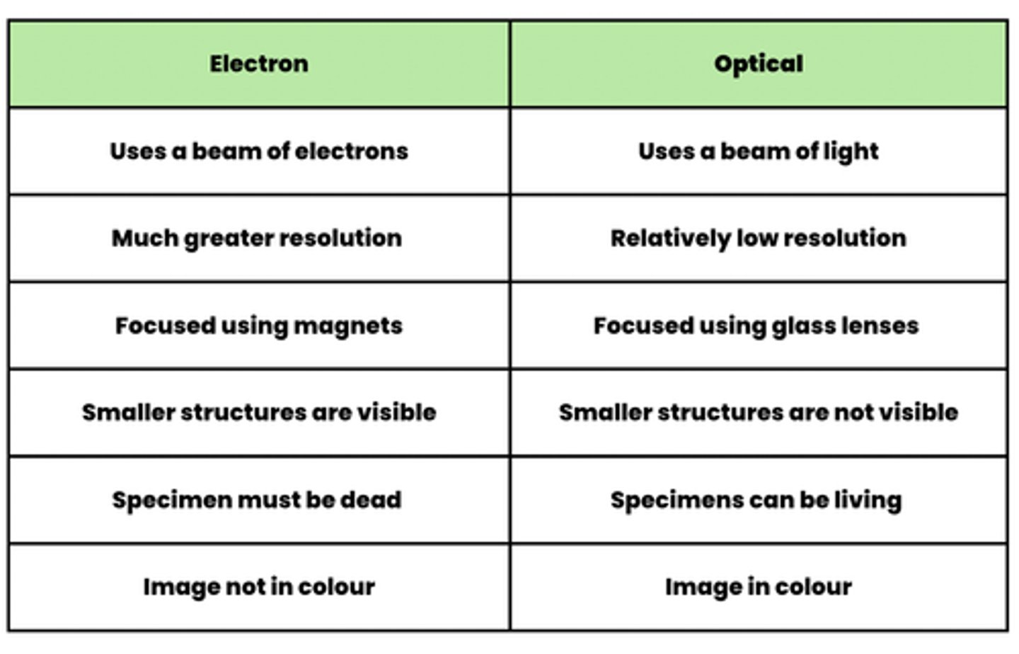
Compare the features of a optical microscope vs an electron microscope
optical- uses a beam of light, relatively low resolution, focused using glass lenses, smaller structures are not visible, specimens can be living, image in colour
electron- uses a beam of electrons, much greater resolution, focused using magnets, smaller structures are visible, specimen not dead, image not in colour
Why do electron microscopes have a higher resolution than optical microscopes? (1)
Electrons have a shorter wavelength
Why is it that/ suggest why optical microscopes generate a coloured image? (1)
Light particles are able to carry information about light, electrons are not able to carry this information
What are laser scanning confocal microscopes? (3)
- Special type of light microscope
- That uses a laser beam to scan a specimen
- Which will be labelled with a fluorescent tag
How does a laser scanning confocal microscope work (in simple terms)? (3)
- When the laser beam hits the fluorescent tag
- It gives off light
- Which is focused through a pinhole to a detector
Draw the structure of an optical microscope (5)
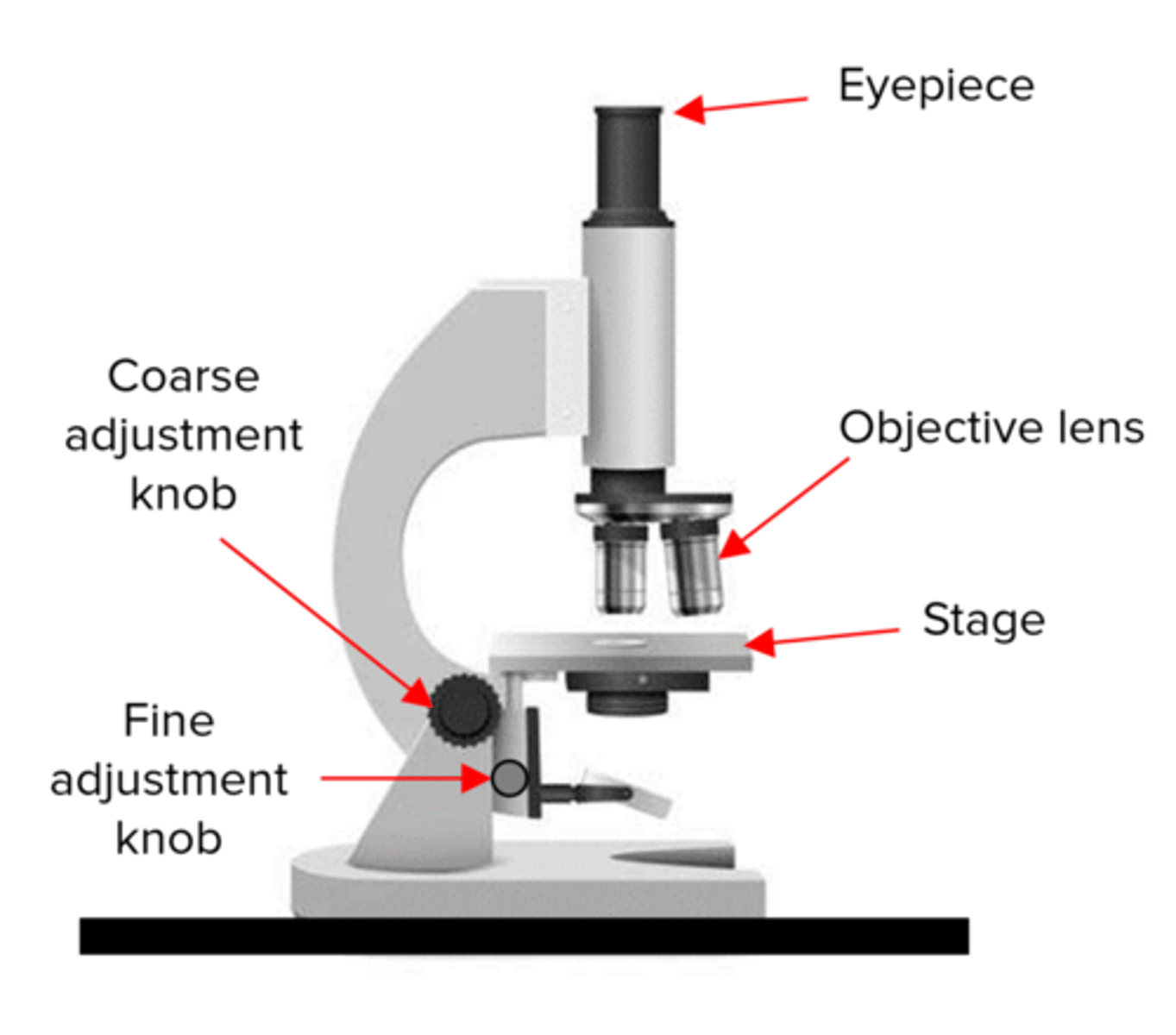
What is the function of the eyepiece lens of an optical microscope? (1)
The lens in which the viewer looks through to see the specimen
What is the function of the objective lens of an optical microscope?
The lenses that are closest to the specimen for magnification
What is the magnification of a low power objective lens? (1)
4x
What is the magnification of a medium power objective lens? (1)
10x
What is the magnification of a high power objective lens? (1)
40x
What is the function of the coarse adjustment knob of an optical microscope? (2)
- Brings the specimen into general focus
- By moving the stage up / down
What is the function of the fine adjustment knob of an optical microscope? (2)
- Tunes the focus
- Increases the detail of the specimen
What is the function of the stage clips? (1)
Holds the slide in place
What is the function of the stage? (1)
The platform where the slide is placed
What is the preparation of a slide using a liquid specimen called? (1)
Wet mount
What is the preparation of a slide using a solid specimen called? (1)
Dry mount
What is the first step in preparing a slide using a liquid specimen? (2)
- Add a few drops of the sample to a slide
- Using a pipette
How do you cover the liquid on a slide to prevent air bubbles? (2)
- With the coverslip
- Gently pressing down to remove air bubbles
What safety precaution should you take when preparing a slide to avoid cross-contamination? (2)
- Wear gloves
- Wash hands regularly
What is the first step in preparing a microscope slide using a solid specimen? (2)
- Use scissors
- To cut a small sample of the tissue
Why should you still wear gloves when preparing a slide with a solid specimen? (2)
- To prevent stain from dyeing your skin
- To protect your hands from sharp objects
How do you obtain a thin layer of cells from a solid tissue sample for slide preparation? (3)
- Peel away or cut
- A very thin layer of cells from the sample
- Using a scalpel or forceps
Why is it important for the tissue sample to be thin when preparing a microscope slide? (1)
So that light from the microscope can pass through the specimen
What should be applied to a tissue sample on a slide before placing the coverslip? (1)
Apply your stain
Describe how a transmission electron microscope produces a micrograph (4)
1. Specimen is stained with electron dense substances e.g. heavy metal salts
2. Beam of electrons are transmitted through the specimen
3. Staining substances deflect the electrons in the beam
4. The pattern that the remaining electrons produce as they pass through specimen is converted into an image
Why is the image generated by TEM, an image of the internal features of the specimen only? (1)
Electrons are able to pass through the specimen
Describe how a scanning electron microscope produces a micrograph (3)
1. Specimen is coated with a thin film of heavy metal e.g. gold
2. Electron beam is scanned to and across the specimen
3. Electrons that are reflected from the surface are collected and produce an image on a viewing screen
Why is the image generated by SEM, an image of the external features of the specimen only? (1)
Electrons are reflected from the specimen
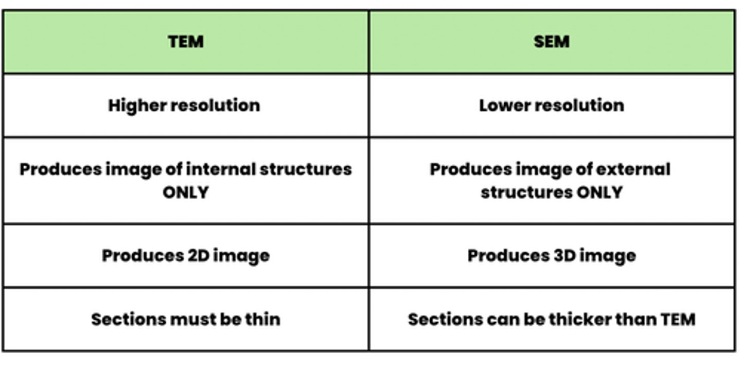
Compare the features of a SEM vs TEM
SEM- lower resolution, produces image of external structures only, produces 3d images, sections can be thicker than TEM
TEM- higher resolution, produces images of internal structures ONLY, produces 2d images, sections must be thin
What is the difference between 'magnification' and 'resolution'? (2)
- Resolution describes how well a microscope can distinguish between two points that are close together
- Magnification describes how enlarged an image is compared to the object
What is the order of magnification and resolution between the 3 microscopes? (1)
(Highest) TEM > SEM > Optical Microscope (Lowest)
How do you calculate magnification of an image? (3)
Image size / Actual size
How do you convert measurements from one unit to another? (6)
metres x1000 millimetres x1000 micrometres x1000 nanometres
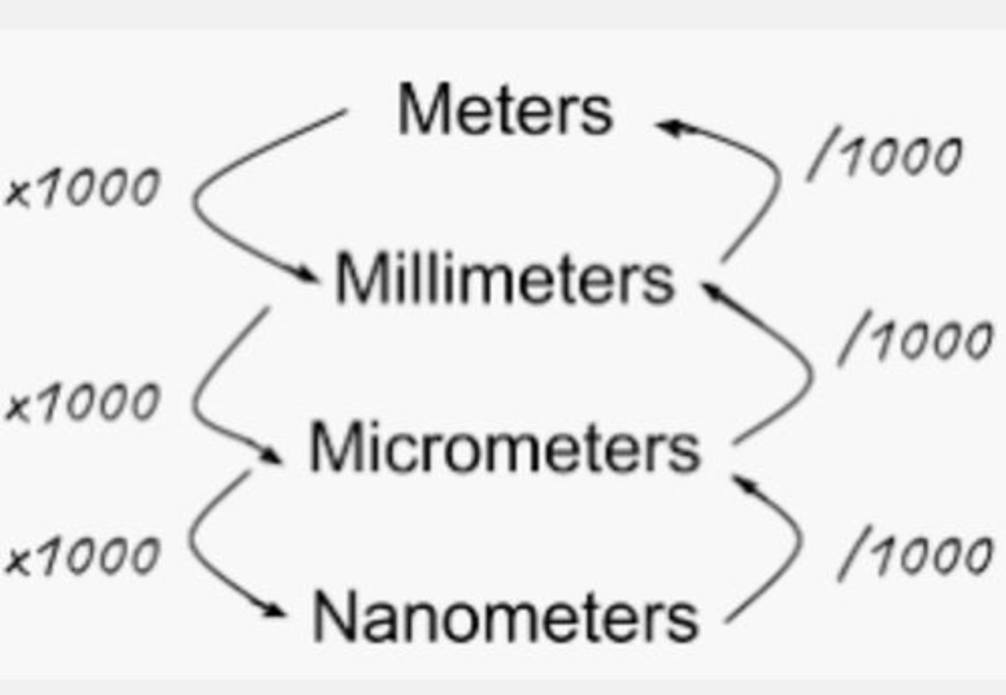
What symbol is used for micrometers? (1)
μm
What symbol is used for nanometers? (1)
nm
What are the rules that must be considered for biological drawings? (5)
- Title must be included
- Use a sharp pencil for drawings and labels
- State the magnification that you are drawing form
- No shadings
- Annotate all cell components
How could you use an eyepiece graticule and stage micrometer to measure the size of a structure? (5)
1. Place micrometer on stage to calibrate the eyepiece graticule
2. Line up the scales on the graticule and micrometer
3. Count how many graticule divisions are in the 100μm on micrometer
4. Length of 1 eyepiece division - 100μm / number of divisions
5. Use the calibrated values to calculate the actual length of structures
What is meant by 'cell ultrastructure'? (2)
- Fine detailed structure of a cell organelles
- Ultrastructure images are usually from TEM
What are the organelles that are present in eukaryotic cells? (12)
-Nucleus
- Endoplasmic reticulum (Both RER and ER)
- Golgi apparatus
- Ribosomes
- Mitochondria
- Lysosomes
- Chloroplasts
- Plasma membrane
- Centrioles
- Cell wall
-Flagella
- Cilia
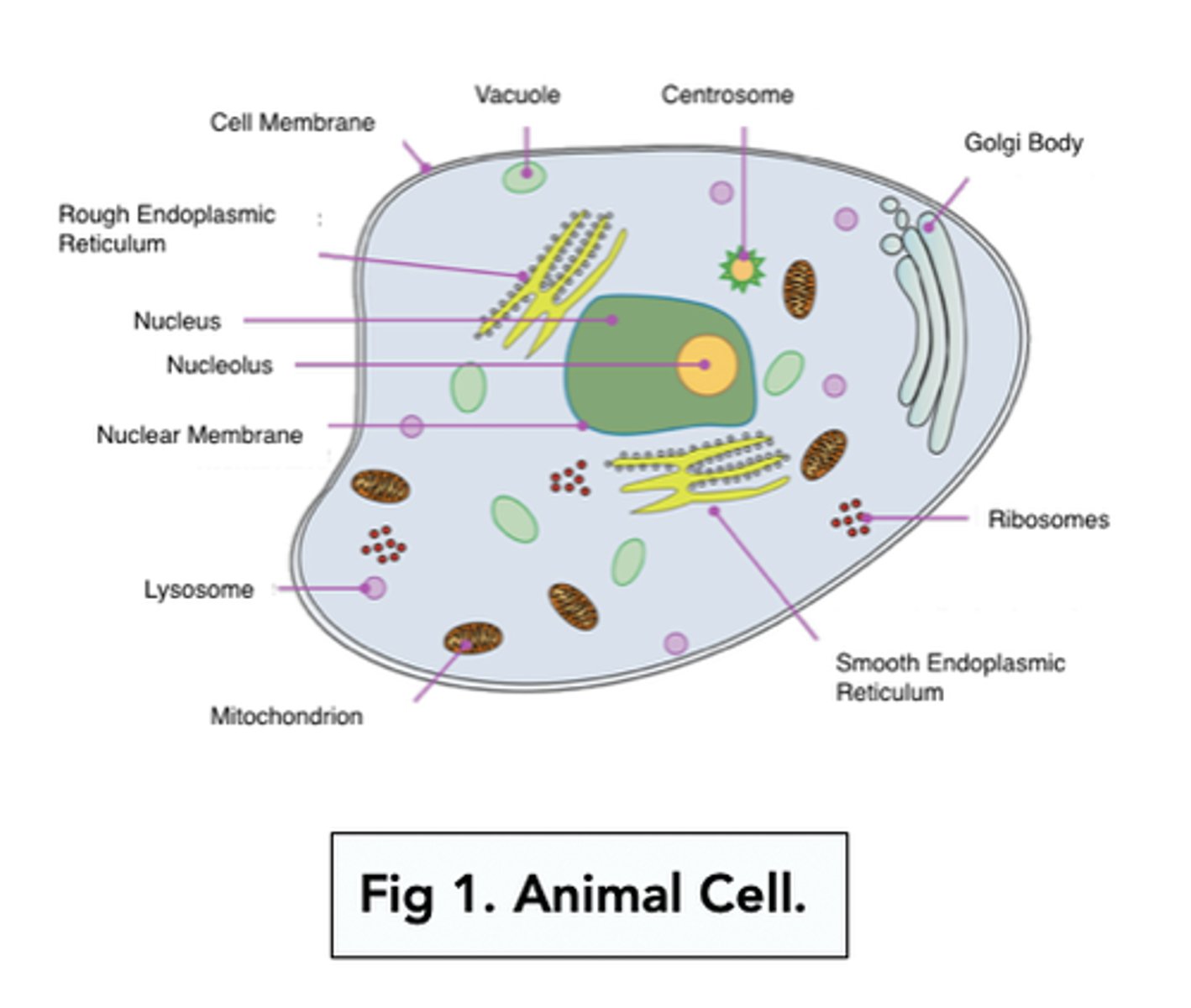
Draw and label the structure of a typical animal cell (7)
Draw and label the structure of a typical plant cell (7)
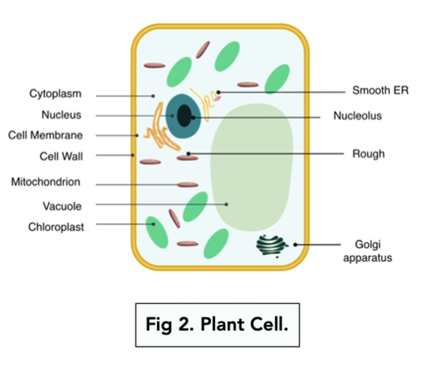
What organelles are only present in plant cells? (3)
- Cellulose cell wall
- Chloroplasts
- Vacuole
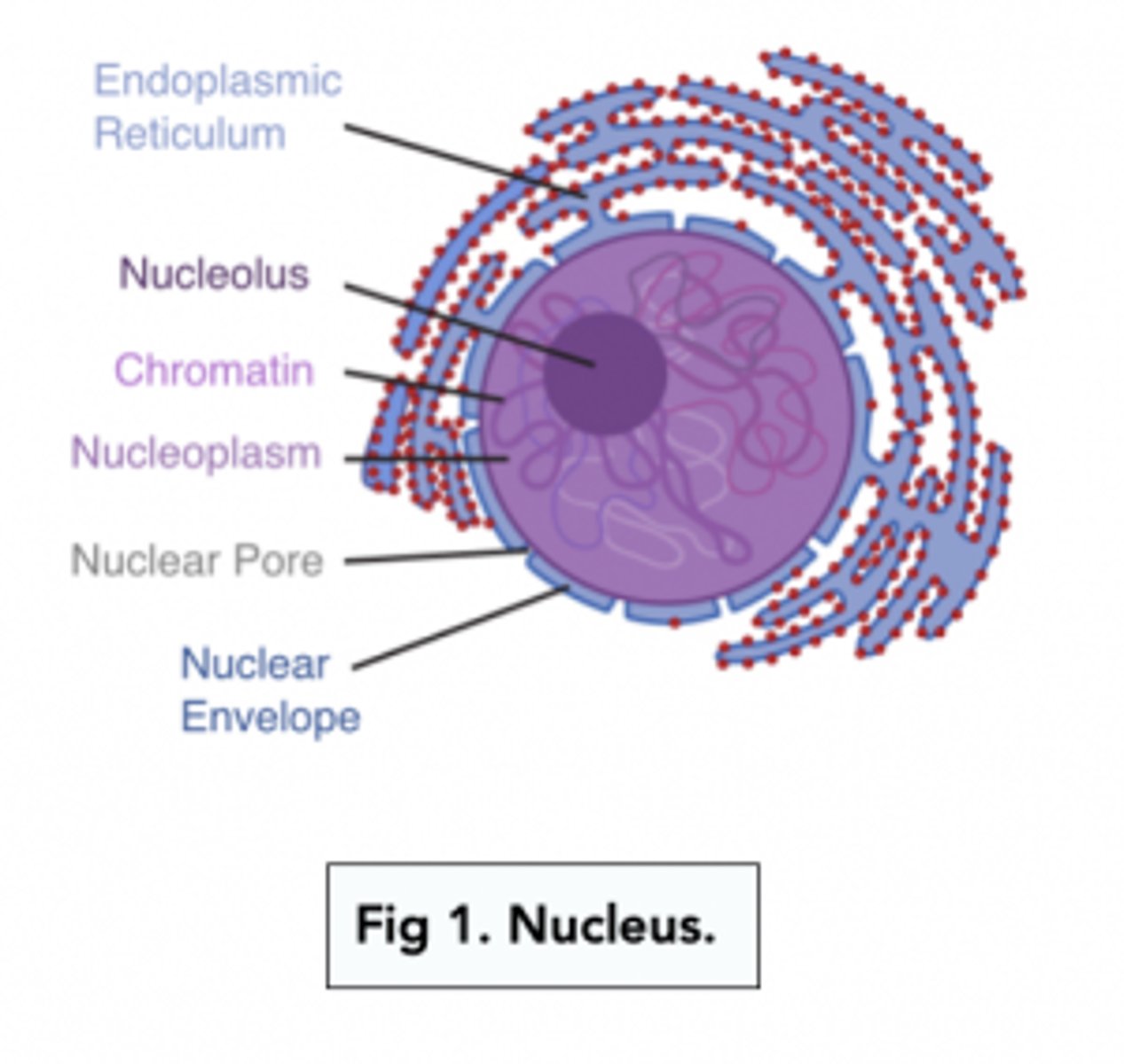
Draw and label the structure of a nucleus (4)
- Nucleolus
- Nuclear envelope
- Chromatin
- Nuclear pores
What does the nucleus contain within its structure to accommodate for its function? (3)
- Histones (protein)
- Linear DNA
- One or more RNA nucleoli
What is the function of the nucleus? (2)
- Contains and produces genetic material (DNA and RNA)
- Controls protein synthesis
- So controls the development and function of the cell
What components make up the structure of ribosomes? (2)
- Proteins
- Ribosomal RNA
What is the function of the ribosomes? (2)
- Site of translation in protein synthesis
- Translating mRNA sequence into a sequence of amino acids
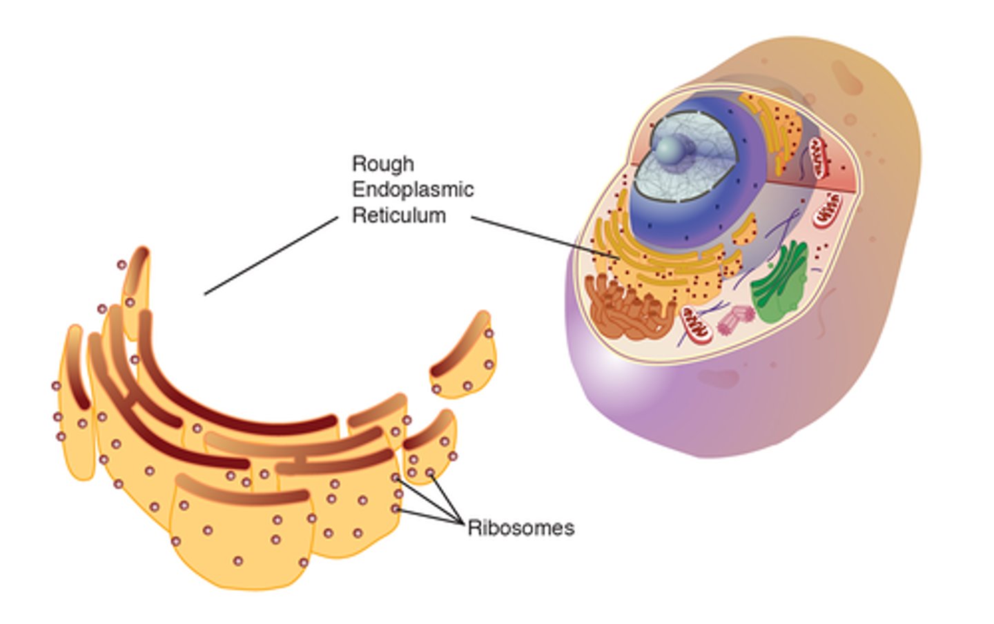
Draw the structure of the rough endoplasmic reticulum (1)
NOTE: you won't need to label each component of the RER at all, but in an exam but you may be asked to recognise it from a micrograph
It looks a lot like a maze with ribosomes on its surface
Describe the function of the rough endoplasmic reticulum (2)
- Has ribosomes on its surface that produce secretory proteins (proteins that are released out of the cell)
- These secretory proteins are sent to Golgi Apparatus for modification and packaging
What is the difference in structure between the RER & SER? (1)
RER contains ribosomes on its surface, whereas, SER lacks ribosomes
Draw the structure of Golgi apparatus (1)
NOTE: you won't need to label each component of the Golgi apparatus at all, but in an exam but you may be asked to recognise it from a micrograph
It looks a lot like a wifi symbol
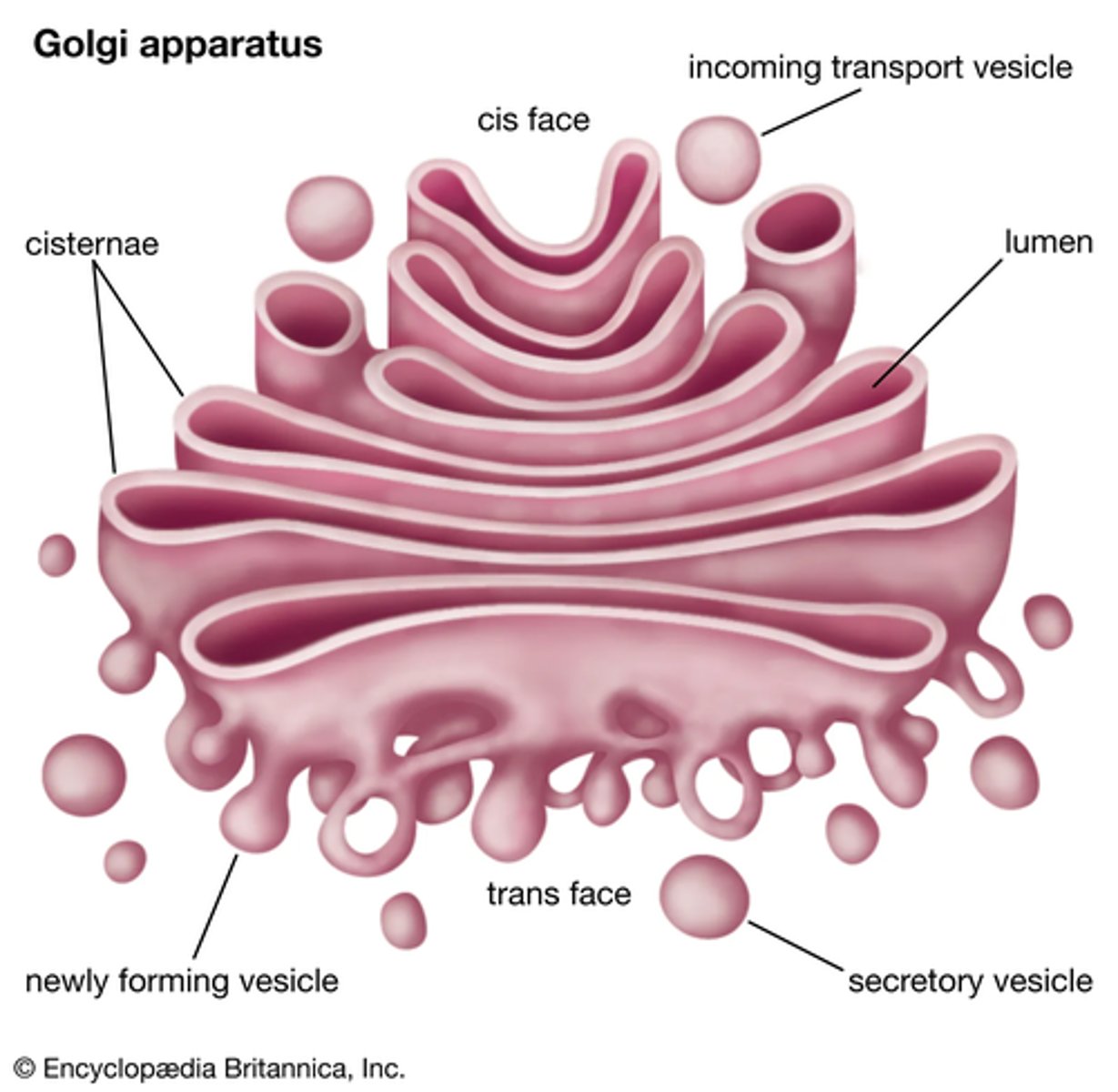
Describe the several functions of the Golgi apparatus (3)
- Adds carbohydrates to proteins received from the RER to form glycoproteins
- Packages proteins/glycoproteins into Golgi vesicles (sacks) for secretion
- Produces lysosomes - a type of Golgi vesicle that releases lysozymes (hydrolytic enzymes)
What is the basic function of the plasma membrane? (1)
Controls the movement of substances into and out of the cell
What are the roles of the organelles that are involved in the production, transport and release of proteins from eukaryotic cells? (5)
1. DNA in the nucleus codes for these proteins
2. Ribosomes in the RER produce these proteins via protein synthesis
3. Mitochondria produces the ATP that is required for protein synthesis
4. Golgi apparatus packages and modifies these proteins into vesicles
5. Vesicles fuse with the cell membrane and release proteins outside of cell
Describe the functions of lysosomes (3)
- Digests material that is taken in by phagocytosis
- Non-functioning organelles within the cell are engulfed and digested by lysosomes
- Releases enzymes outside of the cell
Draw and label the structure of the mitochondria (8)
- Matrix
- Enzymes
- Inner membrane
- Cristae
- (Circular) DNA
- Inter-membrane space
- Ribosomes (70S)
- Outer membrane
What is the basic function of the mitochondria? (2)
- Involved in aerobic respiration
- Which produces ATP
Why may some cells have lots of mitochondria and could you give an example of a cell? (2)
- To provide energy to cells that require a large amount of ATP
- E.g. muscle cells
What is the basic function of the cell wall? (1)
Provides support, strength and shape to the cell
What is the basic function of the chloroplasts? (2)
- Contains chlorophyll
- Which absorbs light energy for photosynthesis (important for the light dependent stage of photosynthesis)
Draw and label the structure of chloroplasts (8)
- Lipid droplets
- Stroma
- Thylakoids
- Free ribosomes (70S)
- Circular DNA
- Starch grains
- Membrane bound chromosomes
- Granum
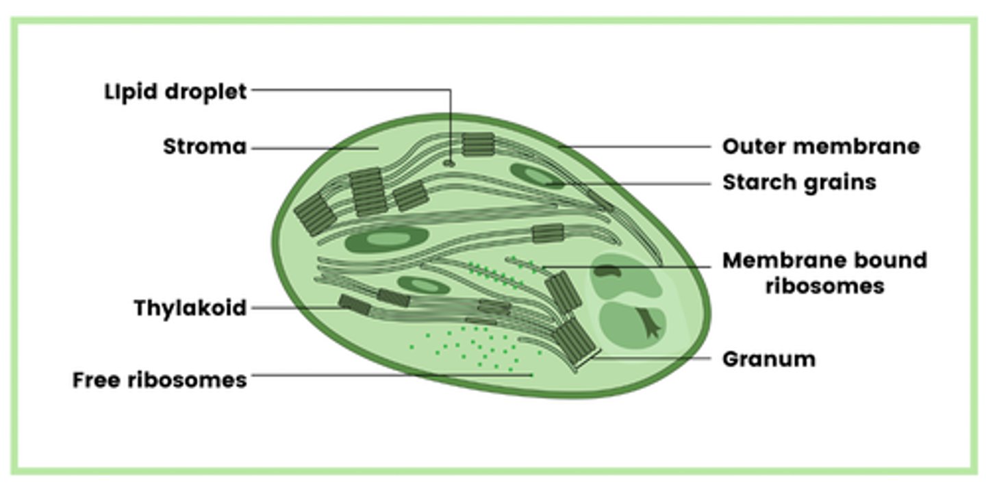
What is the granum that is found in chloroplasts? (1)
Stacked thylakoids
How do granum join together to form grana (plural for granum)? (1)
Link together by thin pieces of membrane - lamellae
What is the basic function of the large vacuole? (1)
Contains soluble sugars, salts and pigments
What features of both chloroplasts and mitochondria demonstrate that they are formerly free-living bacteria that were absorbed into a larger cell? (2)
- They both contain circular DNA
- They both contain 70S ribosomes
What are centrioles? (2)
- Cylindrical structures found in animal cells
- Component of cytoskeleton
Draw the structure of centrioles (2)
- Composed of microtubules
- 9 microtubules organised into triplets
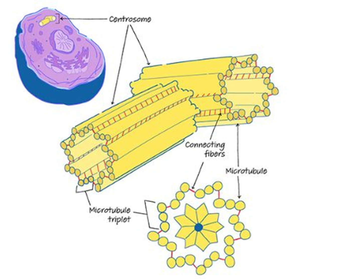
What is the function of the centrioles? (2)
- Two centrioles at right angles to each other form a centrosome
- This organises the spindle fibers during cell division
Draw the structure of cilia (2)
- Hair-like projections
- Made of microtubules
What is the function of cilia? (2)
Allow the movement of substances over the cell surface
Draw the structure of flagella (2)
- Similar structure to cilia
- Made of longer microtubules

What is the cytoskeleton? (1)
Network of protein filaments and tubules in the cytoplasm of many living cells
What is features form the importance of the cytoskeleton? (4)
- Provides mechanical strength to cells
- Aids in the movement of molecules within the cell
- Maintains the shape of cells
- Forms both cilia and flagella
What structures of the cytoskeleton allow it to carry out its function? (1)
Microtubules
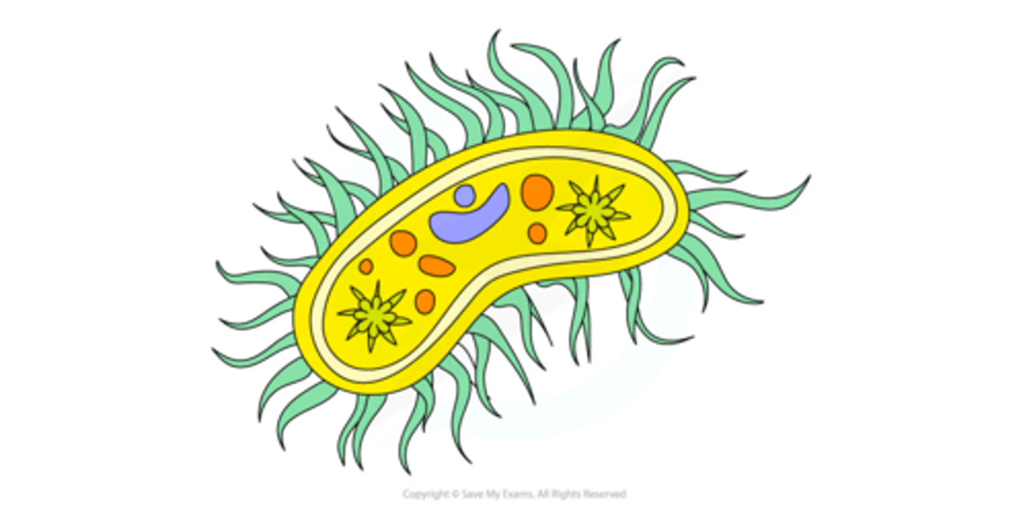
Draw and label the structure of a typical prokaryotic cell (8)
NOTE: you only need to know the structures we have outlined, you do not need to know any of the functions (expect of course the ones that we have made separate flashcards for)
- Cell wall (made of murein - a glycoprotein)
- Cell surface membrane
- Free circular DNA molecule in cytoplasm
- Ribosomes (70S ribosomes)
- Cytoplasm
- Capsule surrounding the cell wall (in some)
- One or more plasmids (in some)
- One or more flagella (in some)
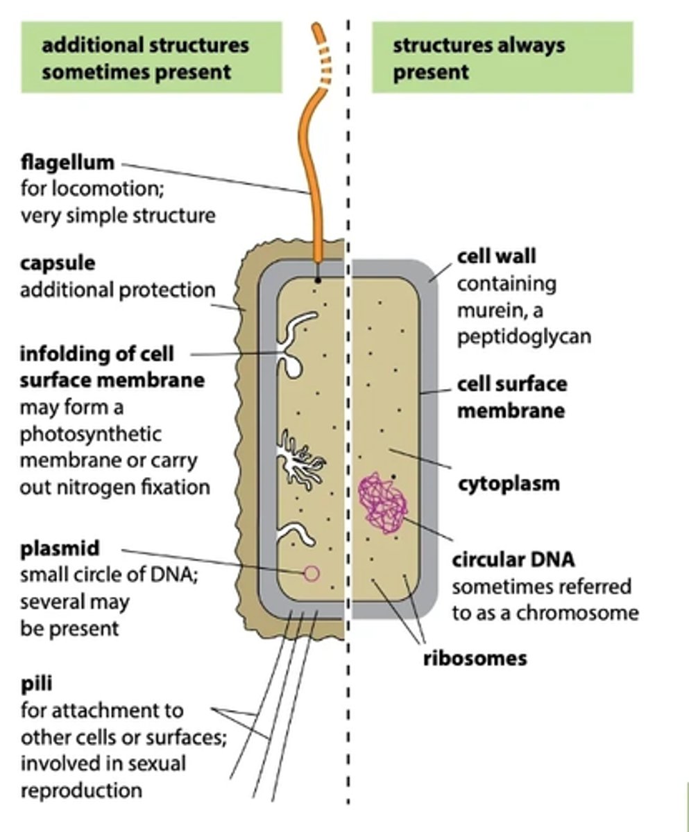
What structures are present in all prokaryotic cells? (5)
- Cell wall (made of murein - a glycoprotein)
- Cell surface membrane
- Free circular DNA molecule in cytoplasm
- Ribosomes (70s ribosomes)
- Cytoplasm
What structures are only present in some prokaryotic cells? (3)
- Capsule surrounding the cell wall
- One or more plasmids
- One or more flagella
Describe the DNA of prokaryotic cells (4)
- No nucleus is present
- DNA is free in the cytoplasm
- Circular DNA
- Not attached to any histone proteins
Describe the properties of plasmids found in prokaryotic cells (3)
- Contain antibiotic resistance genes (amongst others)
- Plasmids are able to be passed between prokaryotes
- Some prokaryotic cells have several plasmids, others have no plasmids
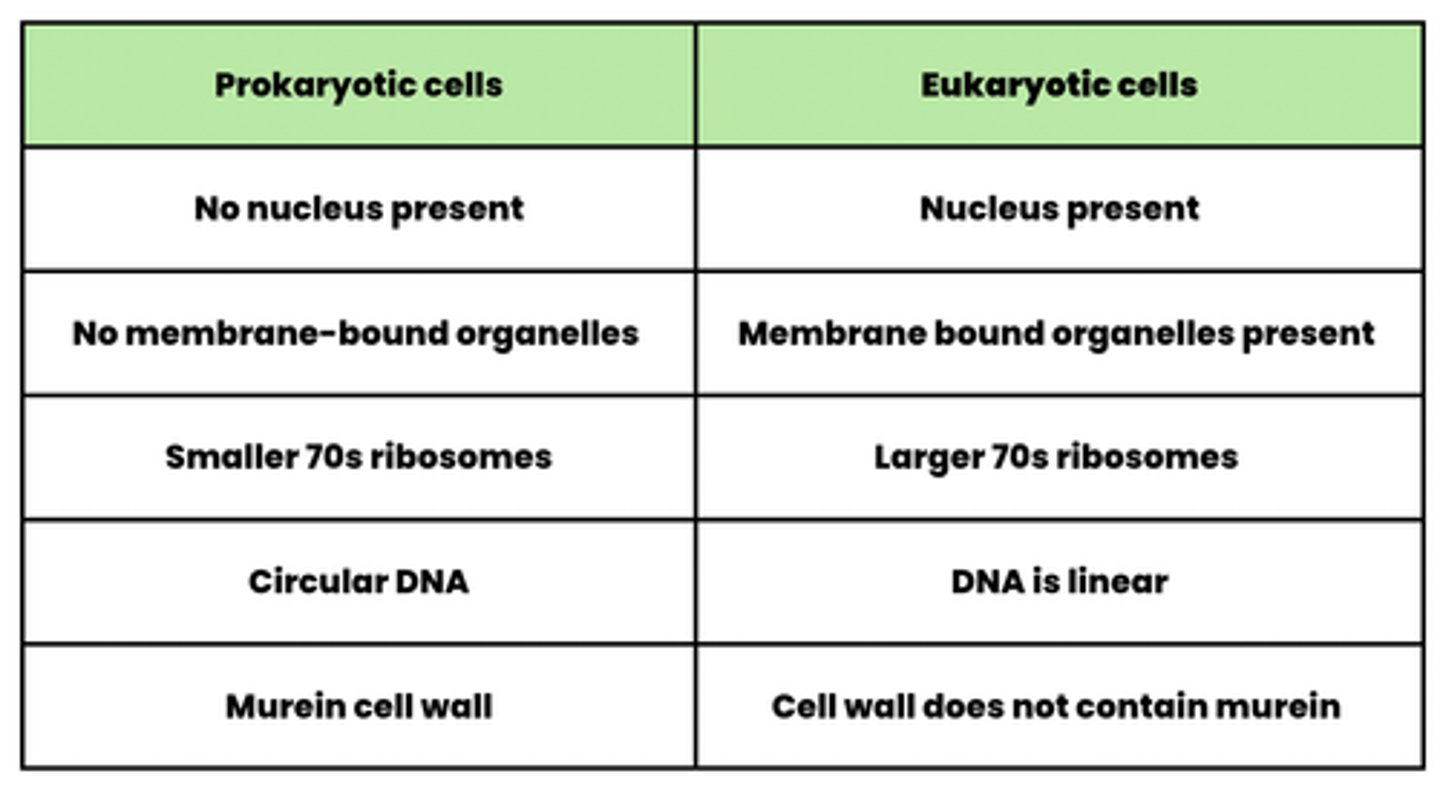
Compare prokaryotic cells and eukaryotic cells (5)
prokaryotic- no nucleus present, no membrane bound organelles, smaller 70s ribosomes, circular dna, murein cell wall
eukaryotic- nucleus present, membrane bound organelles present, larger 70s ribosomes, dna is linear, cell wall doesn't contain murein
What is the difference in the type of ribosome in bacterial and human cells? (2)
Bacterial cells have 70S ribosomes whereas, human cells have 80S ribosomes