GI Images Histology
1/147
Earn XP
Name | Mastery | Learn | Test | Matching | Spaced | Call with Kai |
|---|
No analytics yet
Send a link to your students to track their progress
148 Terms
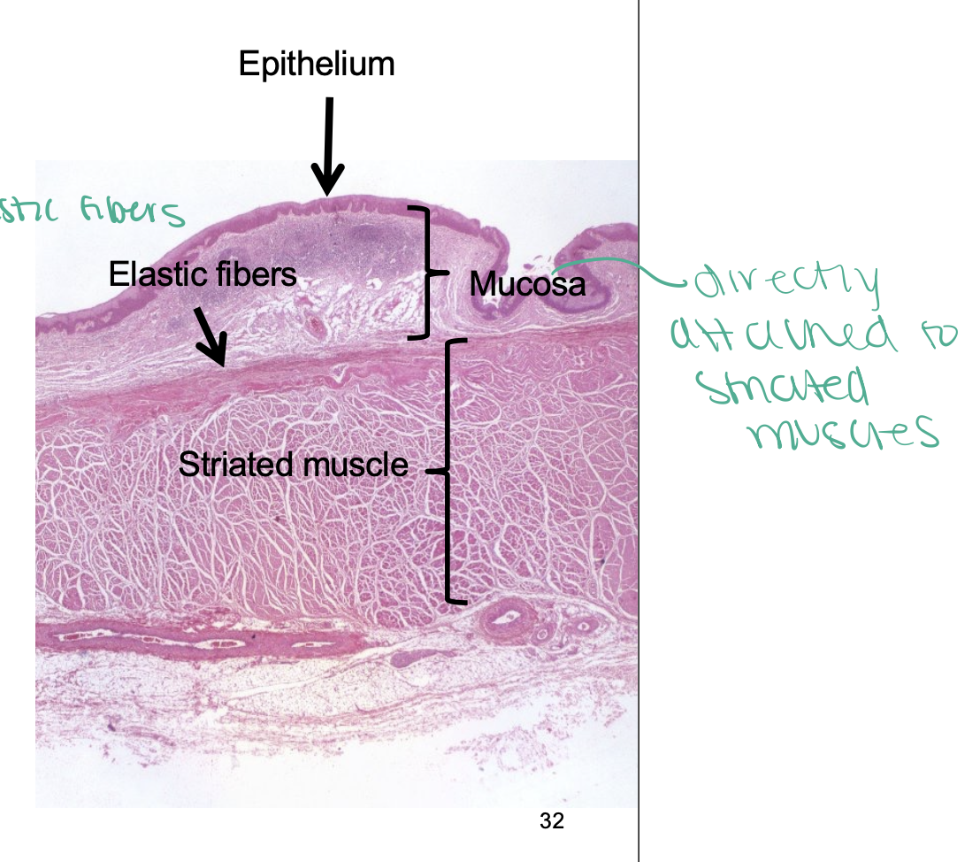
Tongue
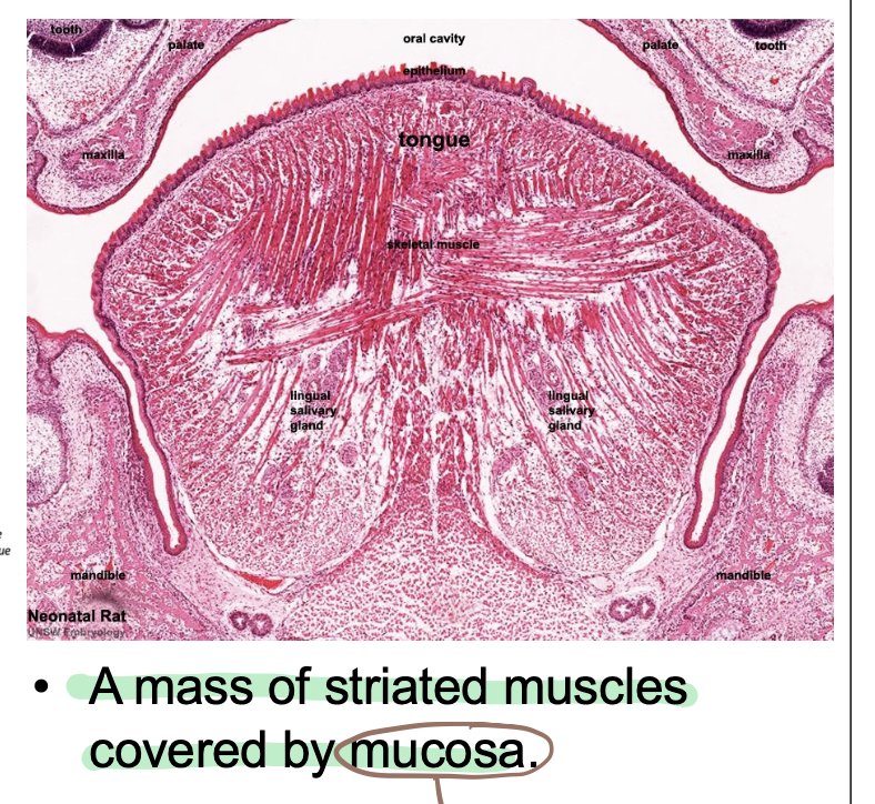
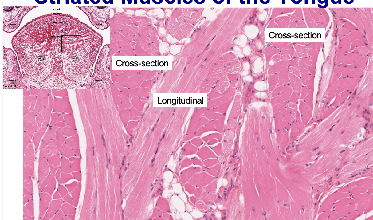
Striated muscles of tongue
arrangement only found in tongue, heart and stomach: has oblique, transverse and longitudinal sections
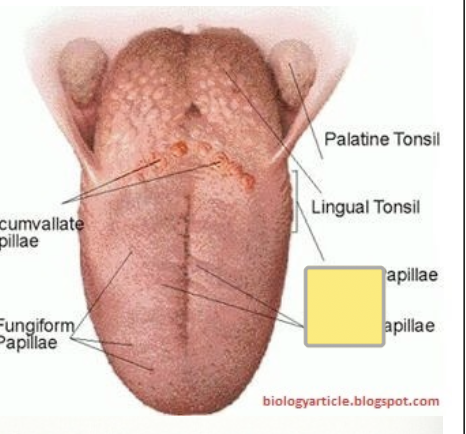
Papillae on physical tongue
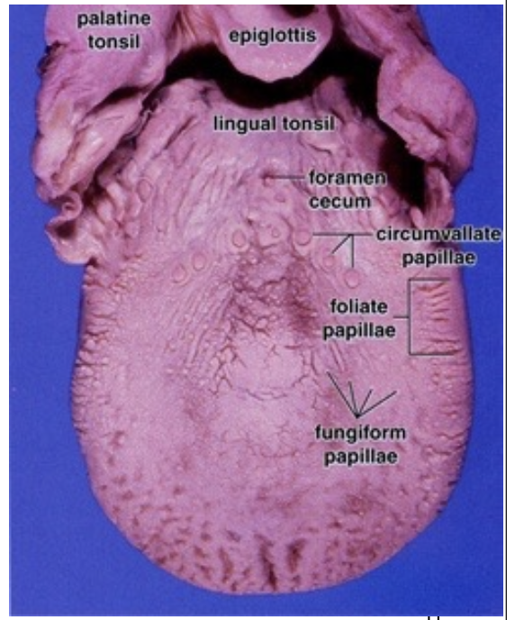
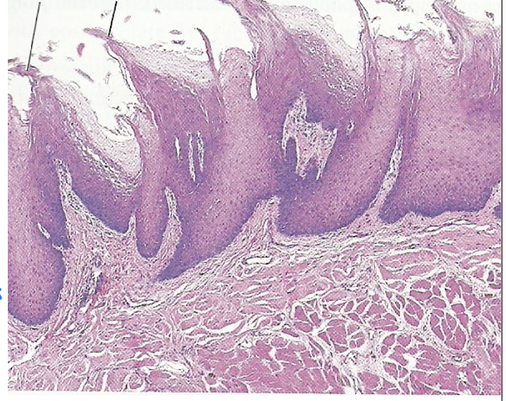
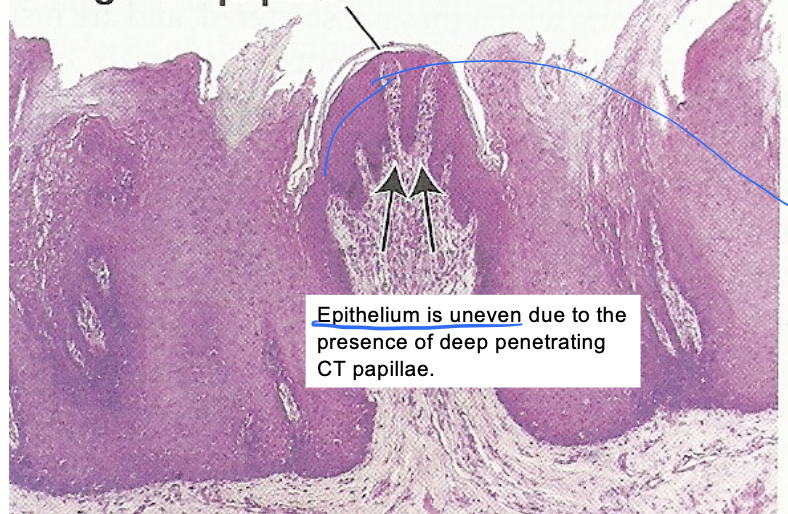
Filiform Papillae
Filiform Papillae Physical Tongue
Fungiform Papillae Physical Tongue
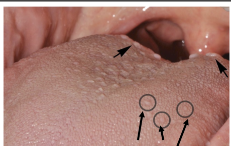
Fungiform Papillae
Circumvallate Papillae Physical Tongue
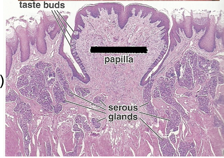
Circumvallate Papillae
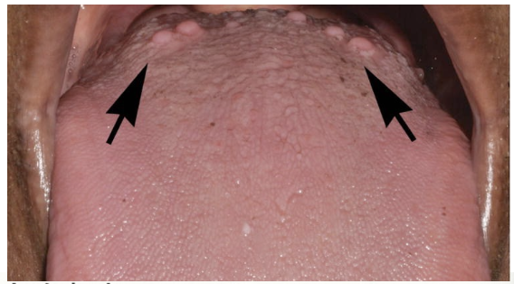
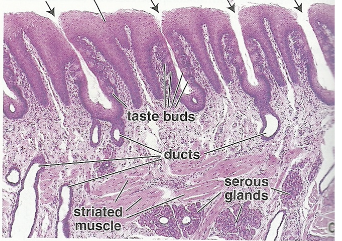
Foliate Papillae
Foliate Papillae Physical Tongue (top)
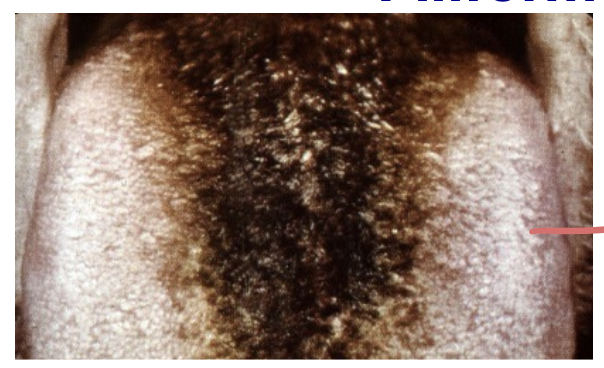
Hairy Tongue Physical
over keratinized of filiform papillae
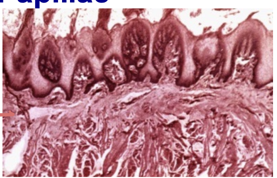
Hairy Tongue Histology
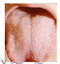
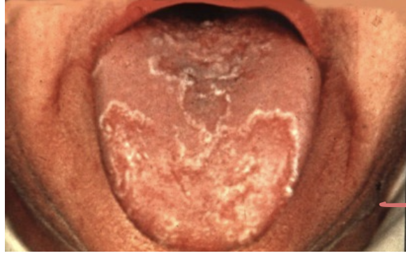
Geographic tongue Physical
Under keratinized of filiform papillae
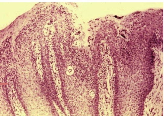
Geographic tongue histology
Under keratinized and loss of papilla
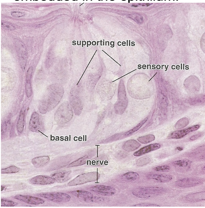
Taste Bud
Beneath the taste buds are nerve fibers that are
also lightly stained
Easily identified by LM 3 cells (1) basal cells which are stem cells (2) supporting cells (3) taste receptor cells of sensory cells and these cells synapse with afferent nerves
Have pore opening and microvilli
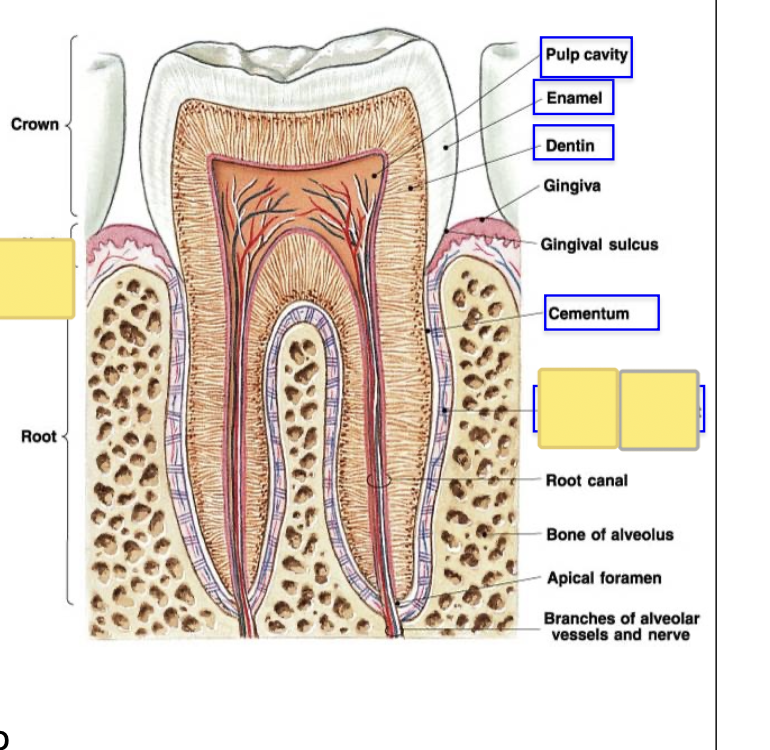
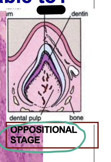
Pulp Cavity
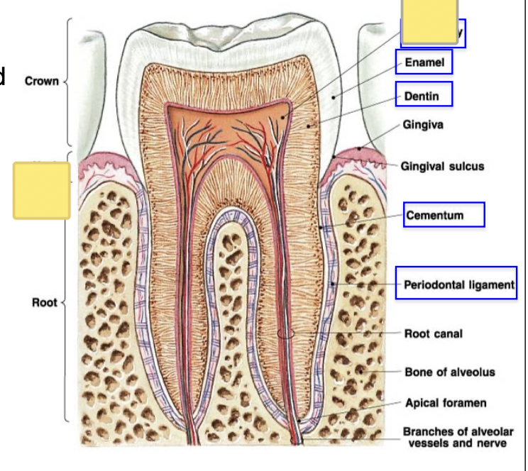
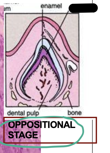
Enamel
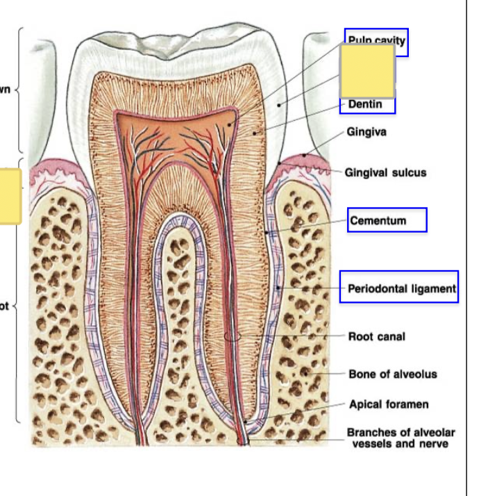
Dentin
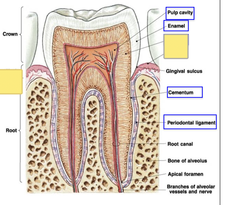
Cementum
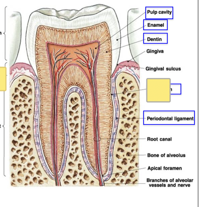
Periodontal Ligament
Tooth formation Bud Stage
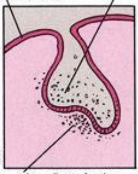
Tooth formation Cap Stage
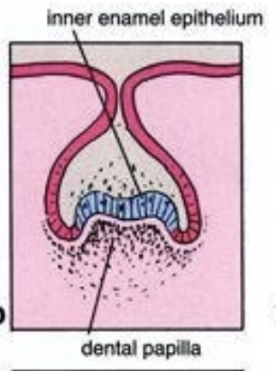
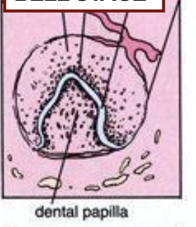
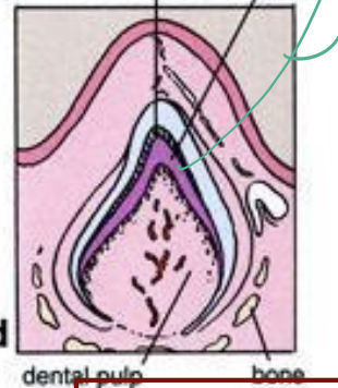
Tooth formation Bell Stage
Tooth formation Oppositional Stage
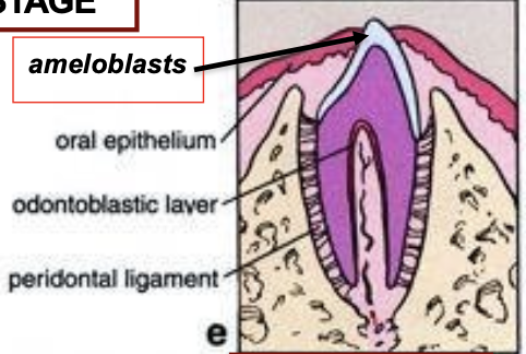
Tooth formation Erruption Stage
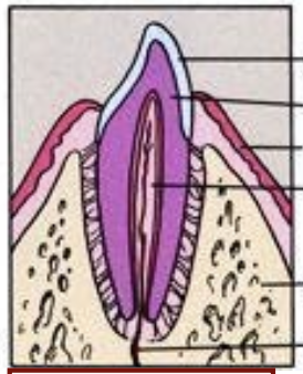
Tooth formation Functional Stage
Ameloblast
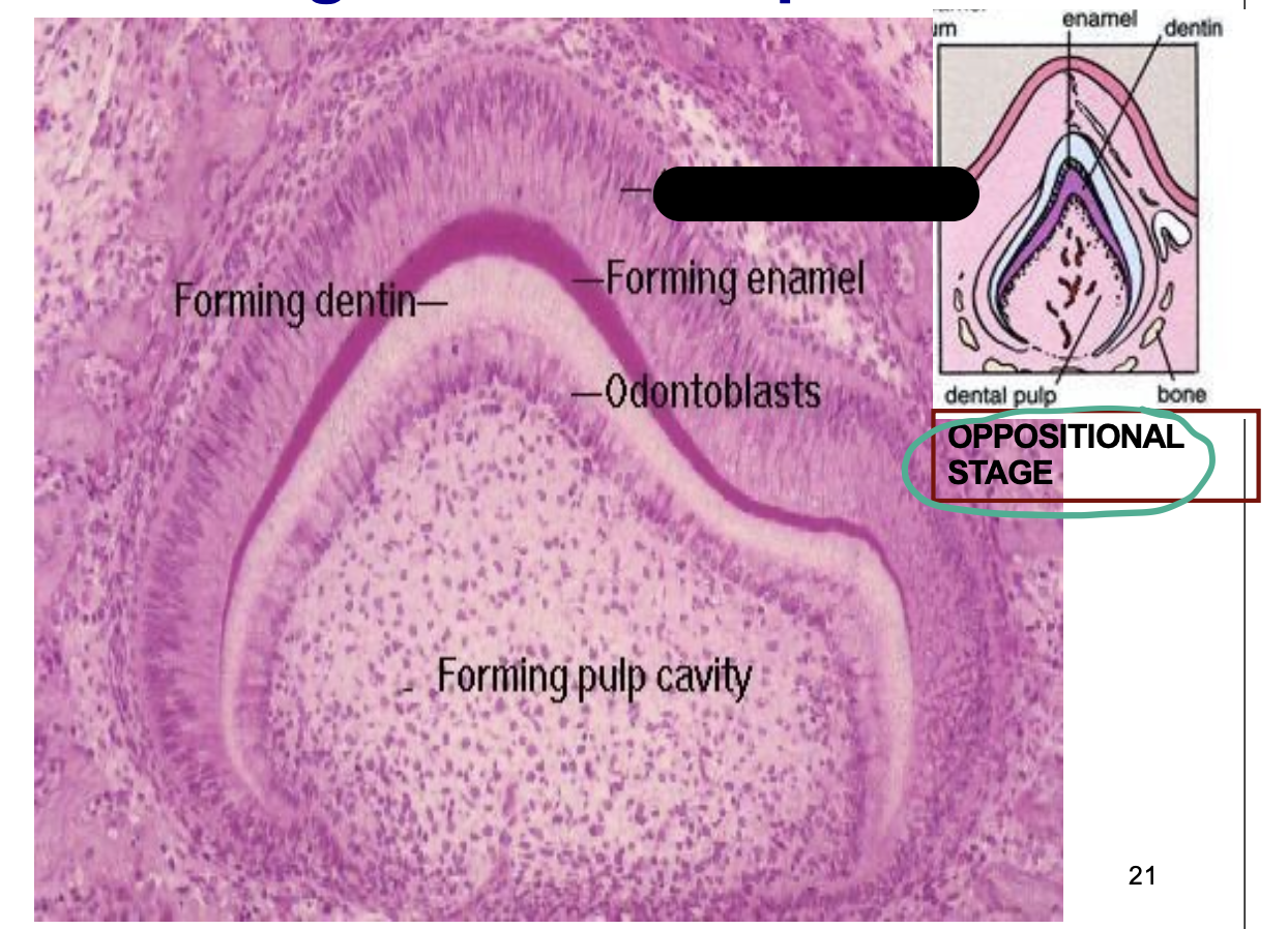
Forming Enamel
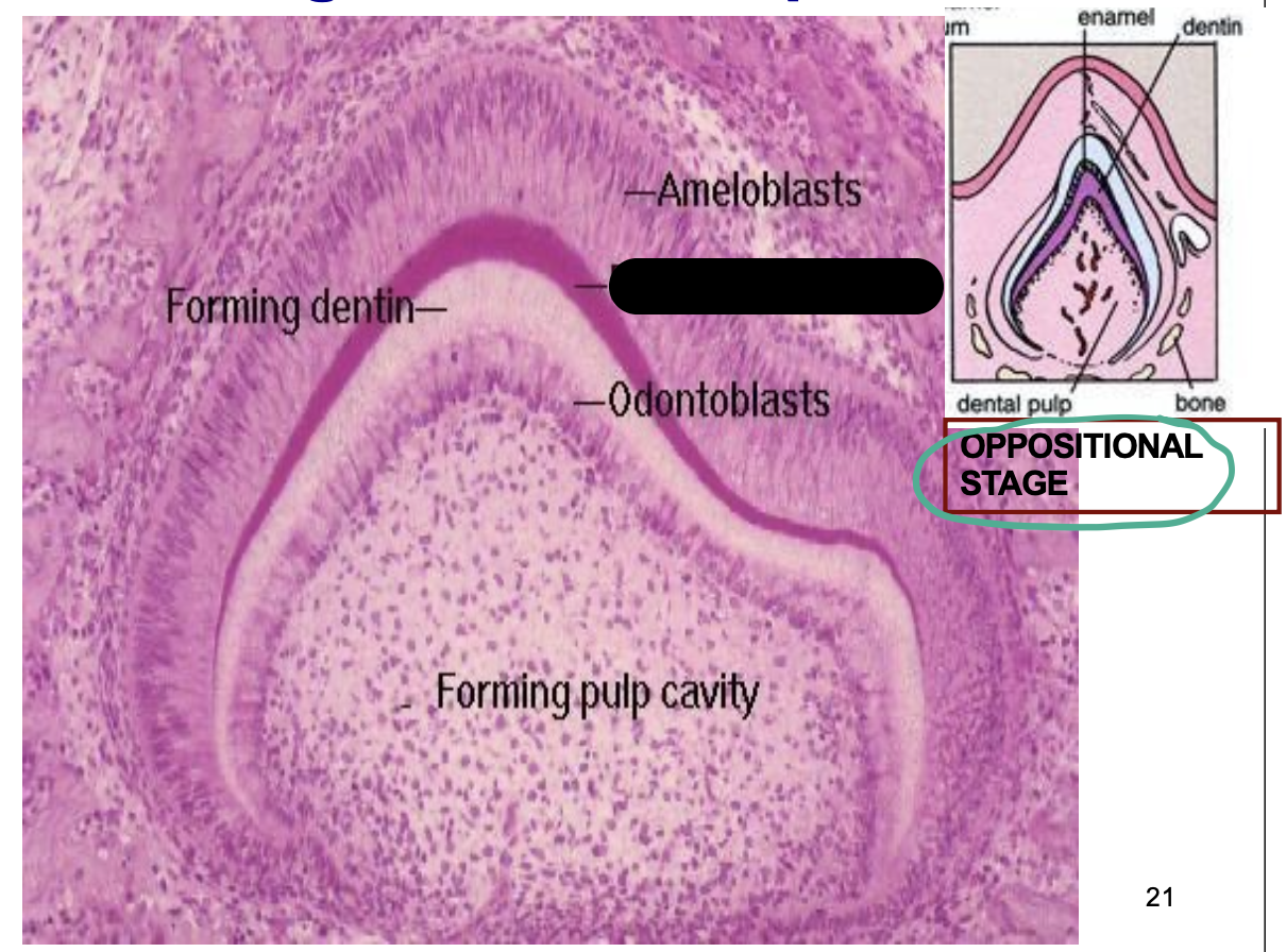
Odontoblast
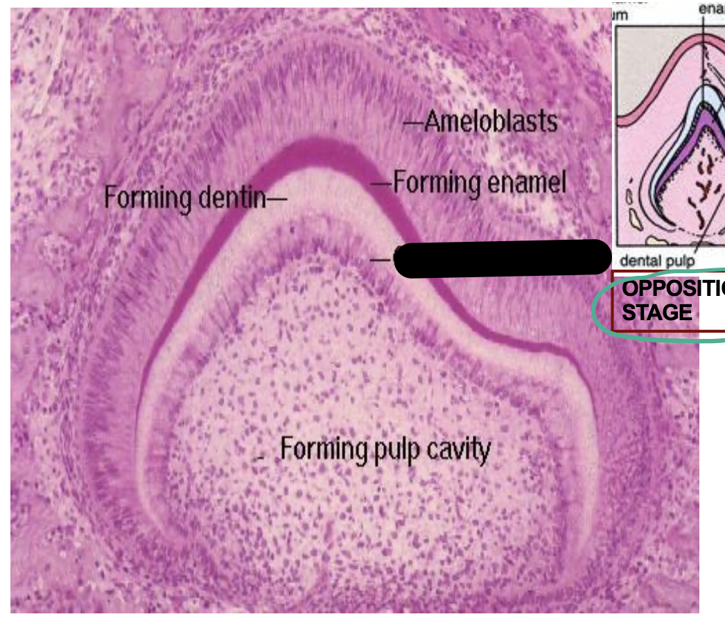
Forming dentin
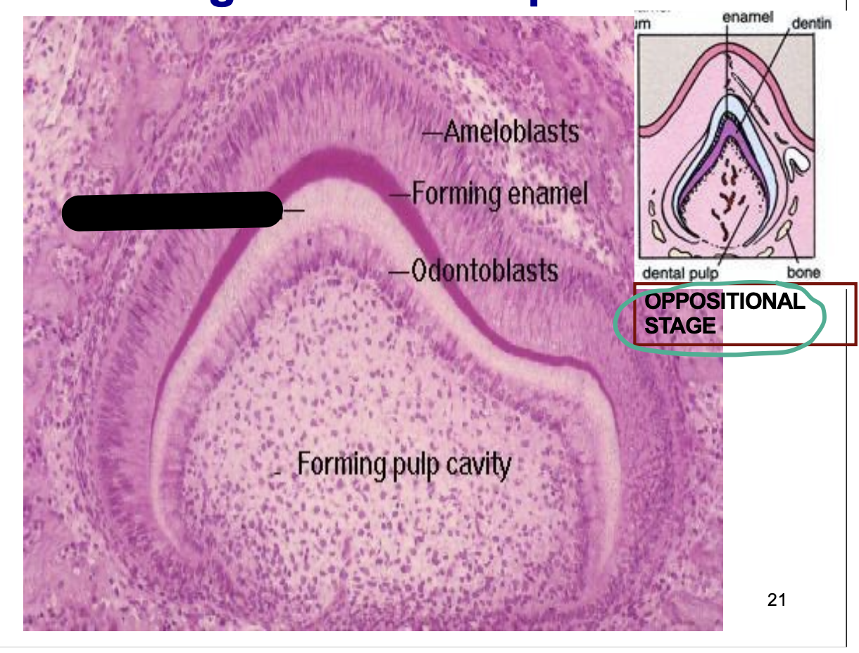
Enamel
Dentin
Major Salivary Gland
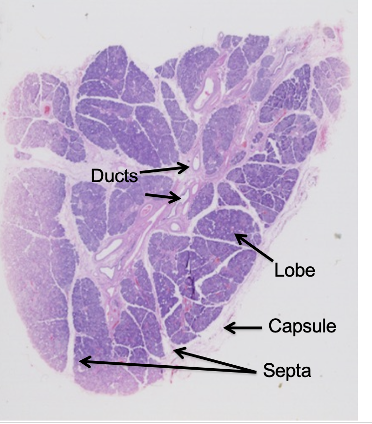
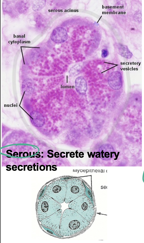
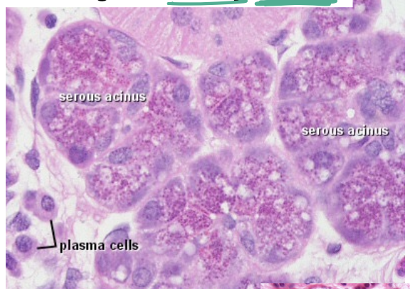
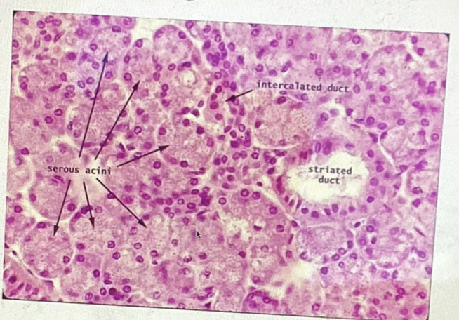
Parotid Gland Histology
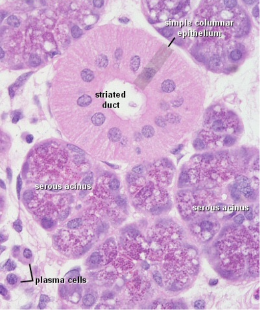
Parotid Gland Serous Acini (pinkish)
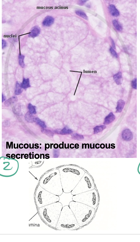
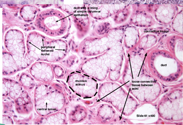
Sublingual Gland Mucous Acini (very light color, nuclei flattened)
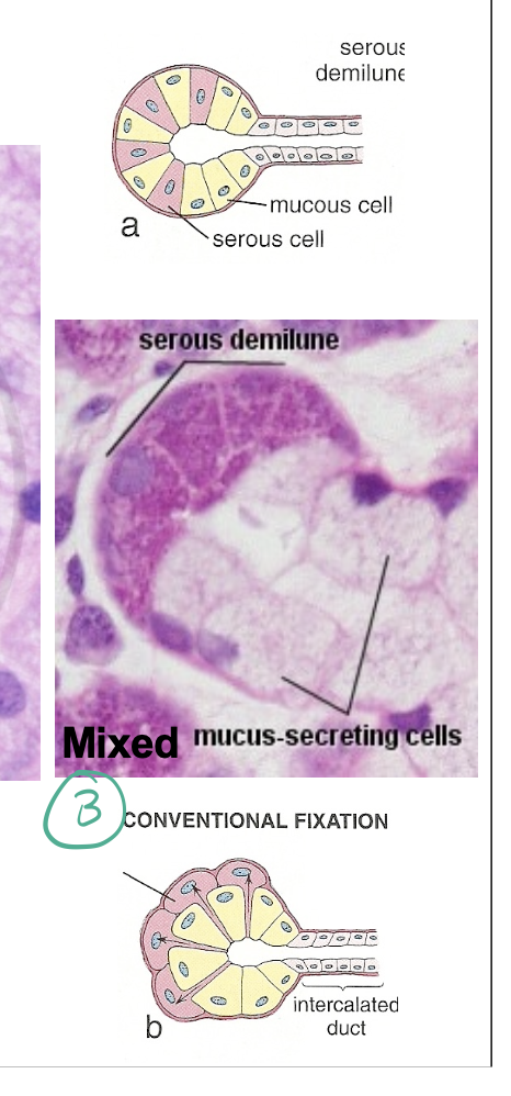
Mixed Acini (serous demilune)
Parotid gland - entirely serous
Submandibular - Mixture (mostly serous)
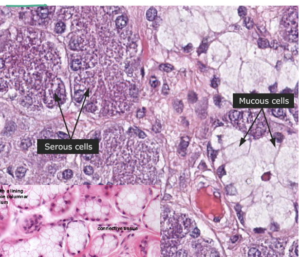
Sublingual- Mixture (mostly mucous)
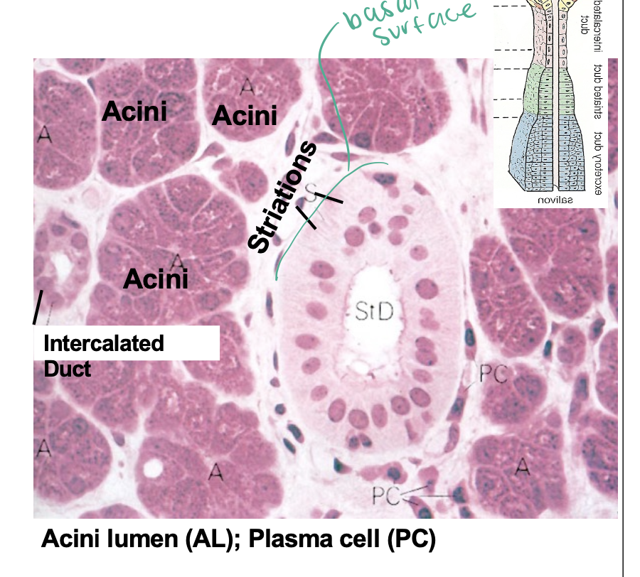
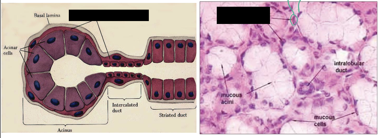
Salivary Gland Duct
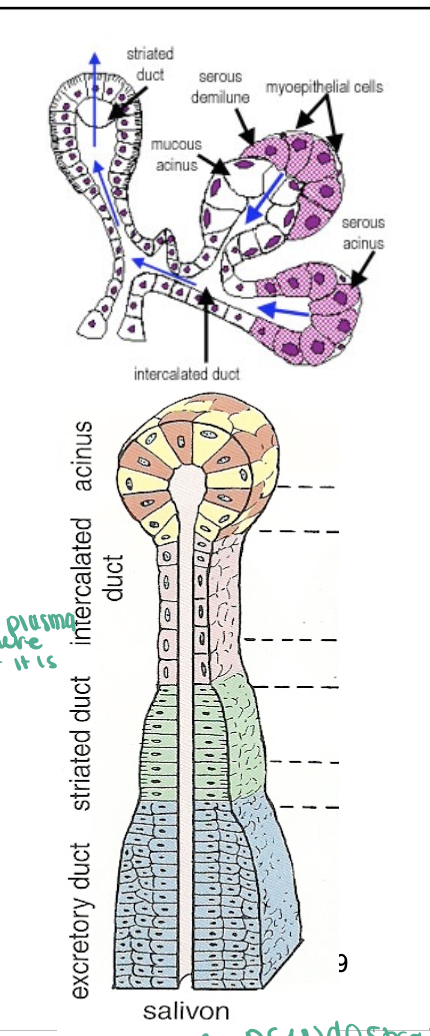
Striated Duct
The basal membrane forms folds that contains numerous long vertical mitochondria characteristic of ion- pumping activity.
Mitochondria give appearance of striated ducts
Myoepithelial Cells
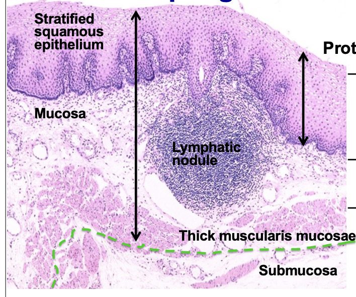
Pharynx
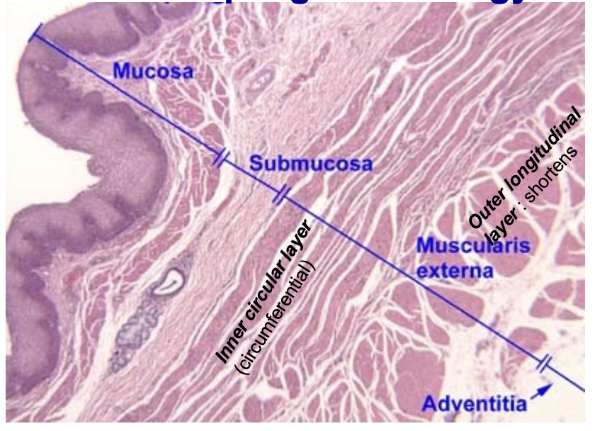
Layer reference
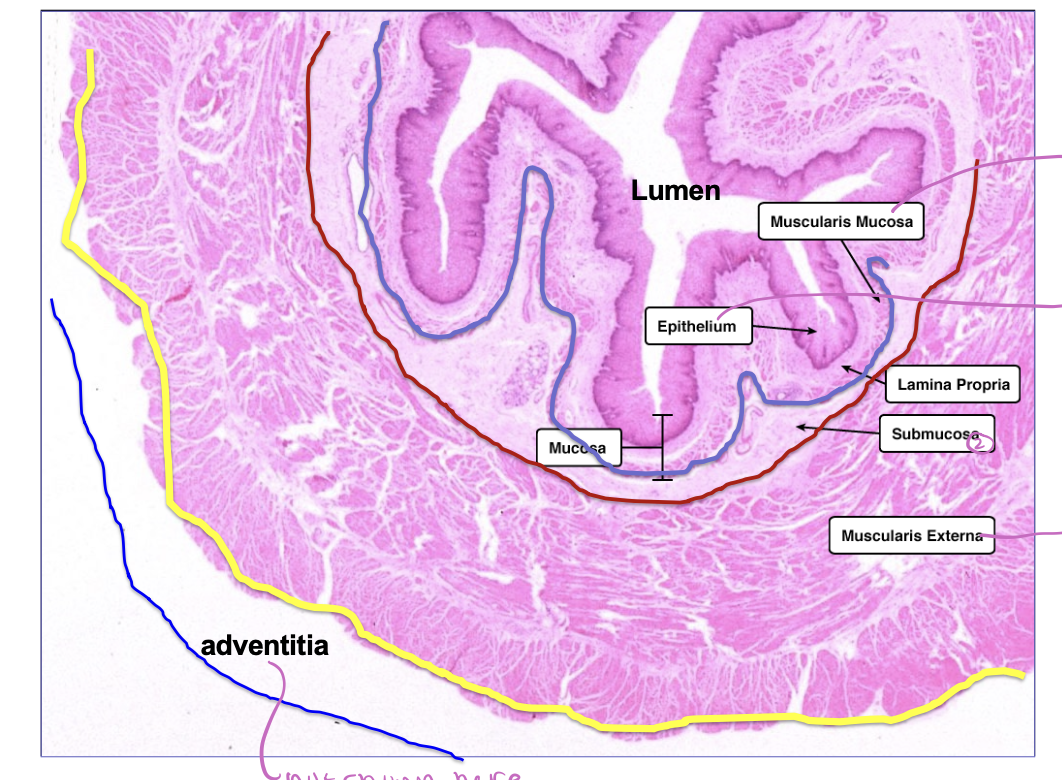
Esophagus Layers
Esophageal cardiac glands
- Present in lower portion in mucosa, produce mucus with neutral pH.
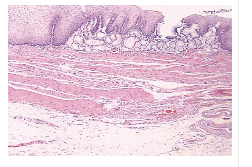
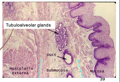
Esophageal glands proper
- Present in submucosa. Produce mucous secretions for lubricating the luminal wall.
Middle Esophagus Muscularis
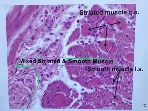
Lower Esophagus Muscularis
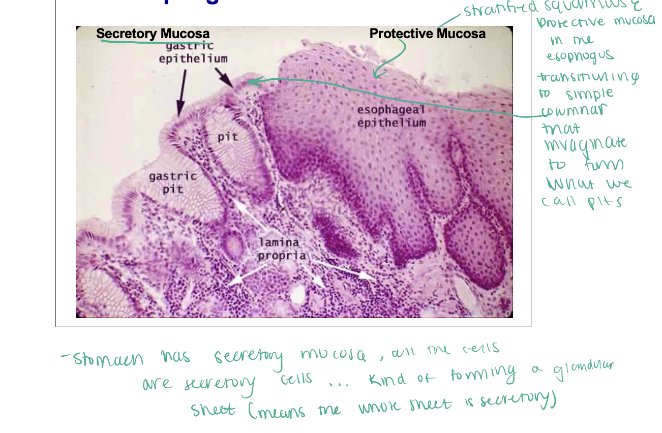
Esophageal Cardiac Junction
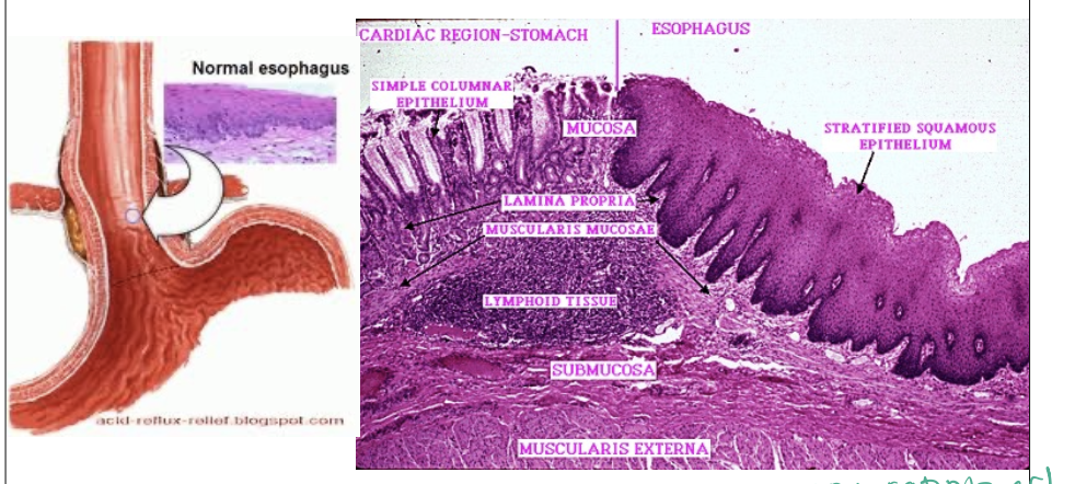
Esophageal Cardiac Junction
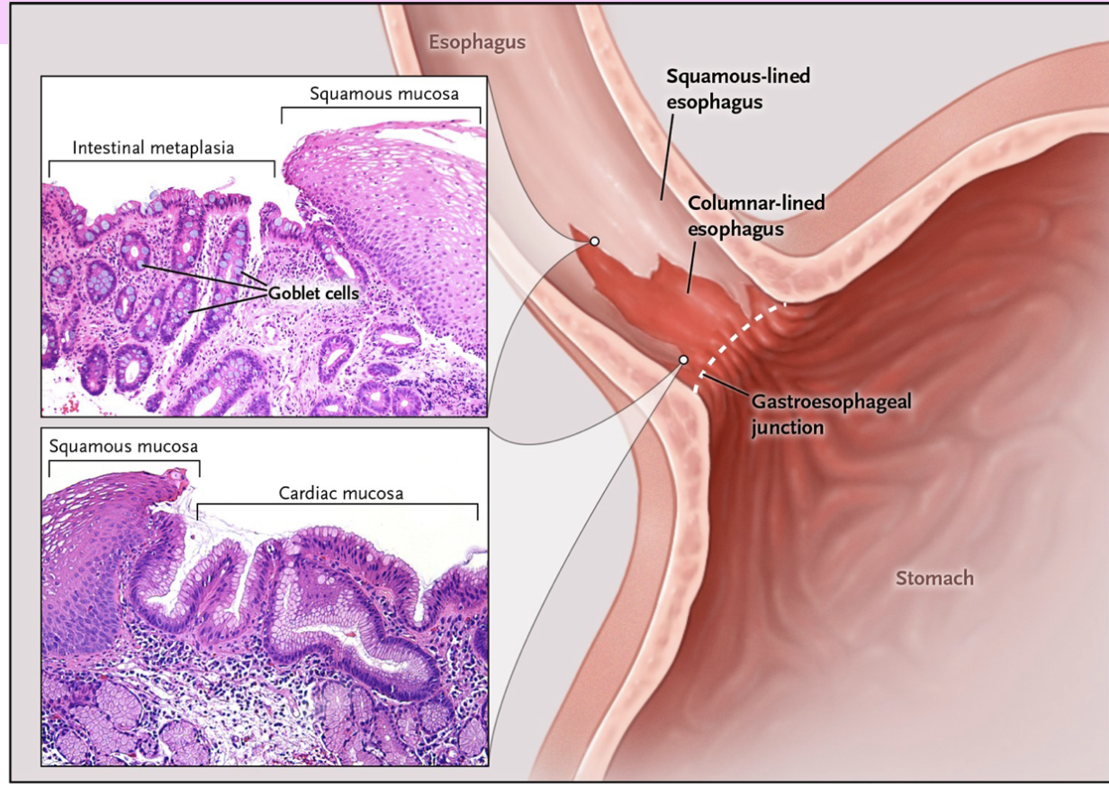
Barrett’s esophagus
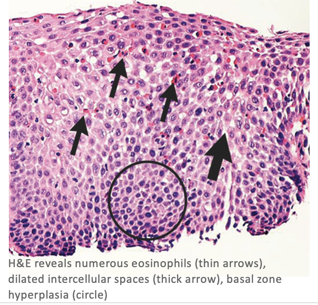
Eosinophilic Esophagitis
Esophagitis
1. Lumen
2. 2.Epithelium-SSE-nonkeratinized
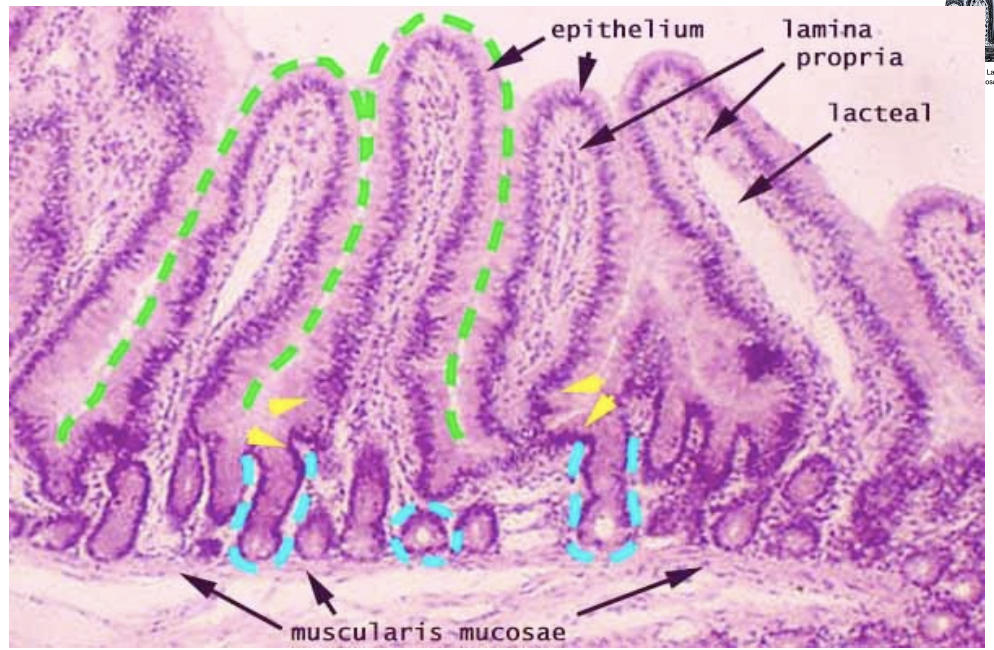
3. Lamina propria
4. Muscularis mucosae
5. Submucosa
6. 6a.Inner circular smooth muscle;
6b.Outer longitudinal smooth muscle
7. Adventitia

Stomach Histology

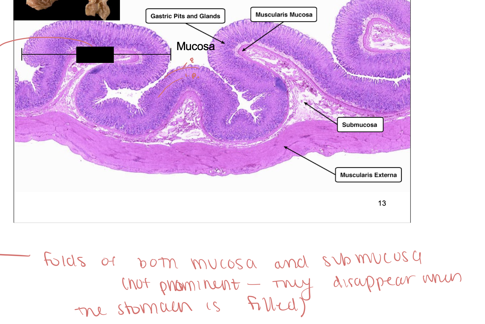
Rugae of stomach
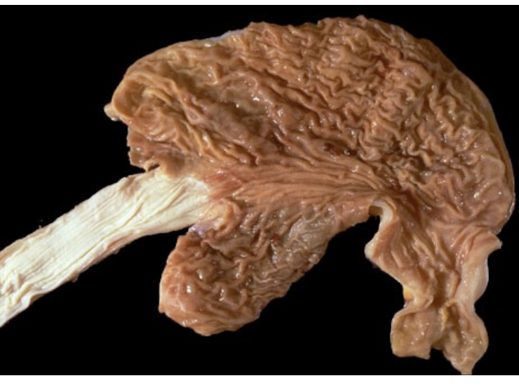
Rugae Histology
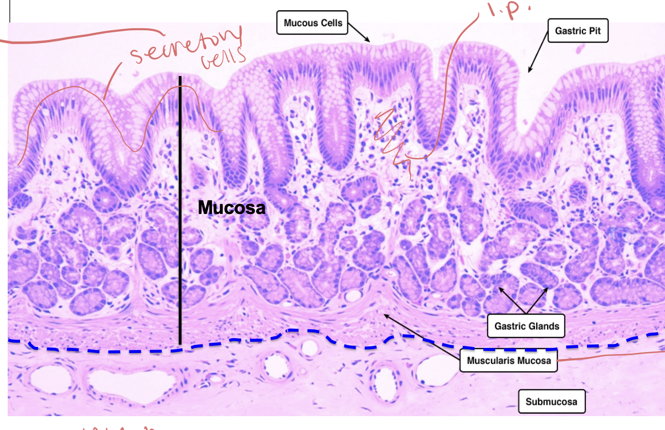
Stomach Secretory
secretory cells form grandular sheet
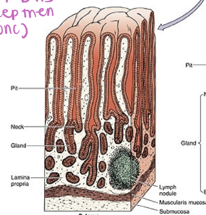
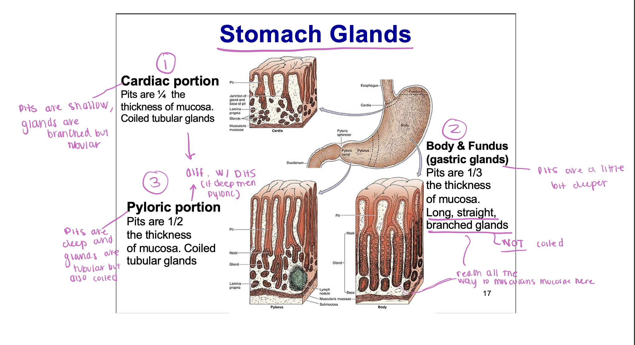
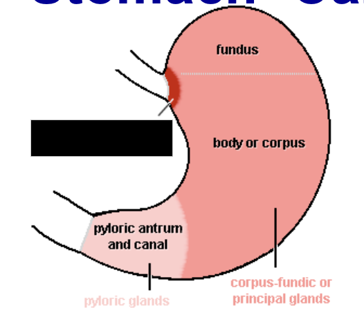
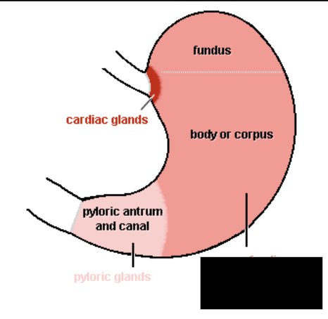
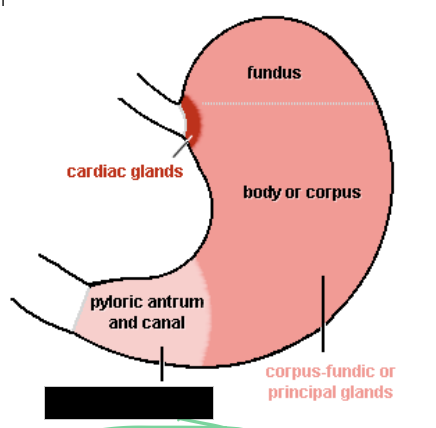
Cardiac portion stomach glands
Pyloric portion stomach glands
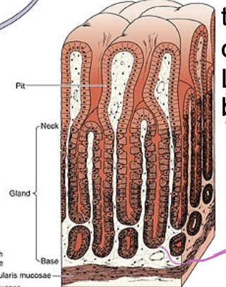
Body and Fundus (gastric glands) portion stomach glands
Stomach Glands
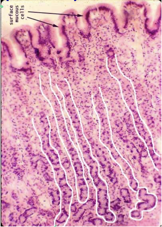
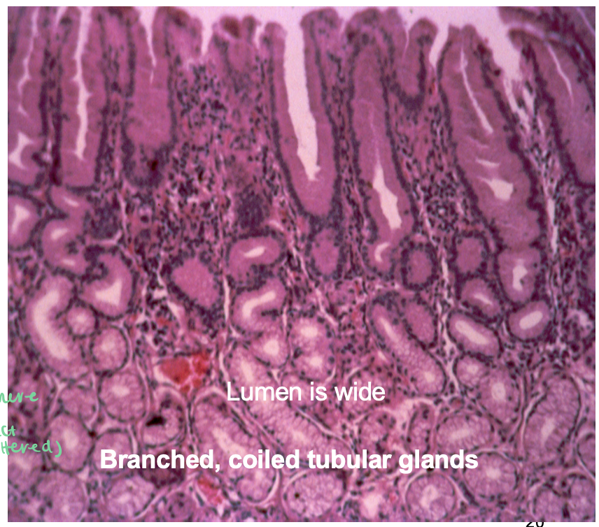
Stomach - Cardiac Glands Histology
Cardiac Gland image
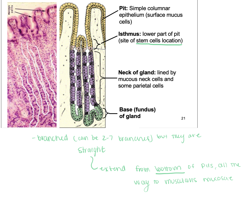
Stomach Body and Fundus (gastric glands)
glands are branched but straight extending from bottom of pit
all the way to muscularis mucosa
Corpus Fundic or Principle Glands
Stomach Pyloric Glands Histology
Pyloric Gland
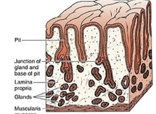
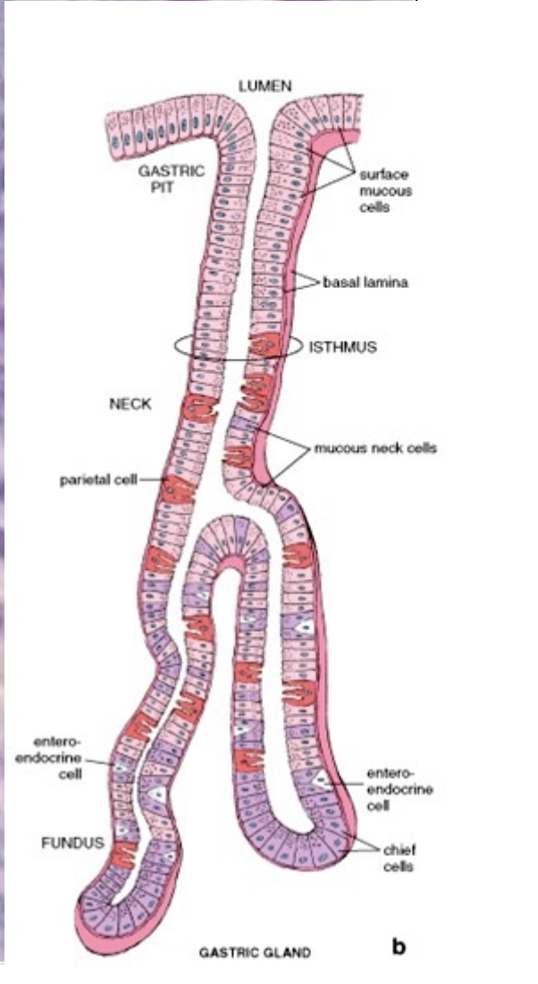
Gastric Gland Overview
Gastric Gland Cell type
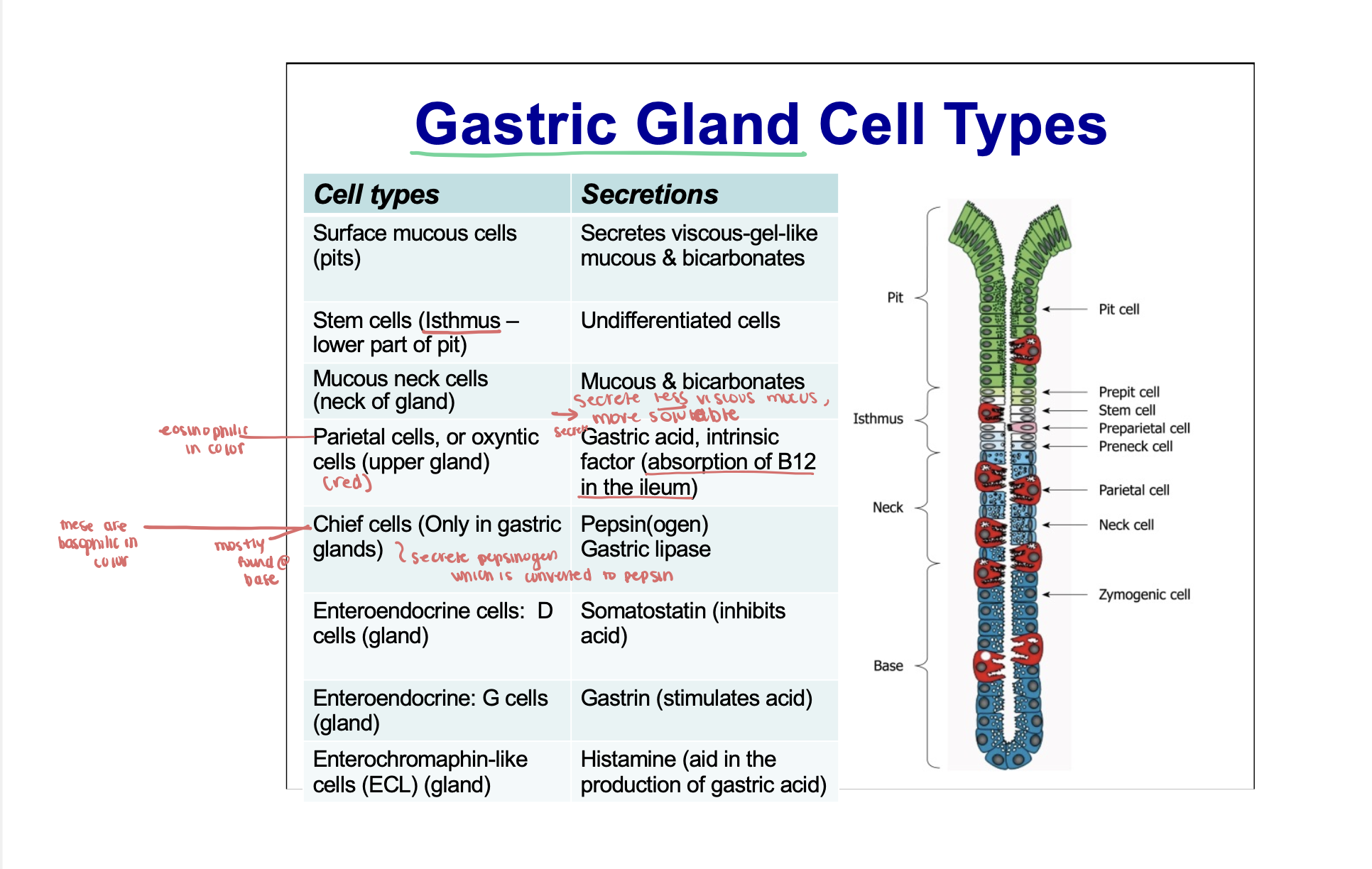
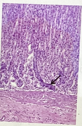
Surface Mucous Cells
Simple columnar epithelium
Mucous Neck Cells
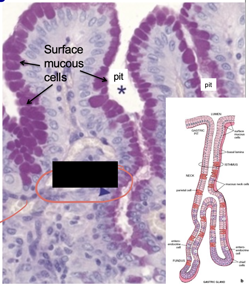
Gastric Gland
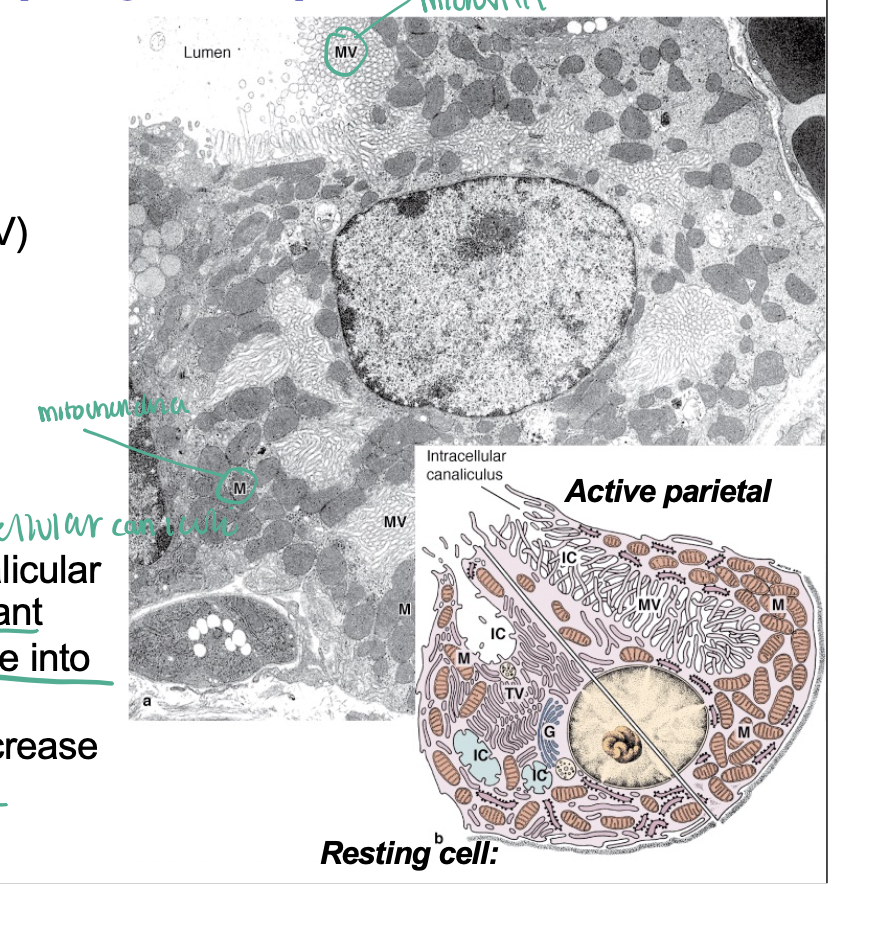
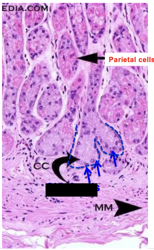
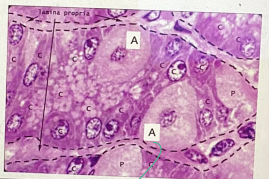
Parietal Cell
Large, eosinophilic (pinkish), fired egg
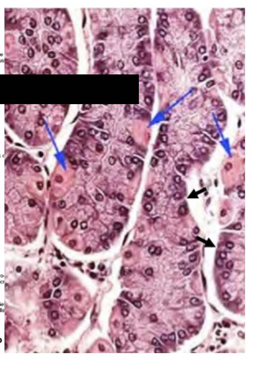
Parietal Cell
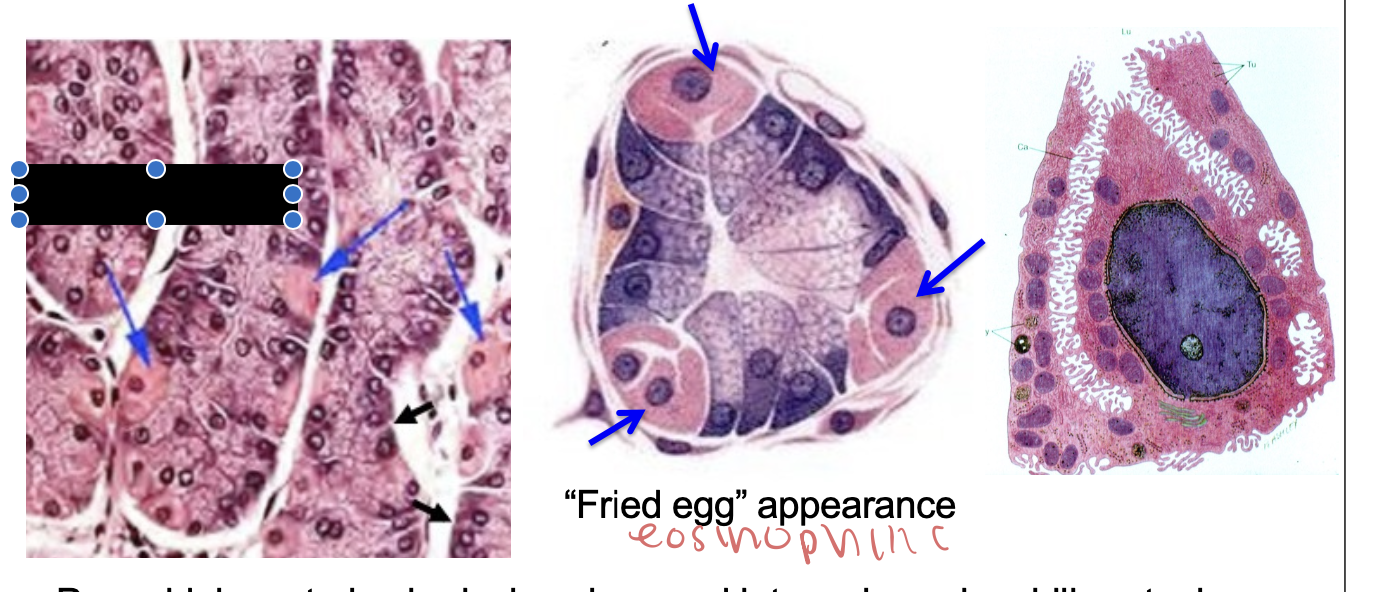
Parietal Cell
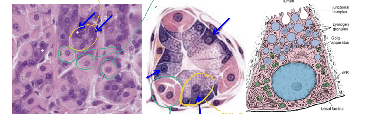
Chief Cells
Appear basophilic (more blue/purple in color)
Yellow = chief cells + last image + blue arrows
Green = Parietal Cell
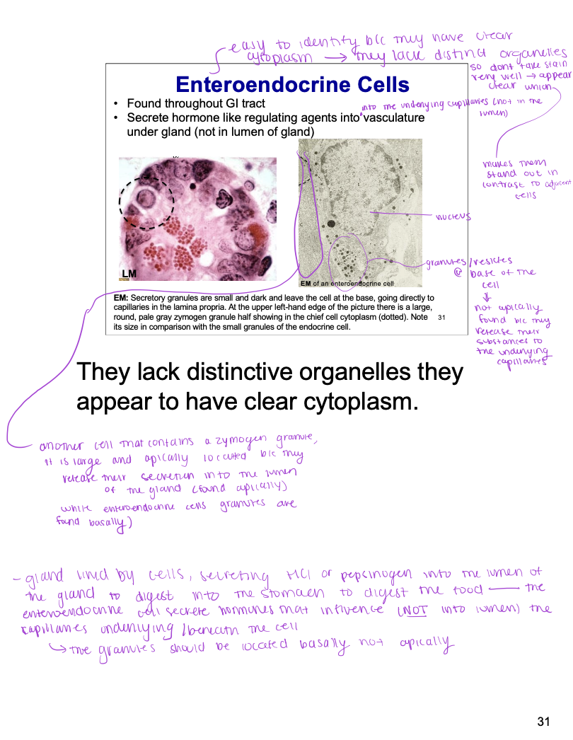
Enteroendocrine cell
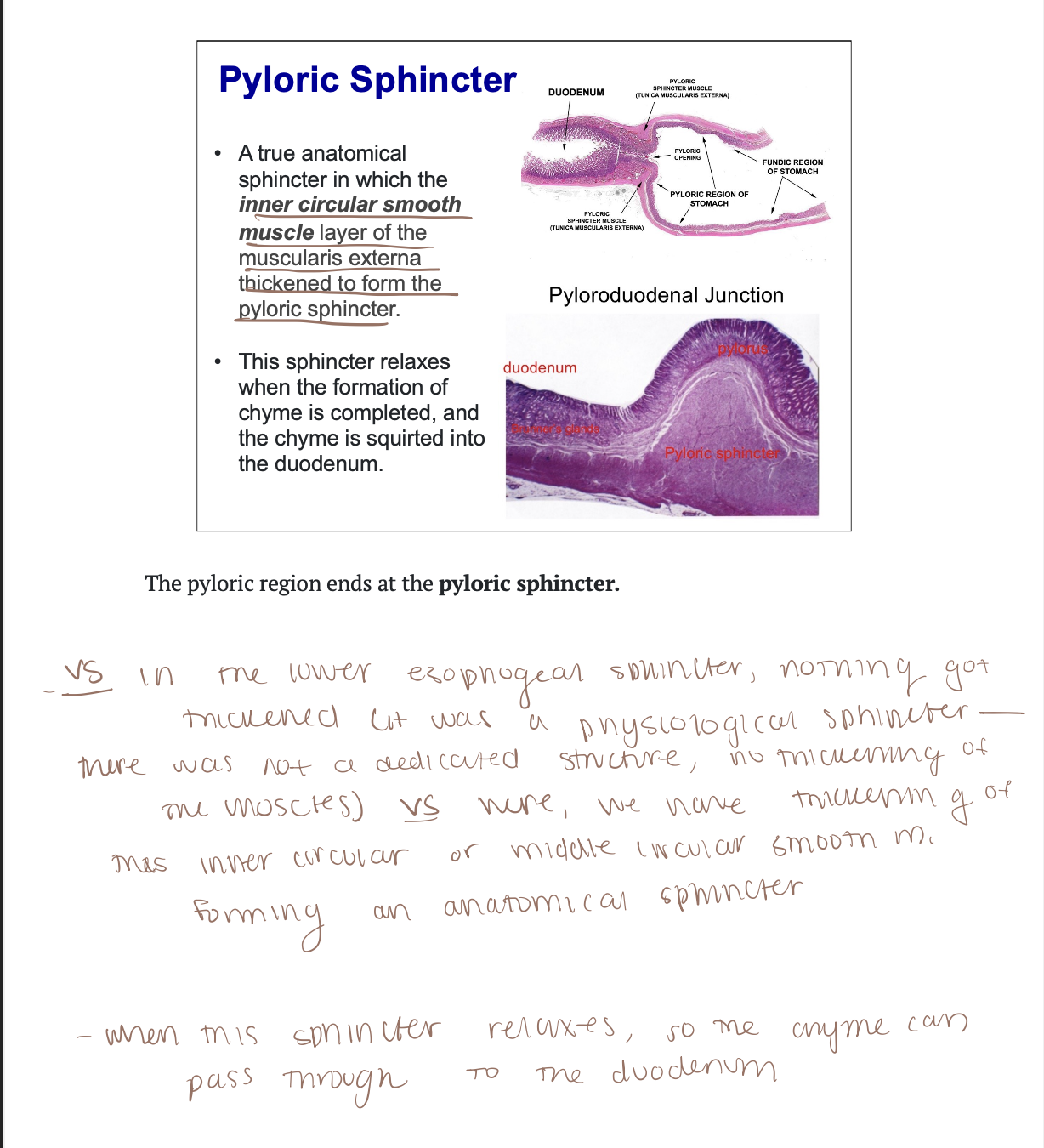
Pyloric Sphincter
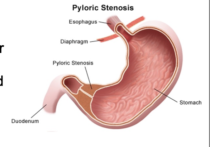
Infantile Hypertrophic Pyloric Stenosis (IHPS)
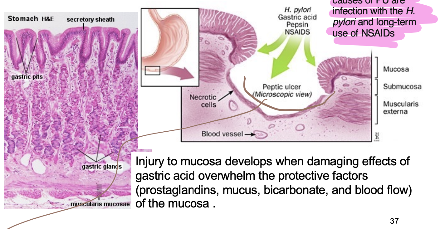
Gastric Ulcer
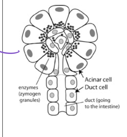
Pancreas
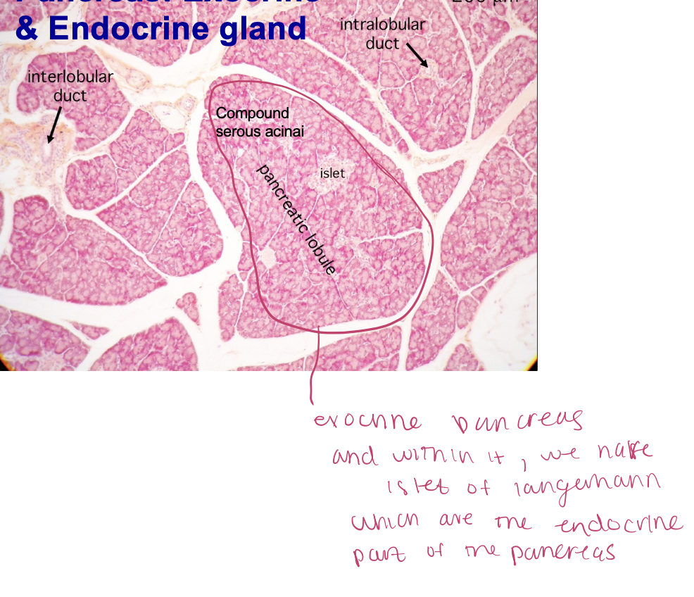
Exocrine pancreas = made up of serous acinar
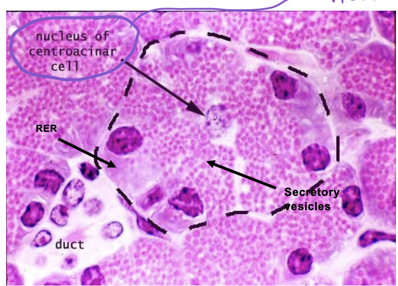
Duct of Pancreas
Pancreas VS Parotid
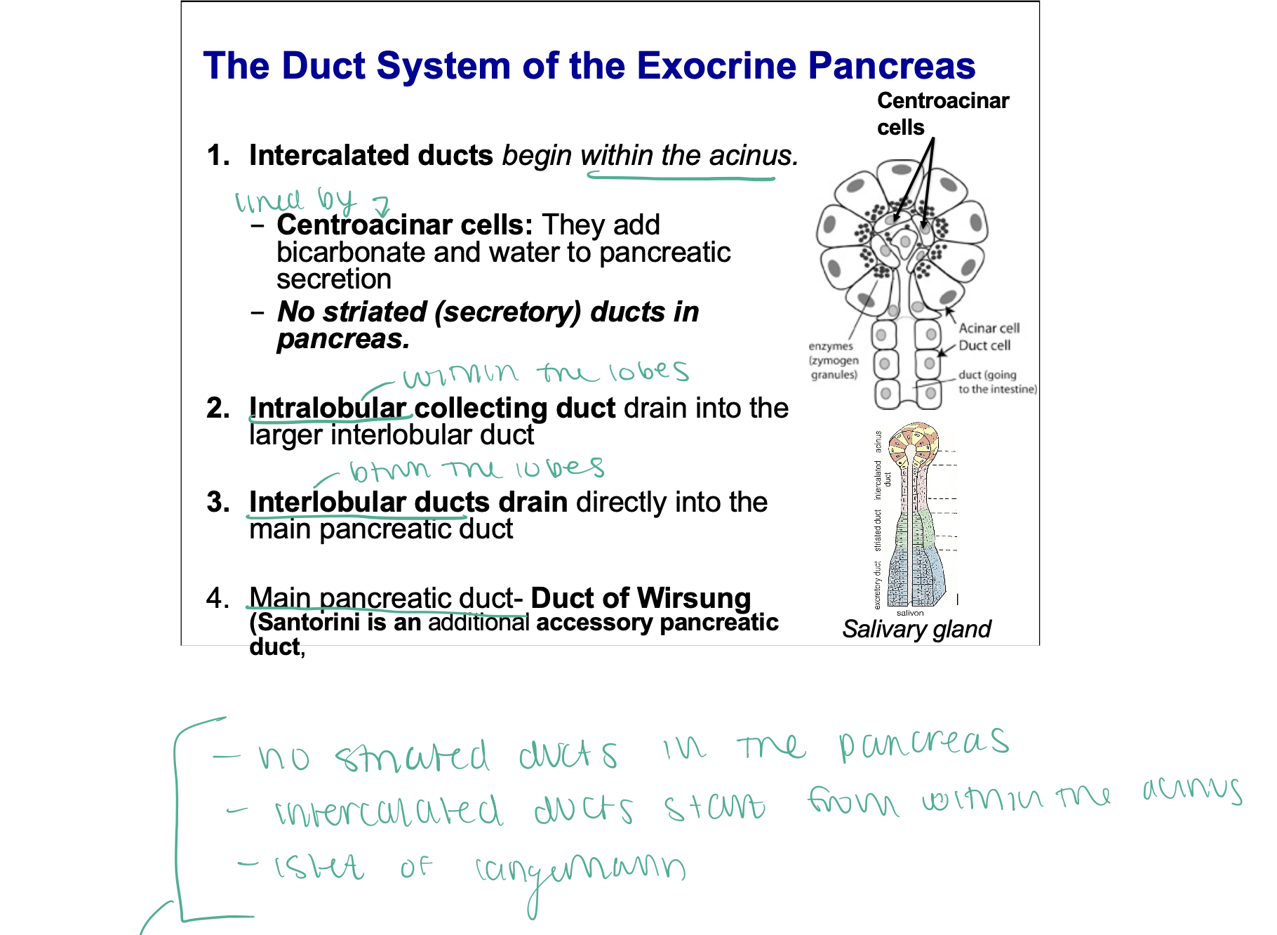
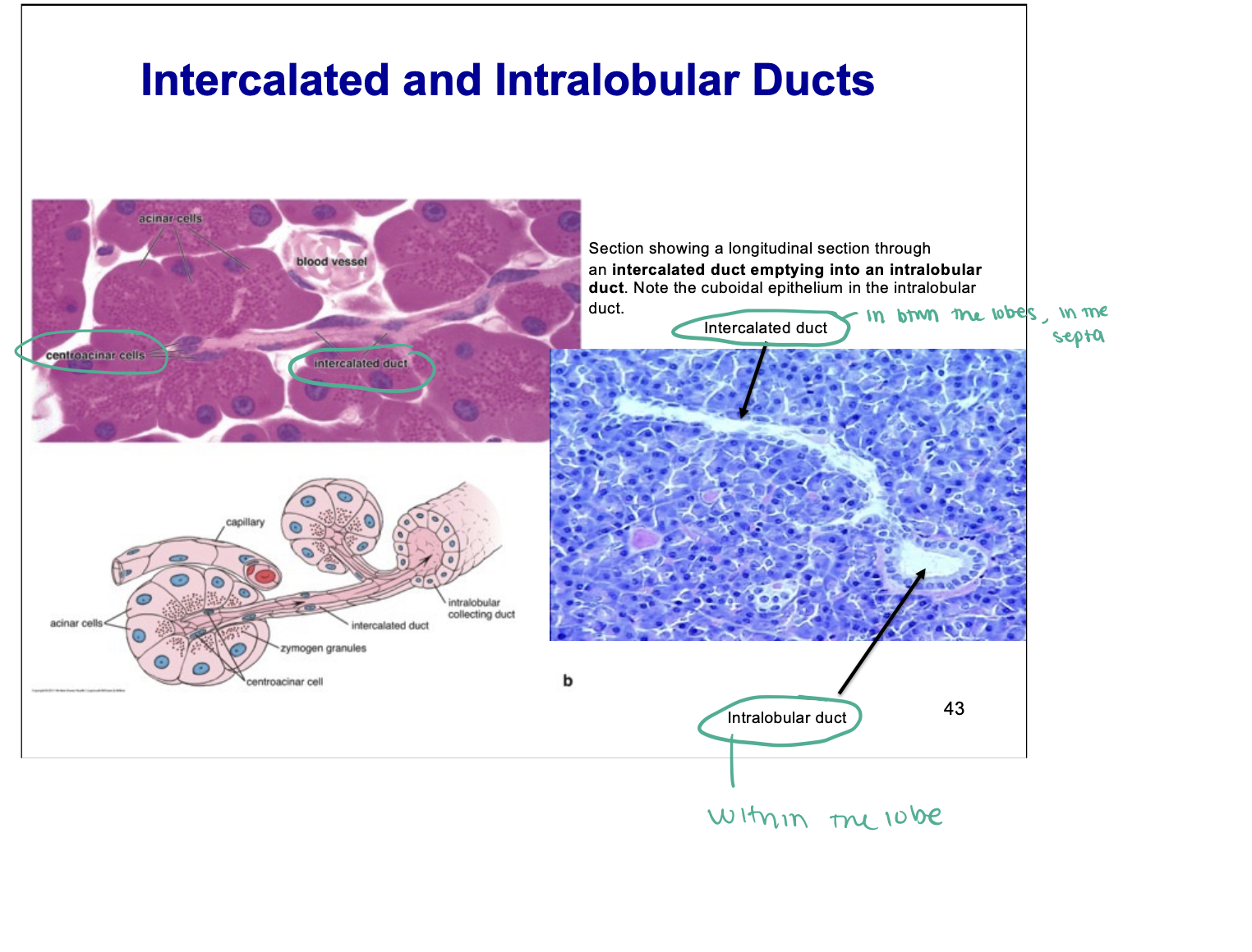
See the intercalated ducts start in the acini (green circled area)
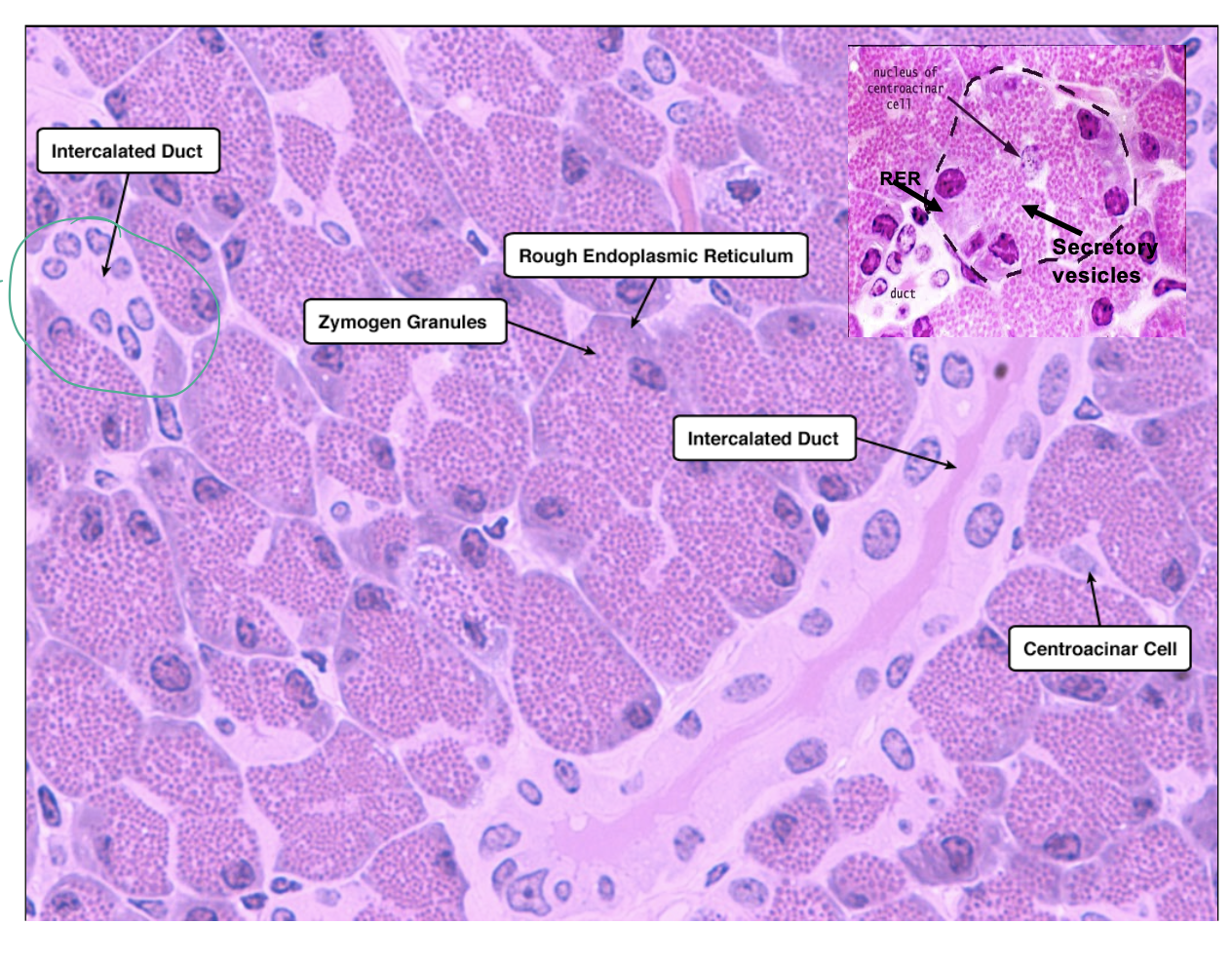
Intercalated and Intralobular Ducts
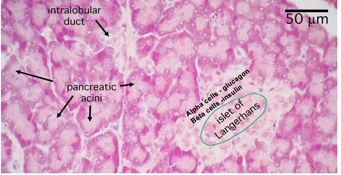
Endocrine pancreas
Esophagus
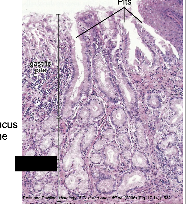
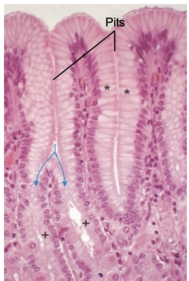
Fundic/Body Region
Surface with pits
Straight glands reaching MM

Parietal Cells
Arrow pointing to Surface Mucous Cells

Chief cells
AT BASE ——- More basophilic (blue/purple)
Parotid Gland
Serous + Striated Ducts
Parietal Cells
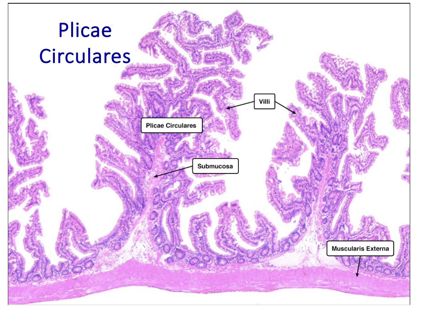
Plicae Circulares
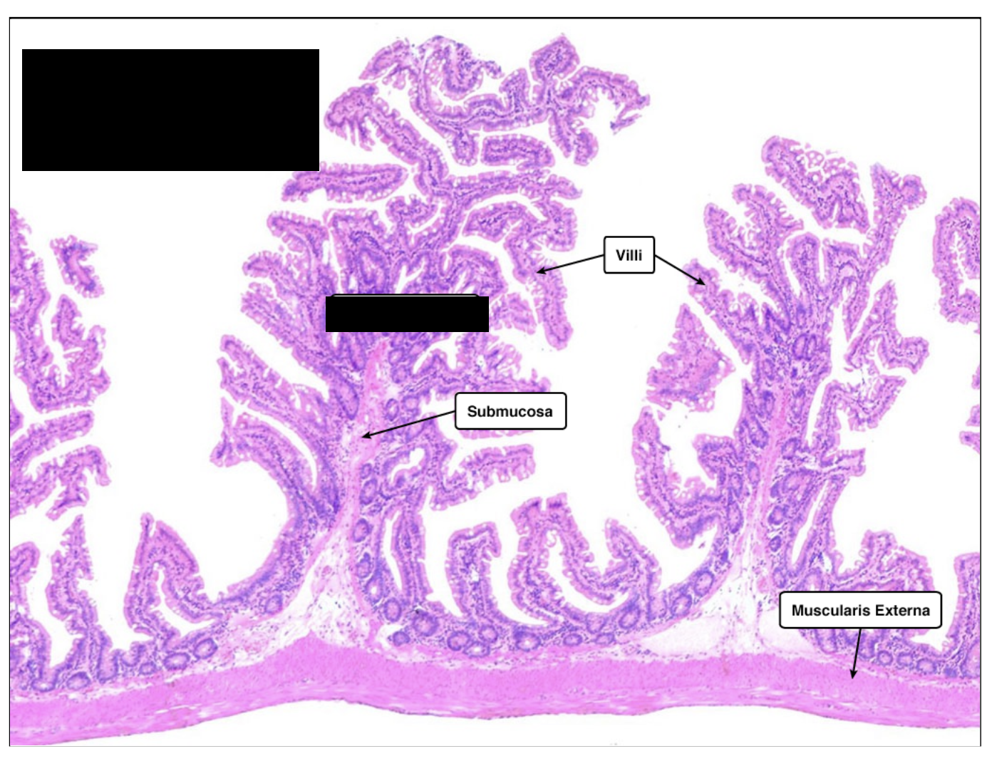
Villi