LCCW Regional Anatomy III (lab) MT1 McBride
1/66
There's no tags or description
Looks like no tags are added yet.
Name | Mastery | Learn | Test | Matching | Spaced |
|---|
No study sessions yet.
67 Terms
(A) Intercostal m.
(B) Innermost Intercostals m.
Name A & B
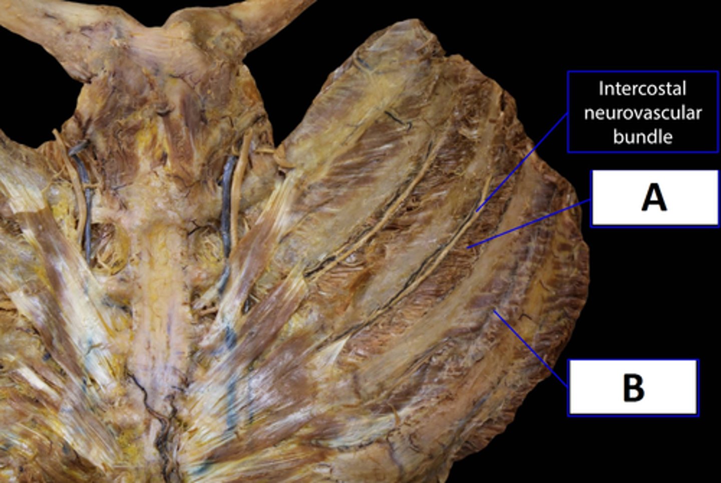
External Intercostal m.
Name Z
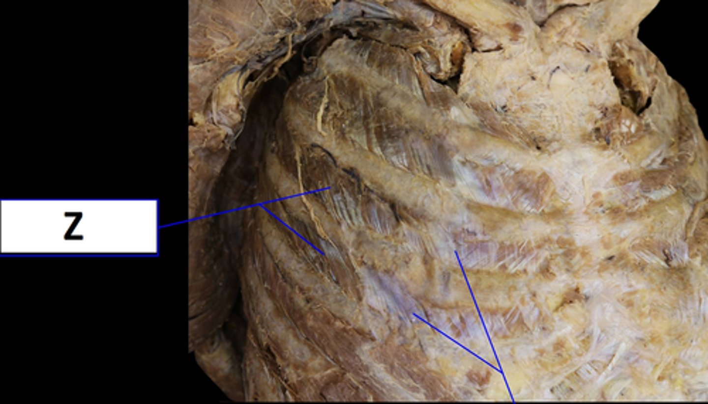
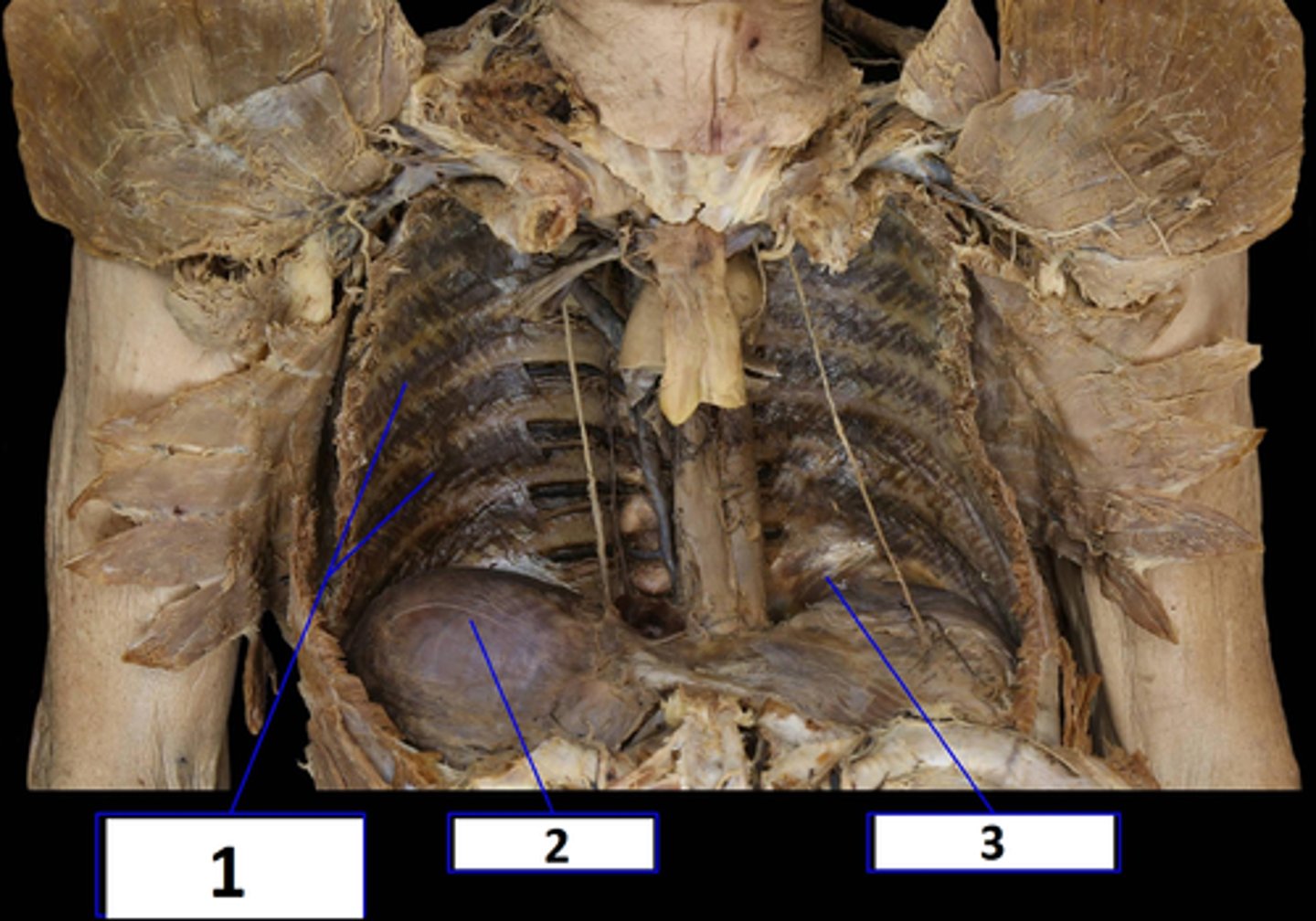
(1) Innermost Intercostal Muscles
(2) Diaphragm
(3) Subcostalis
Name 1, 2, & 3
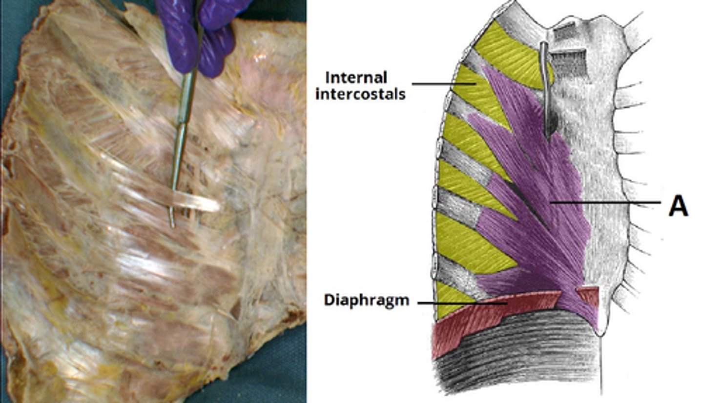
Transversus thoracis (sternocostal)
Name A
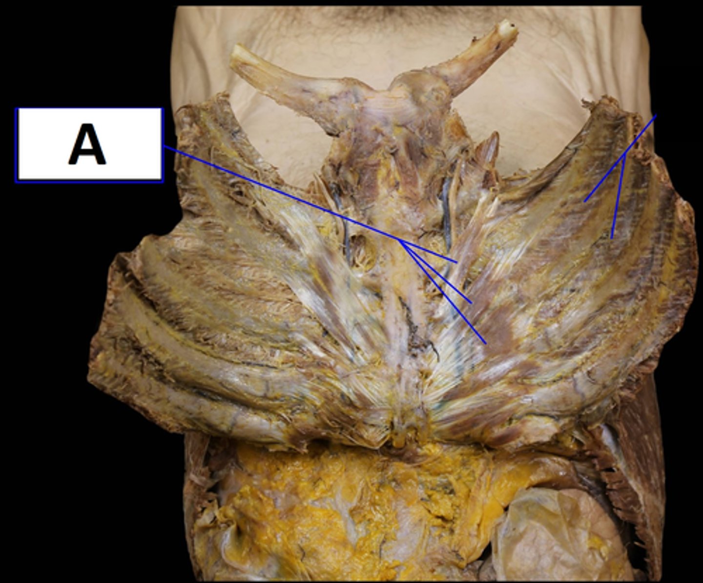
Transversus thoracis (sternocostal)
Name A (view II)
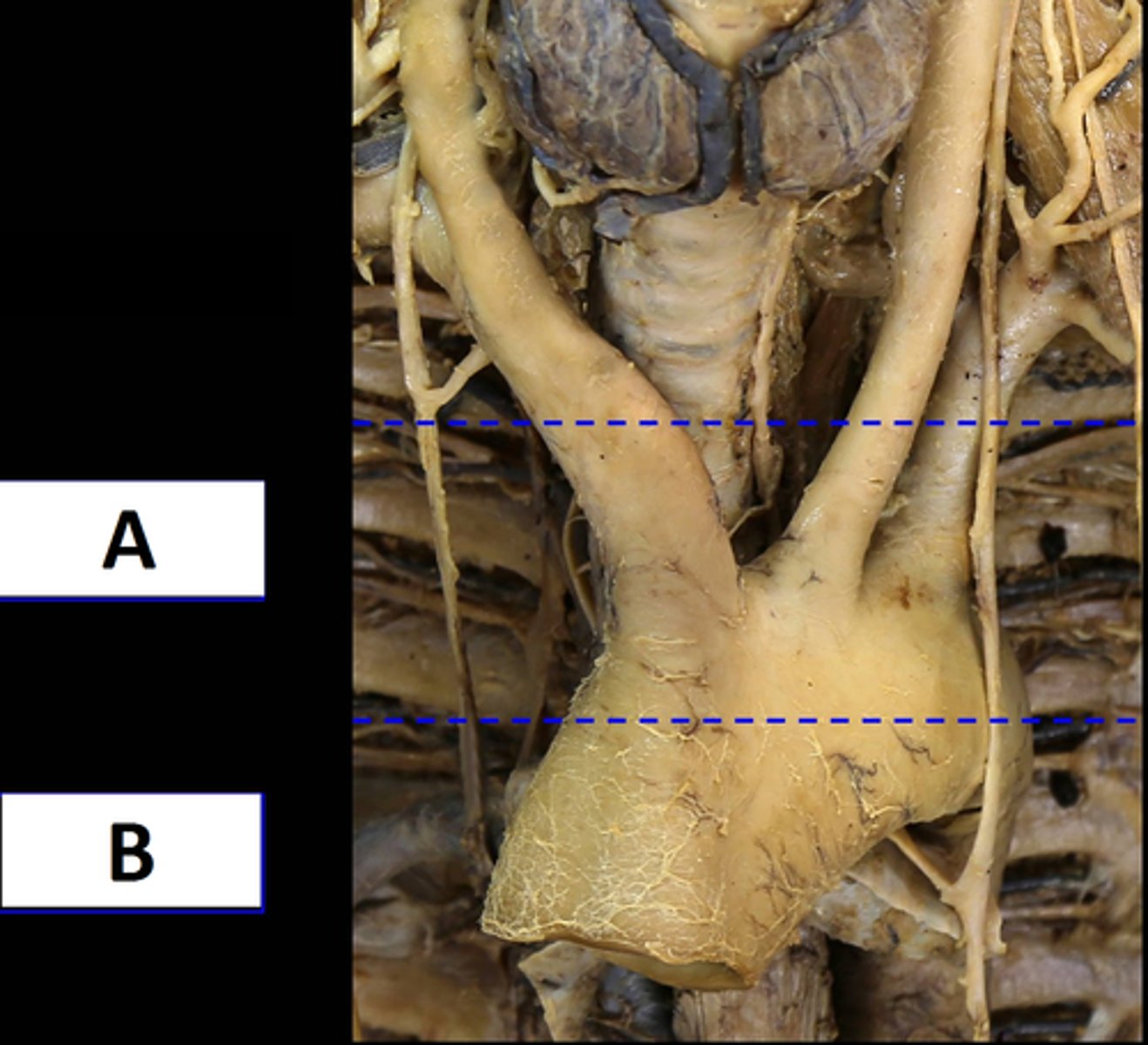
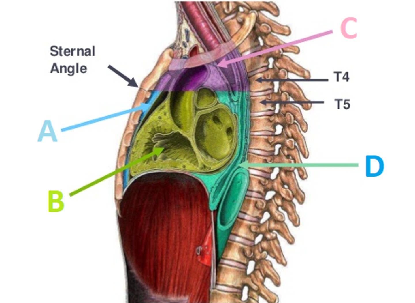
(A) Superior Mediastium
(B) Inferior Mediastium
Mediastium Divisions:
Name Layers A & B
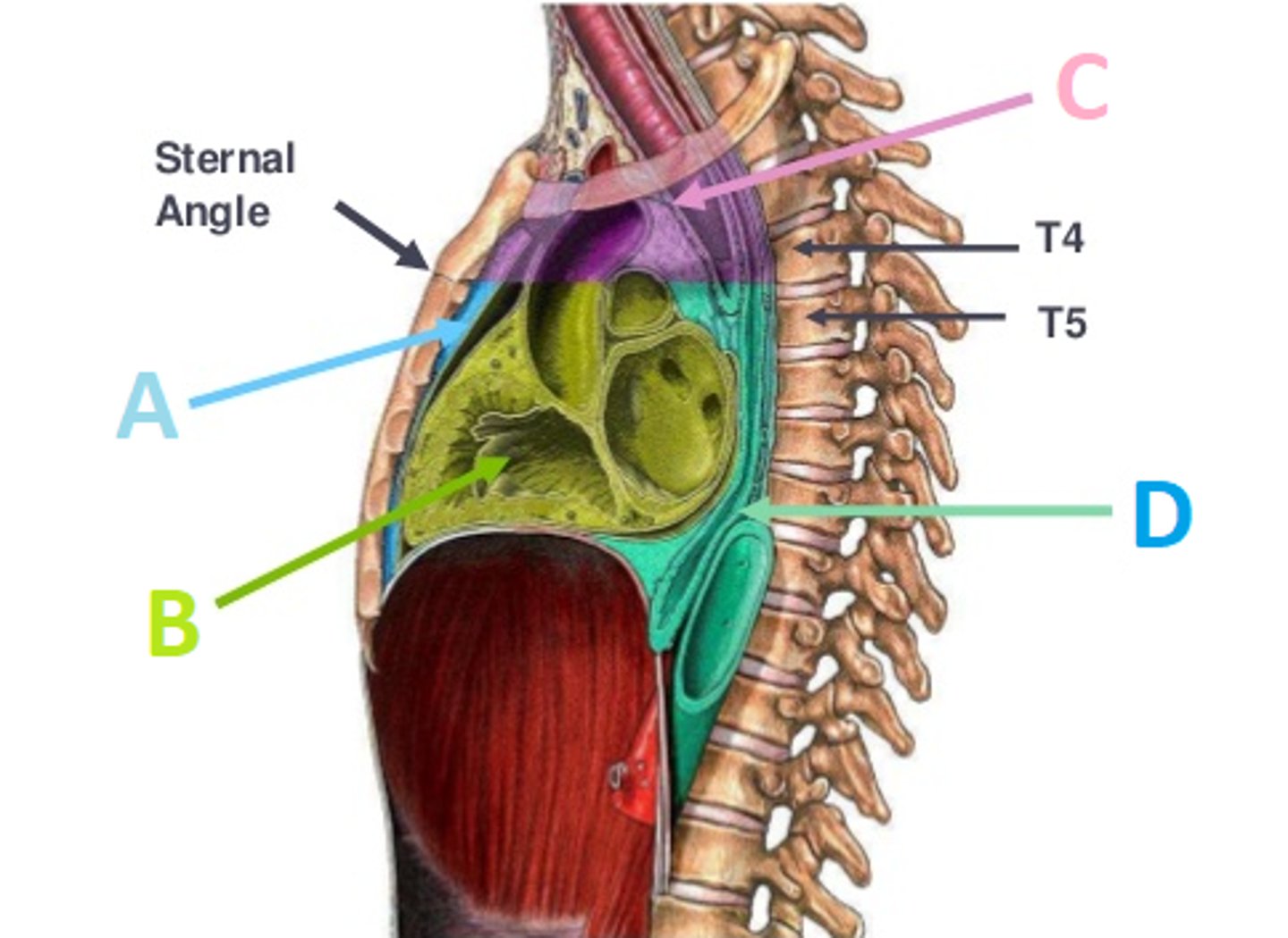
Anterior Mediastium
Mediastium Divisions:
Name A
Middle Mediastium
Mediastium Divisions:
Name B
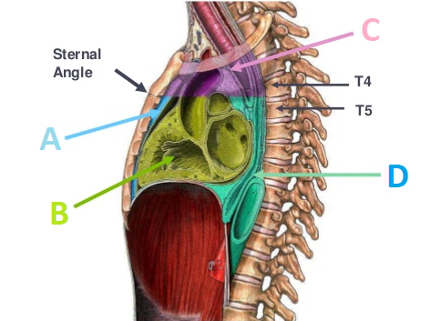
Superior Mediastium
Mediastium Divisions:
Name C
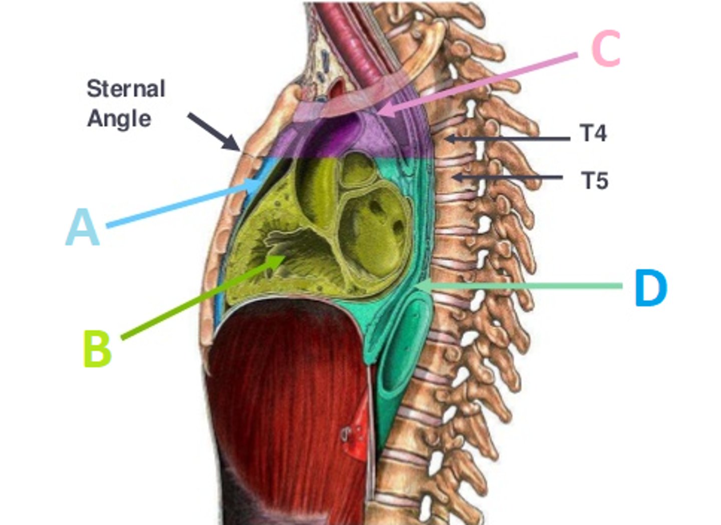
Posterior Mediastium
Mediastium Divisions:
Name D
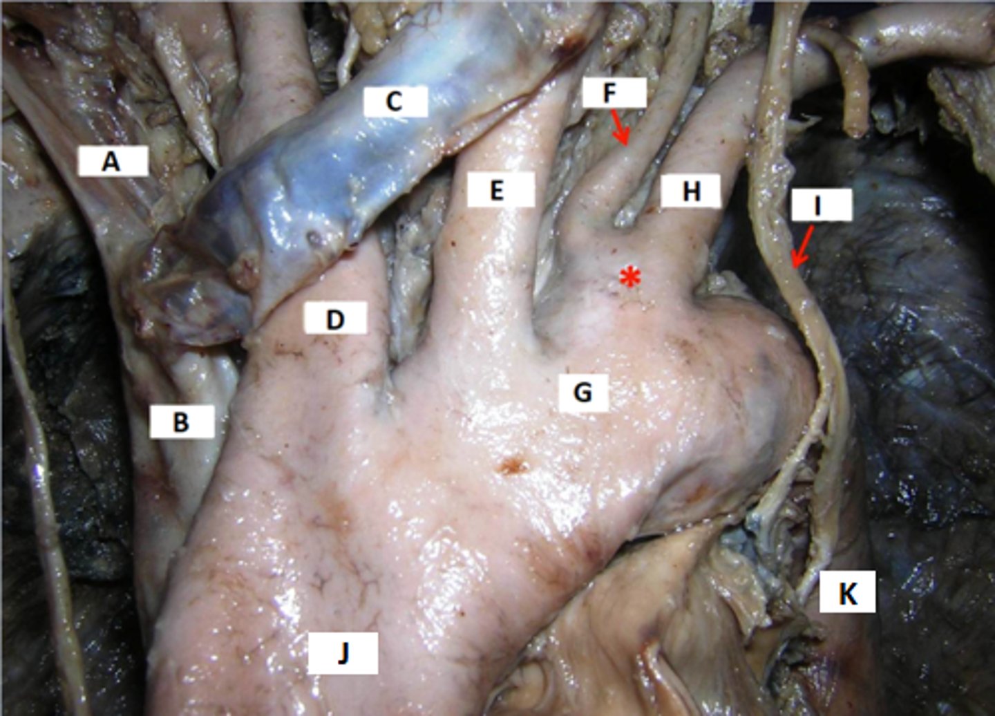
(A) Right Brachiocephalic Vein
(B) Superior Vena Cava
(C) Left Brachicephalic Vein
(D) Brachiocephalic Trunk
(E) Left Common Carotid Artery
(F) Left Vertebral Artery
(G) Aortic Arch (transverse section)
(H) Left Subclavian Artery
(I) Left Vegas Nerve
(J) Ascending Aorta
(K) Descending Thoracic Aorta
Name A - K
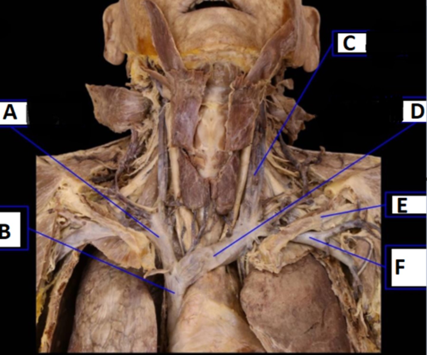
(A) R. Brachiocephalic Vein
(B) Superior Vena Cava (vein)
(C) Internal Jugular
(D) Left Brachiocephalic Vein
(E) subclavius mm. (clavicle removed)
(F) Left. Subclavian Vein
Name A - F
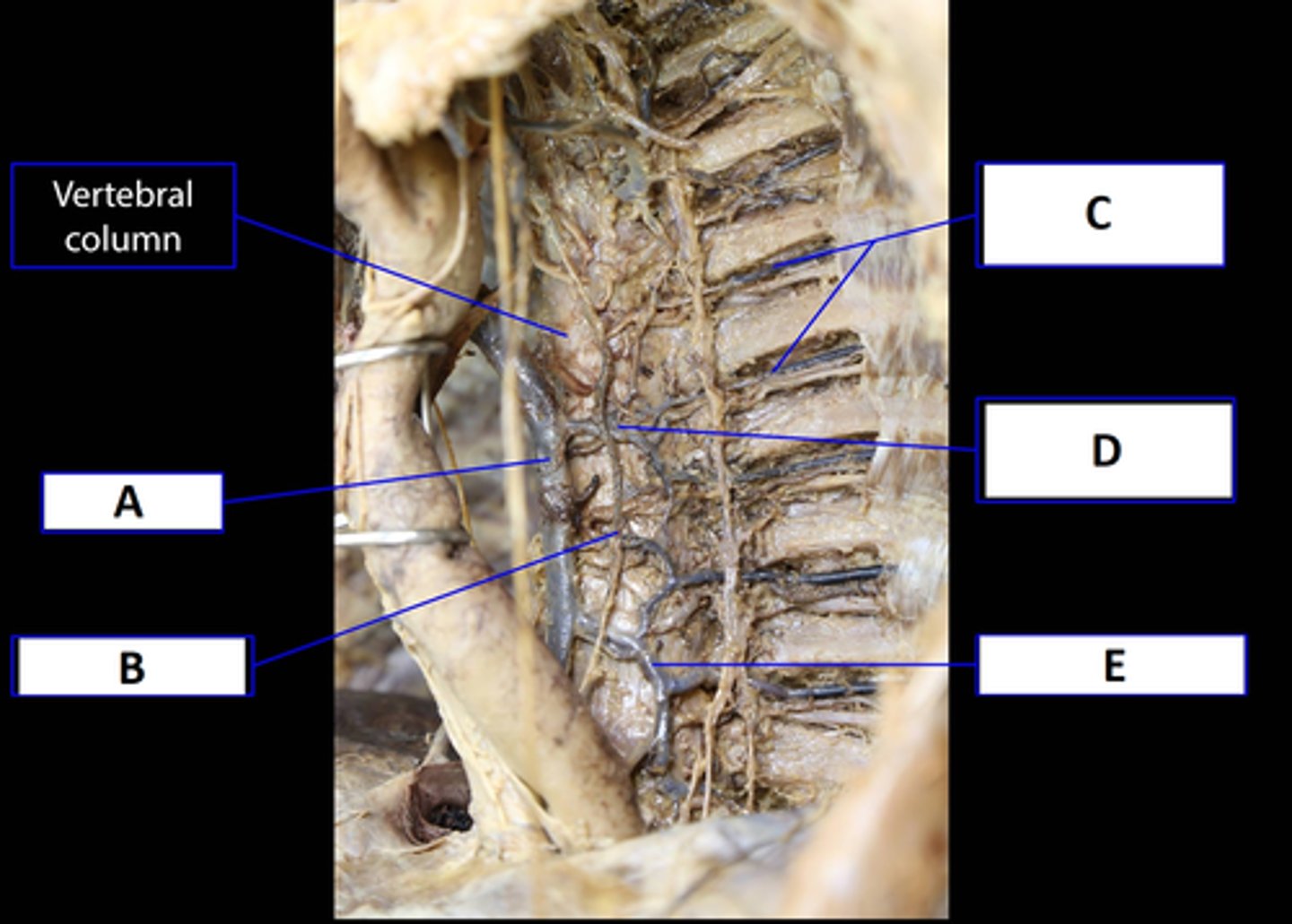
(A) On right side: azygos vein (drains into the Superior Vena Cava)
(B) Thoracic Duct
(C) Posterior Intercostal V.A.N.
(D) Left side Ascending - *Accessory hemi-azygos vein
(E) Left side Descending- Hemi-azygos
The Descending Aorta (thoracic and abdominal portions) supply blood to the Posterior Intercostal V.A.N.S
Name A - E
What gives blood supply to (C)?
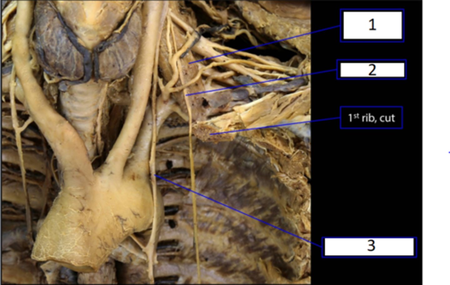
(1) L. Anterior Scalene
(2) L. Phrenic Nerve
(3) L. Vegas Nerve
Name 1, 2, & 3
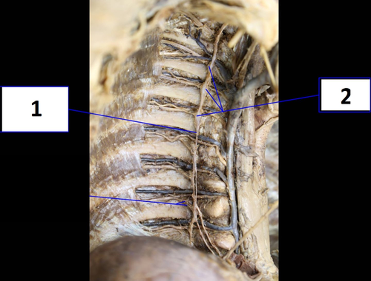
(1) Thoracic Sympathetic Trunk
(2) Thoracic Ganglia
Name 1 & 2
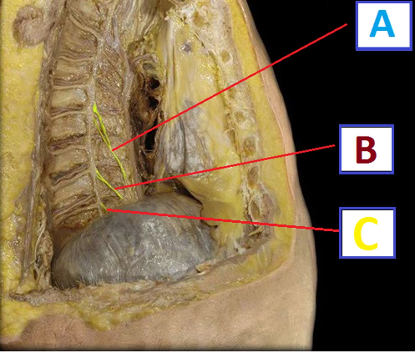
(A) Greater splanchnic App.x T5-T9
(B) Lesser splanchnic App.x T9-T12
(C) Least splanchnic App.x T12-L2
Name A, B & C
Approx. what vertebral levels do they run?
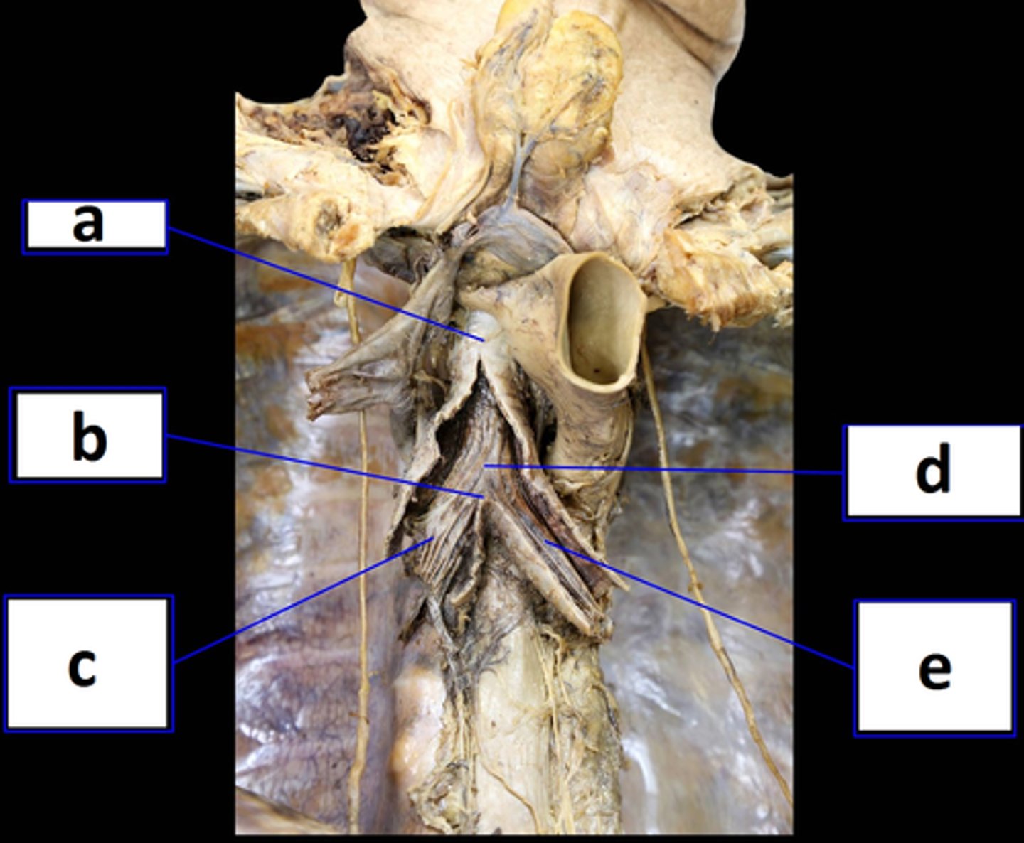
(a) Trachea
(b) Carina of Trachea (@ bifurcation)
(c) Right Primary Bronchi
(d) Tracheal Bifurcation
(e) Left Primary Bronchi
Name a - e
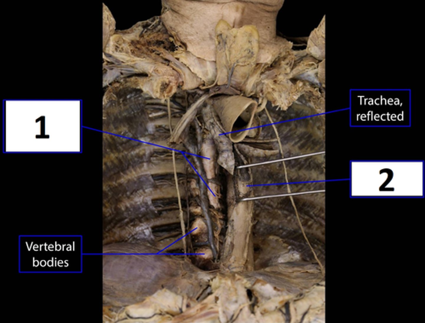
(1) Anterior Longitudinal Ligament (ALL)
(2) Esophagus (pulled to right side, sits anterior to ALL)
Name 1 & 2
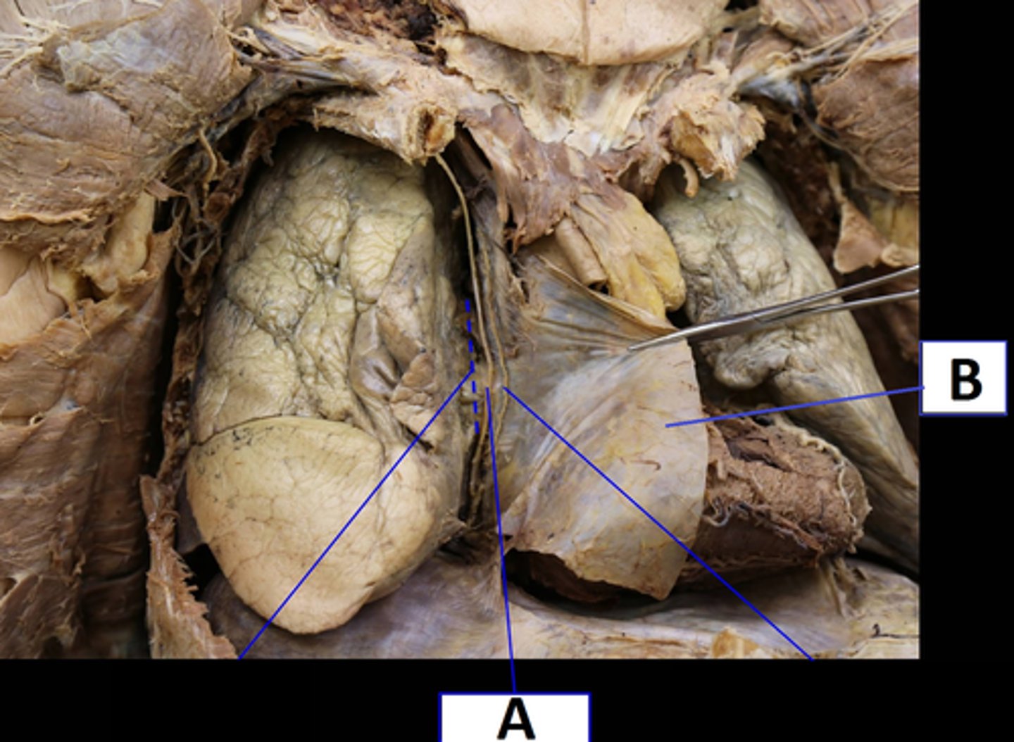
(A) Phrenic Nerve
(B) Parietal Pleura
Name A & B
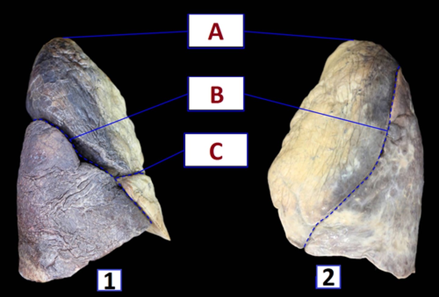
(1) Right Lungs (has 3 lobes)
(2) Left Lung (has 2 lobes)
(A) Apices of Lungs
(B) Oblique Fissures
(C) Horizontal Fissure (right side only)
Name 1 & 2
What differentiates 1 form 2 ?
Name A,B, & C
R - 3 lobes and 2 fissures
L - 2 lobes and 1 fissure
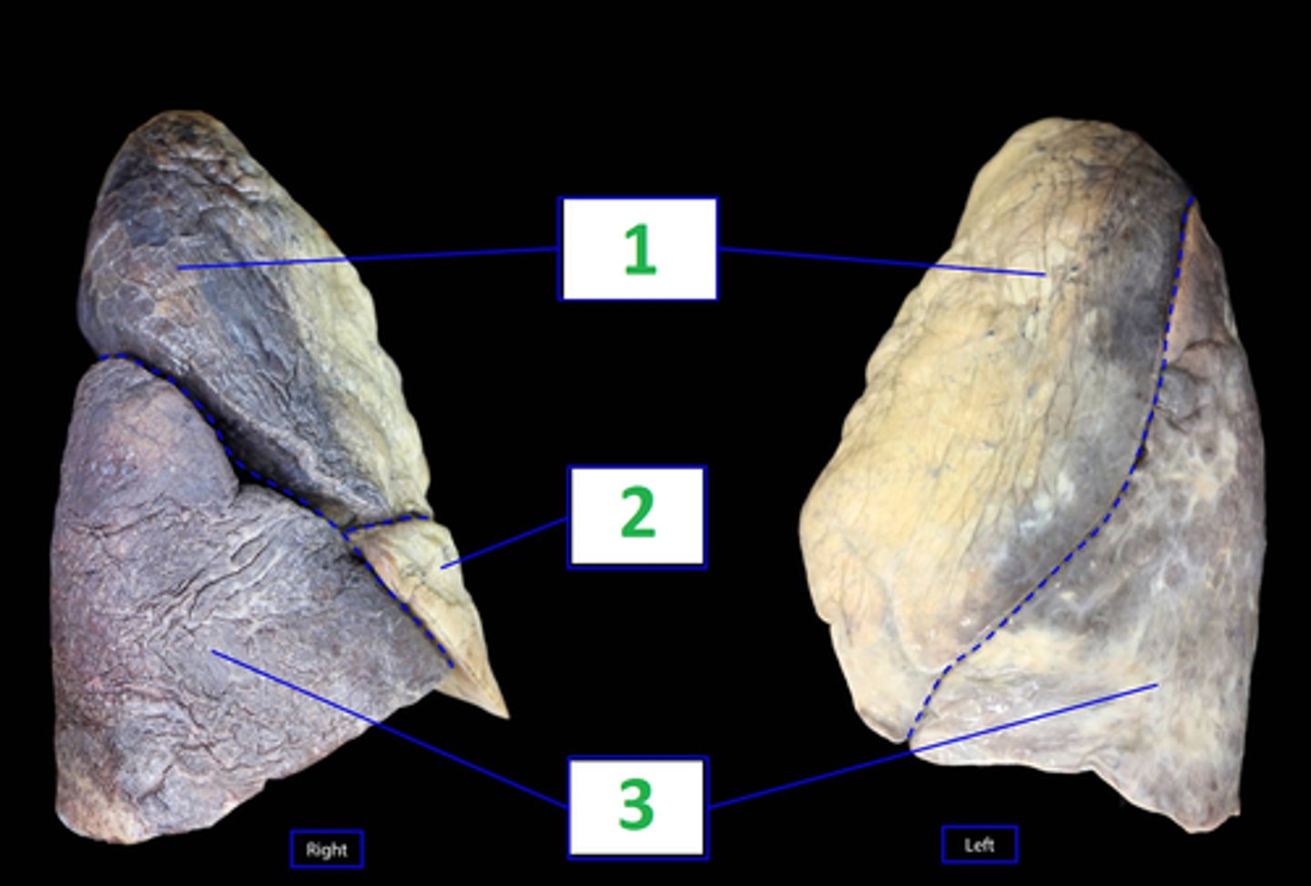
(1) Superior Lobes
(2) Middle Lobe (right lung only)
(3) Inferior Lobes
The Right lung has ____ Lobes and ___ fissures.
The Left Lung has ____ lobes and ___ fissure.
Name 1, 2, & 3
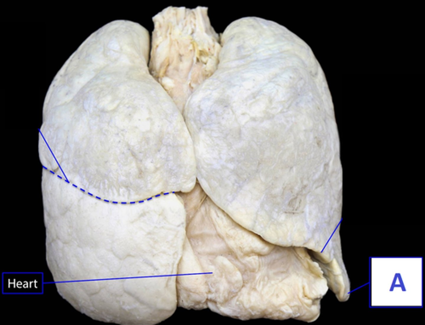
(A) Lingula
Note: Think LLL*
- Left, Lung, Lingula
Because the Left Lung does not have a middle lobe, what unique structure does it have instead (A)?
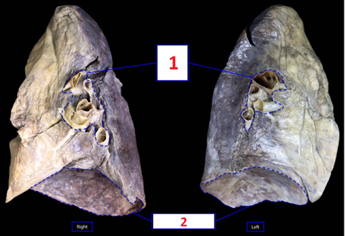
(1) Hila of Lungs
(2) Bases of Lungs (aka Diaphragmatic Surface)
The Hila/Hilum contains the *Roots of the Lungs, which are the:
- Primary Bronchial Tubes
- Pulmonary Arteries and Veins
Name 1 & 2
What structure contains the Root of the lungs? (1)
What structures are contained in this structure (1)?
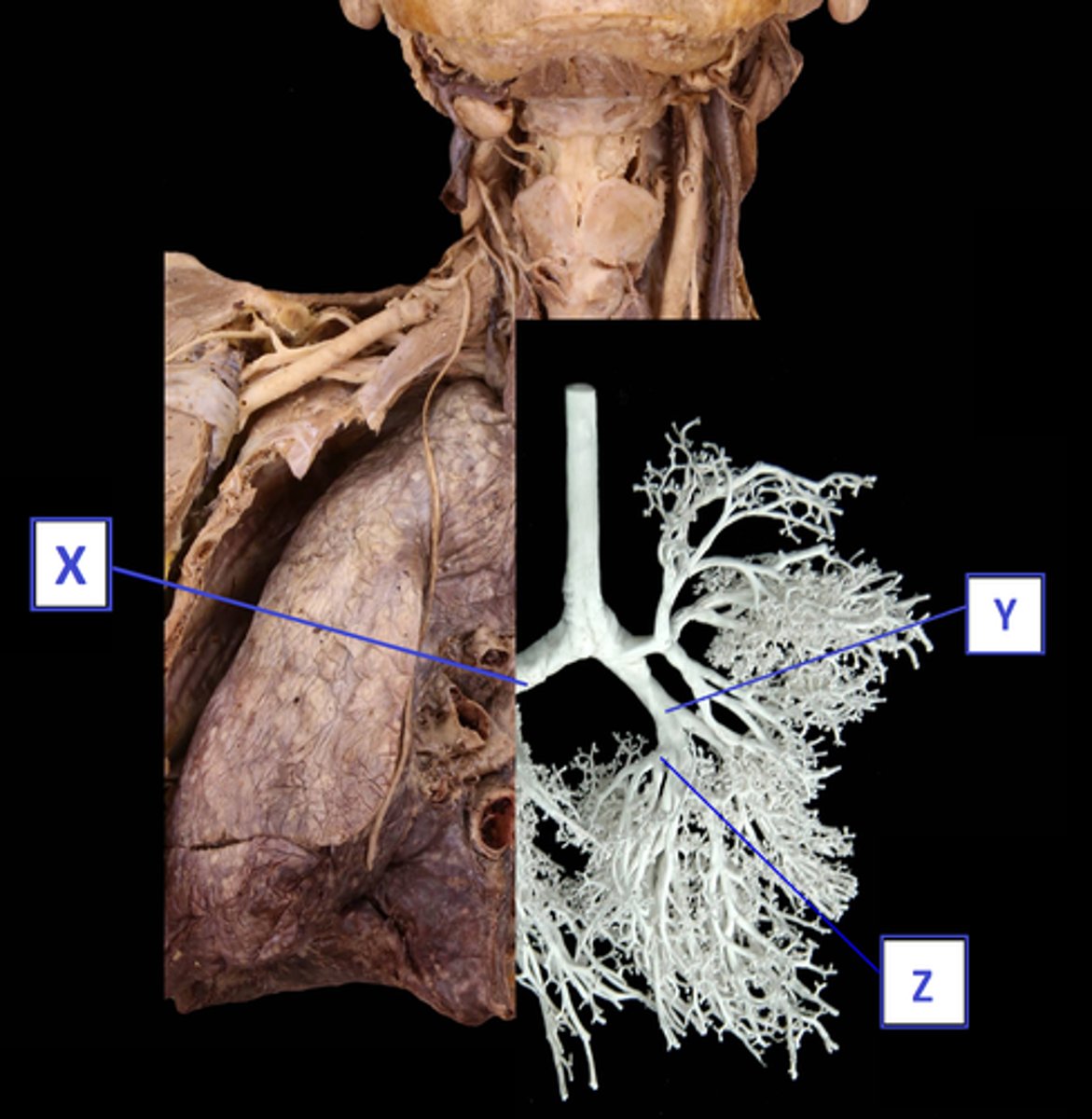
(X) Primary "principle" Bronchi
(Y) Seccondary "lobar" Bronchi
(Z) Teriary "segmental" Bronchi
Name X, Y & Z
(A) Myocardium
(B) Pericardium
(C) Endocardium
Layers of Heart:
Name A, B, & C
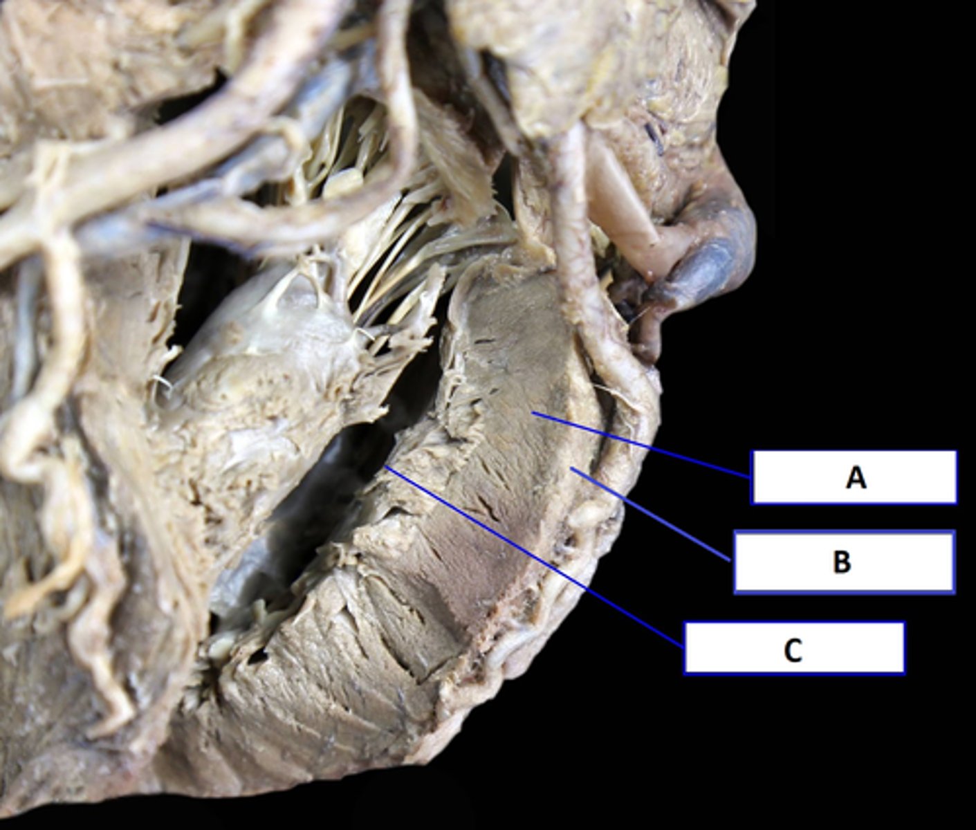
(A) Superior Vena Cava
(B) Ascending Aorta
(C) Serous Pericardium (Parietal Layer) Internal
(D) Pericardial Sac/ Cavity
(E) Serous Pericardium (Visceral Layer) External
(F) Pulmonary Trunk
Name A-F
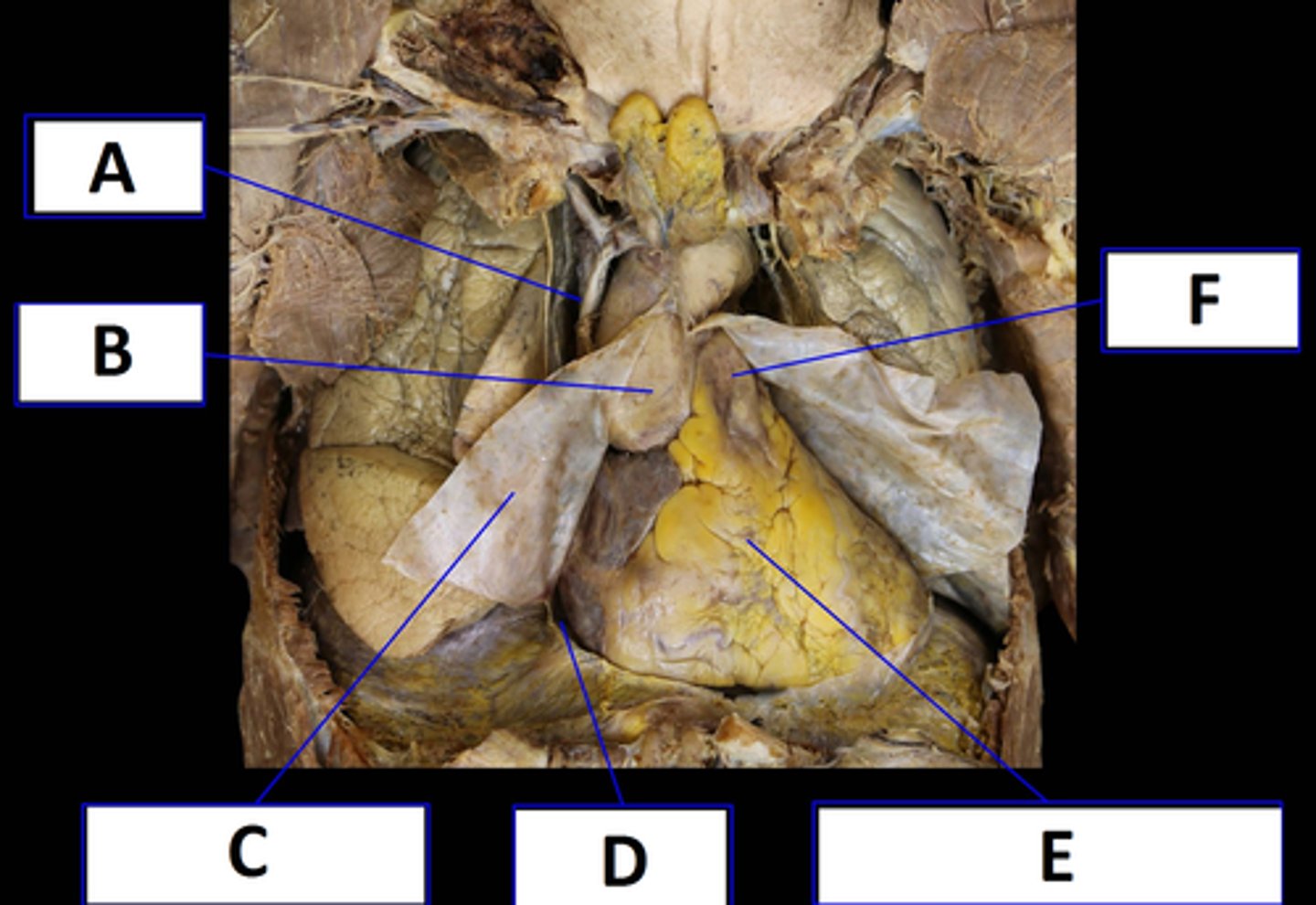
(1) Ascending Aorta
(2) Right Auricle
(3) Right Atrium
(4) Coronary Sulcus
(5) Right Ventricle
(6) Pulmonary Trunk
(7) Anterior Interventricular Sulcus
(8) Left Ventricle
Name 1 - 6
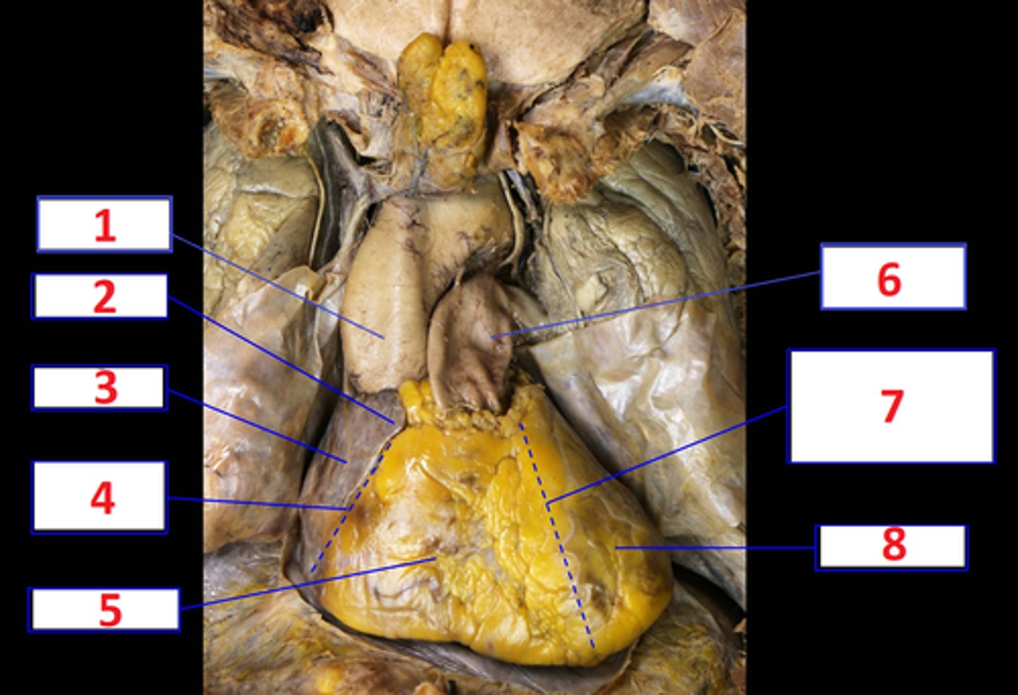
Ligamentum arteriosum
(remnant of the ductus arteriosus in fetal life)
What is the name of the structure outlined in Red?
What is significant about this structure?
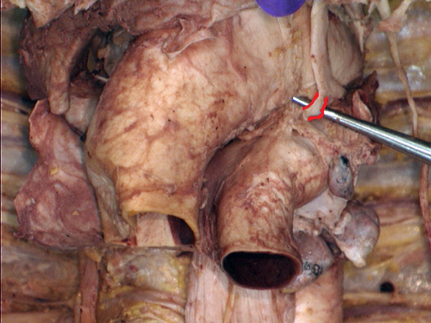
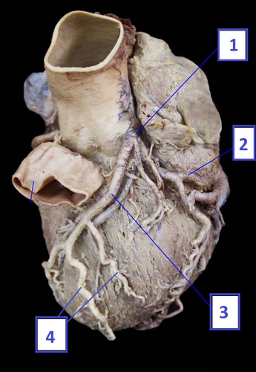
(1) Left Coronary Artery
(2) Circumflex Coronary
(3) Anterior Interventricular Coronary
(AKA: left anterior descending Coronary
(4) Diagonal Artery
Arteries of the Heart: Left Main Coronary
Name 1, 2, 3 & 4
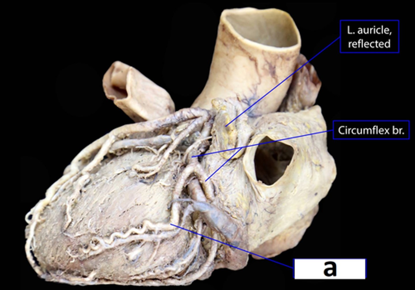
Left Marginal Coronary
Arteries of the Heart: Left Main Coronary
Name a
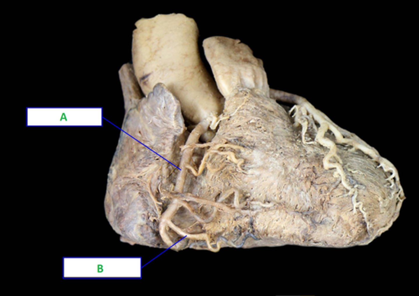
(A) Right Coronary Artery
(B) Right Marginal Coronary Artery
Arteries of the Heart: Right Main Coronary
Name A & B
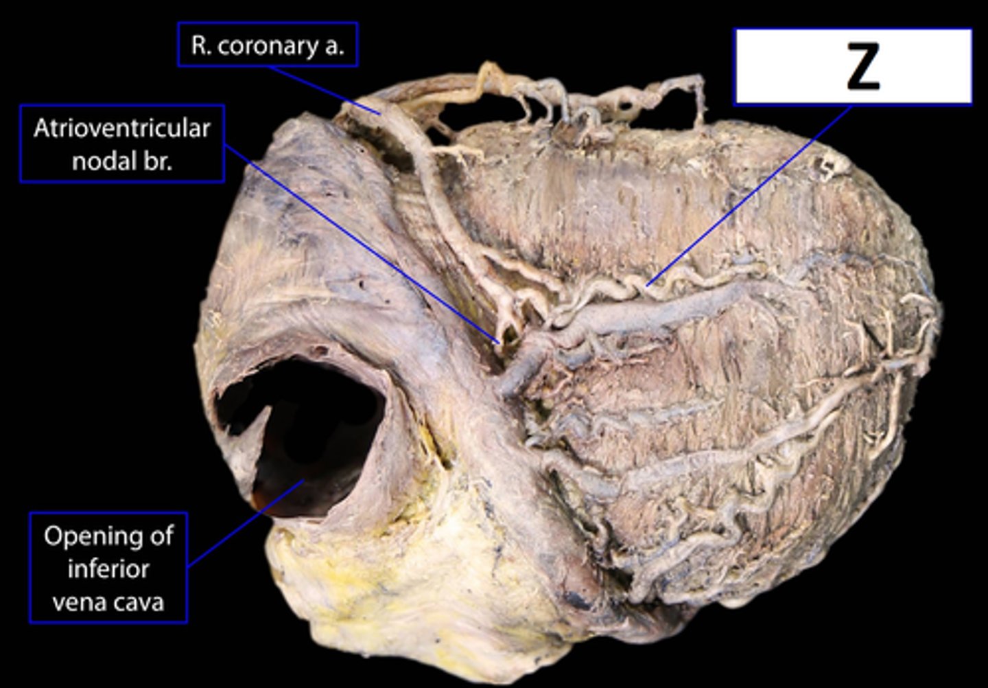
Posterior interventricular Coronary Artery
(Right posterior descending Coronary)
Arteries of the Heart: Right Main Coronary
Name Z
Interatrial Septum
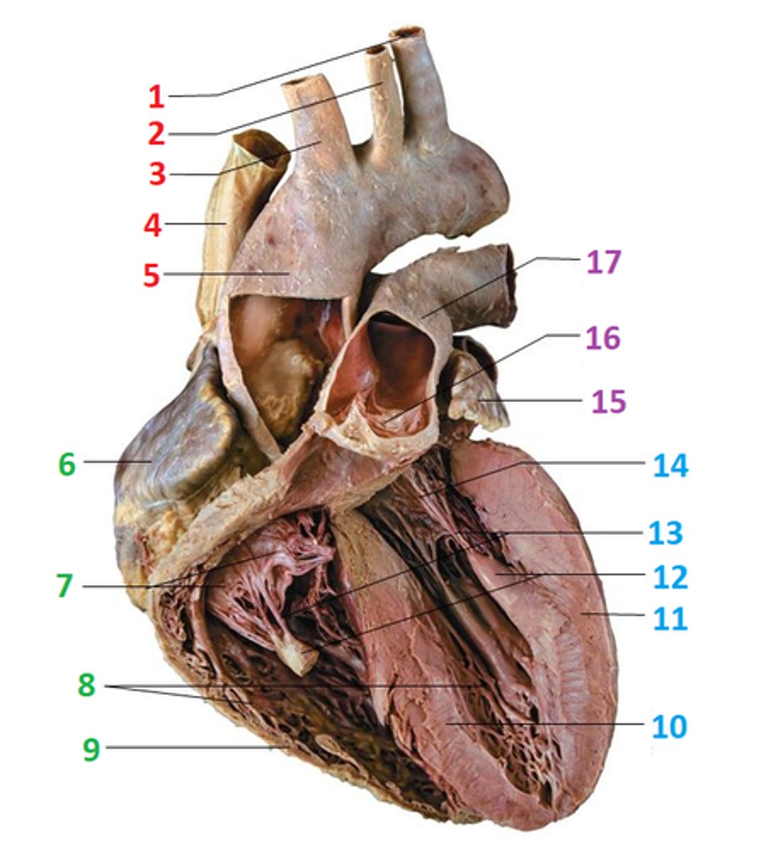
Name the structure outlined in Red
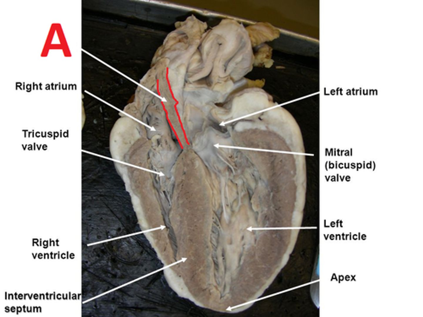
(1) Left Subclavian
(2) Left Common Carotid
(3) Brachiocephalic Trunk
(4) Superior Vena Cava
(5) Ascending Aorta
Name 1 - 5
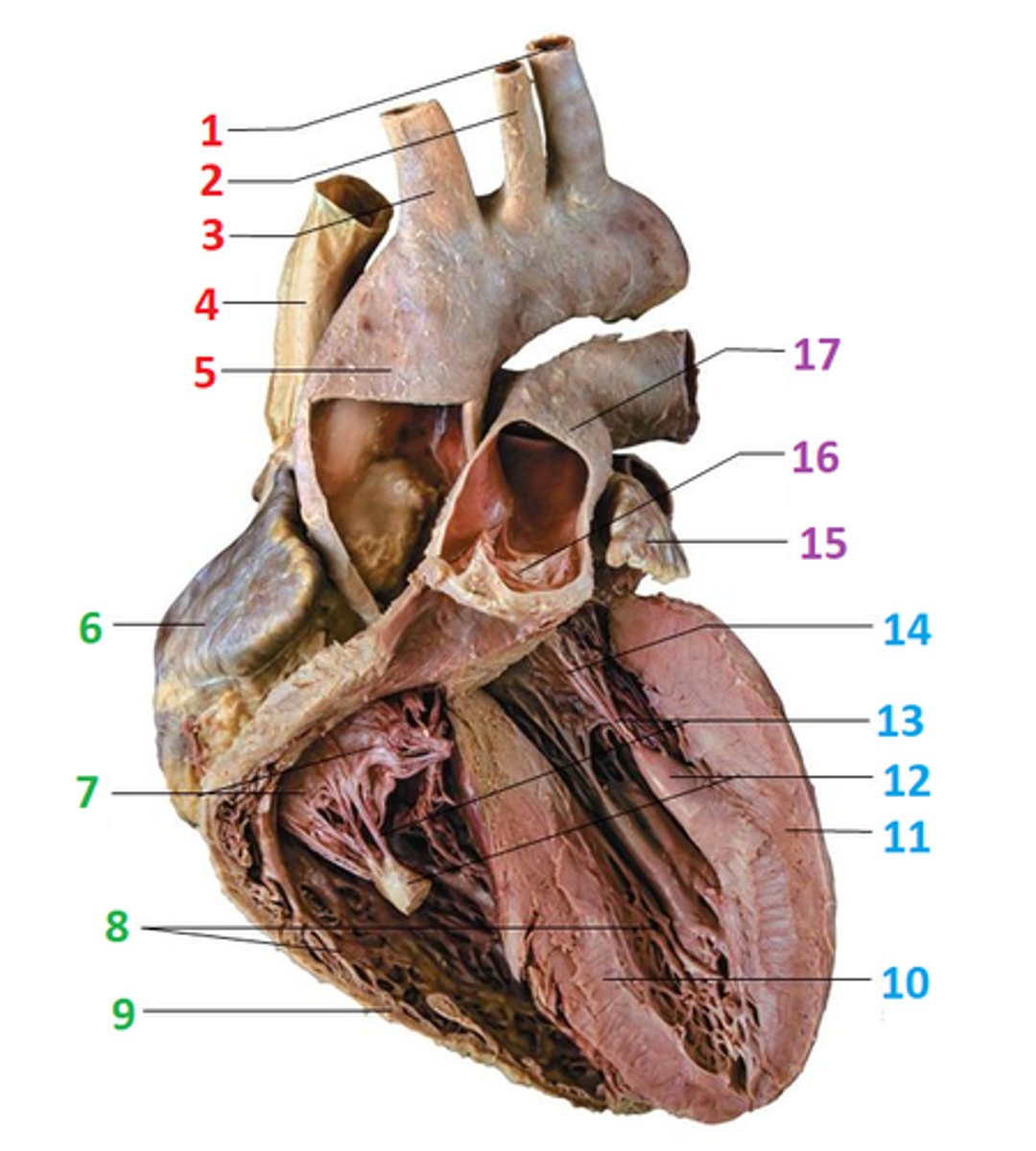
(6) Right Atrium
(7) Cusp of Right AV Valve (Tricuspid)
(8) Trabecula Carnae
(9) Right Ventricle
Name 6 - 9
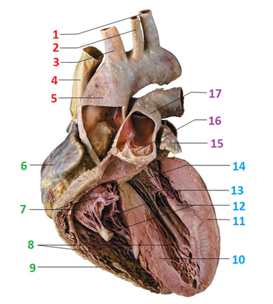
(10) Interventricular septum
(11) Left Ventricle
(12) Papillary Muscles
(13) Cordae Tendinae
(14) Cusp of Left AV Valve (Bicuspid)
Name 10 - 14
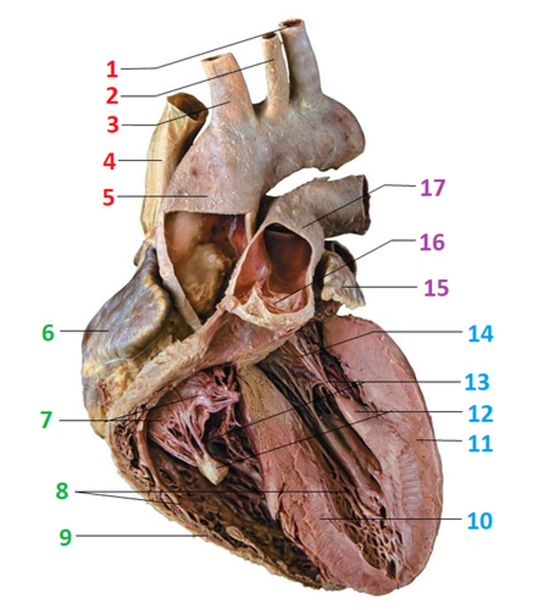
(15) Left Auricle
(16) Cusp of Pulmonary "Semi-Lunar" Valve
(17) Pulmonary Trunk
Name 15 - 17
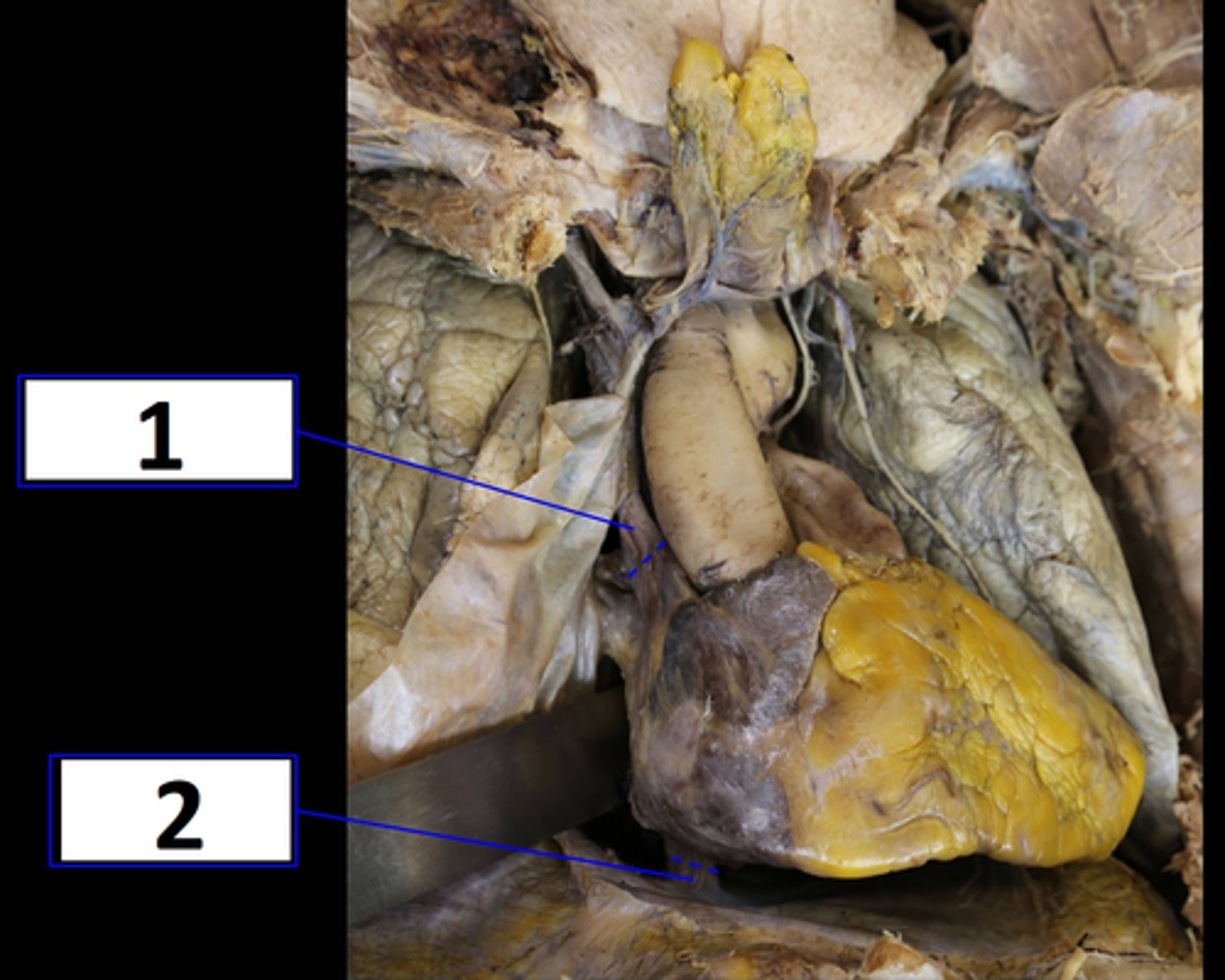
(1) Superior Vena Cava
(2) Inferior Vena Cava
Veins of the heart:
Name 1 & 2
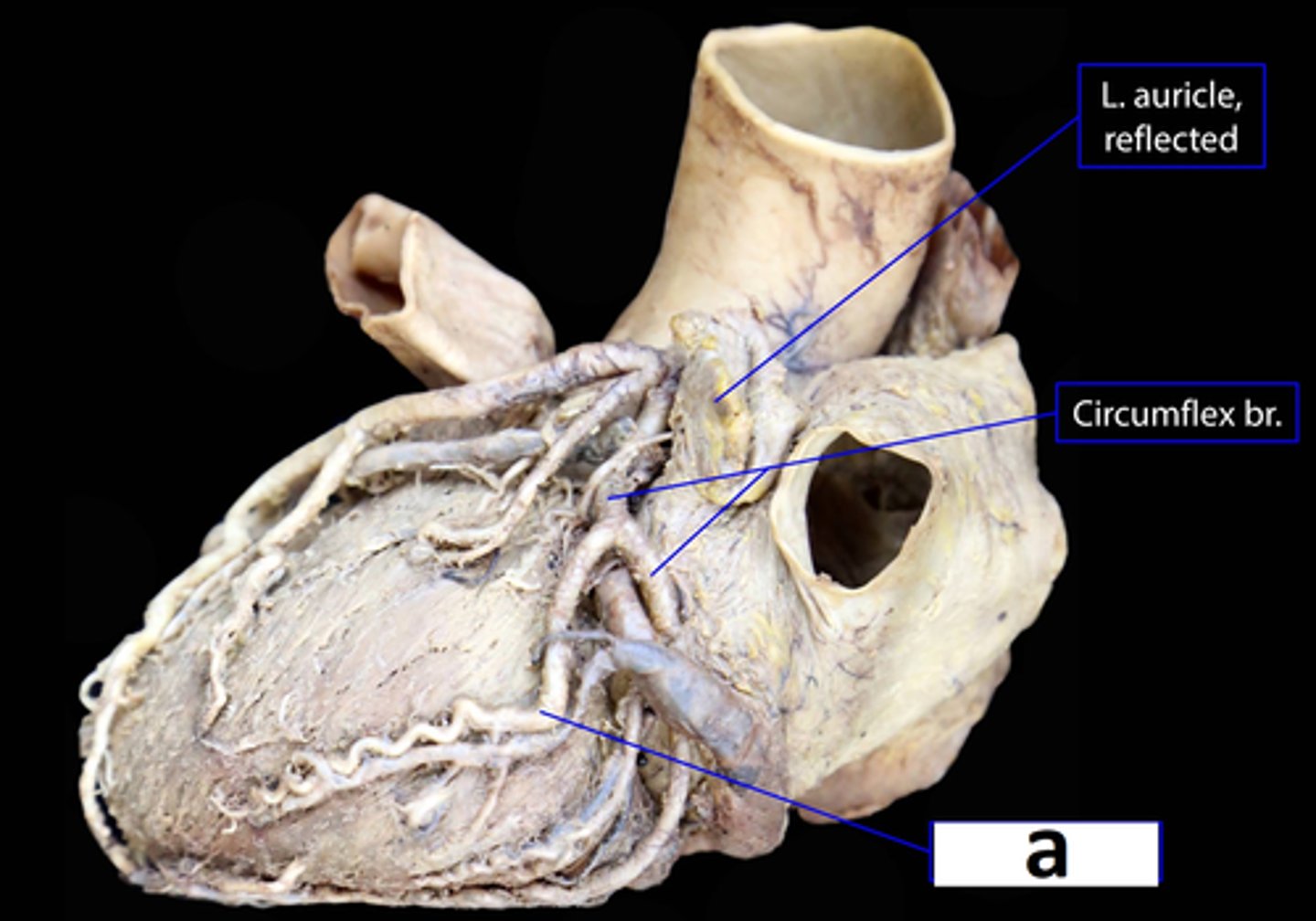
Left Marginal Vein
Veins of the heart:
Name a
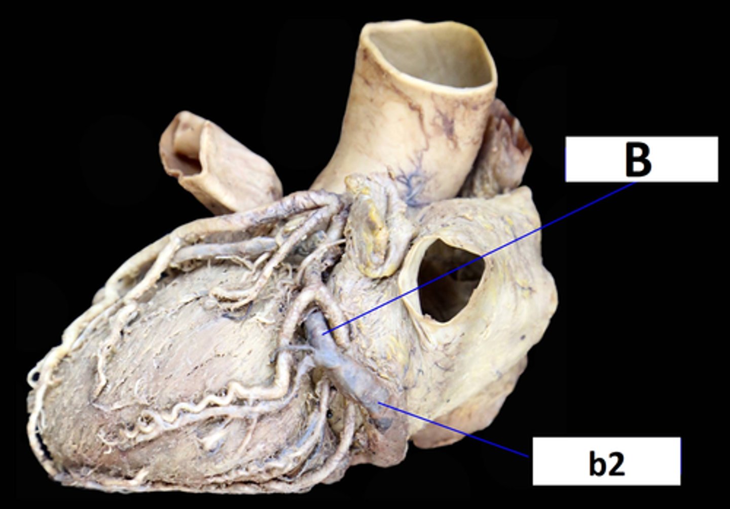
(B) Great Cardiac Vein
(b2) Coronary Sinus
Veins of the heart:
Name B and b2
Middle Cardiac Vein
Veins of the heart:
Name c
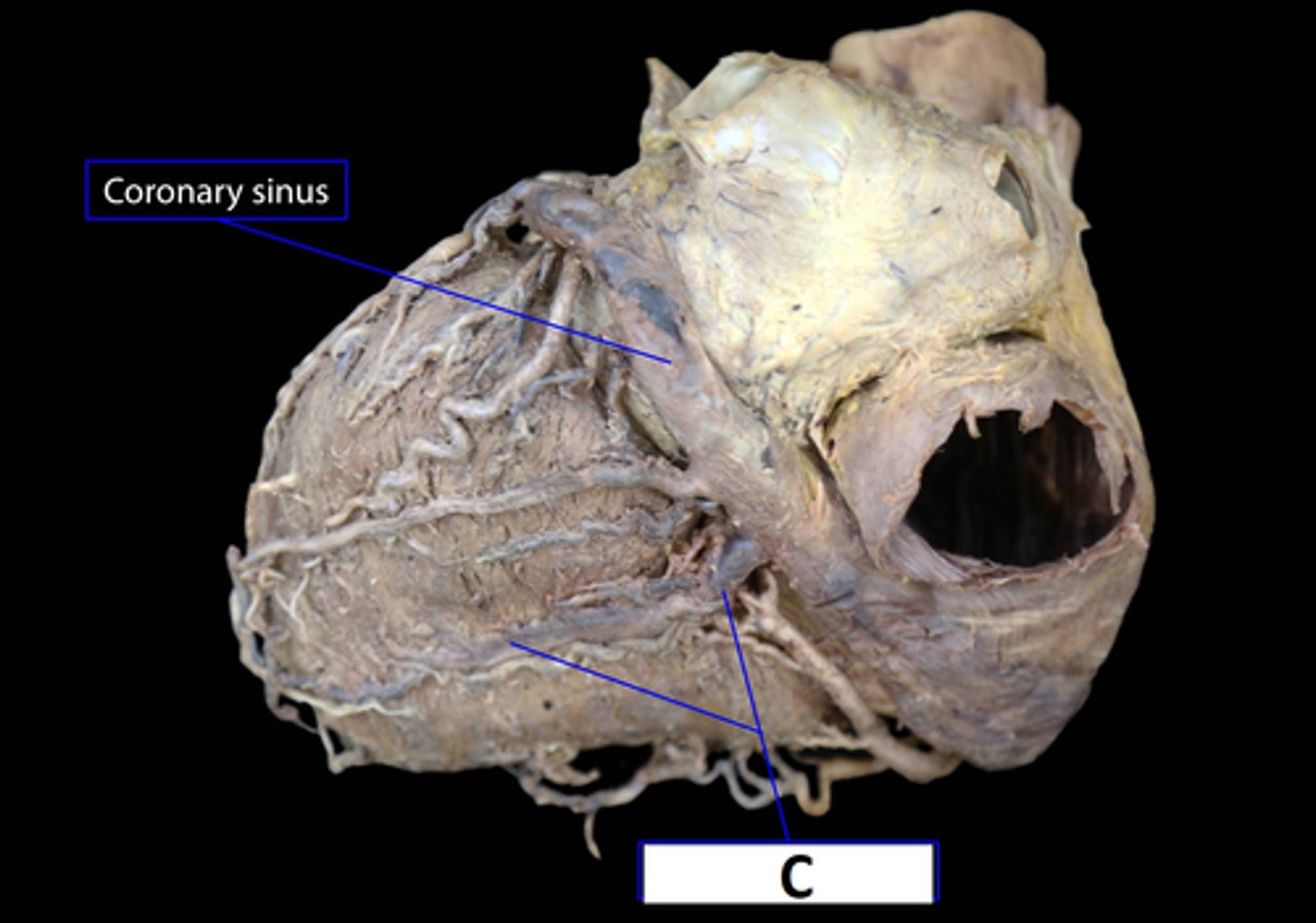
Small Cardiac Vein
Veins of the heart:
Name D
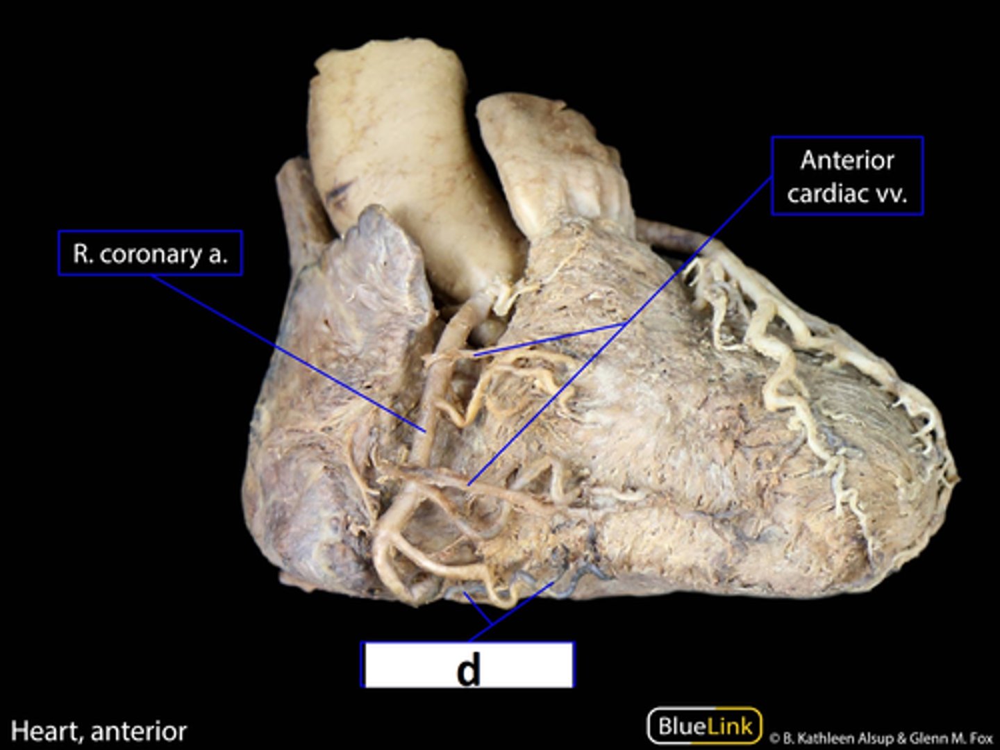
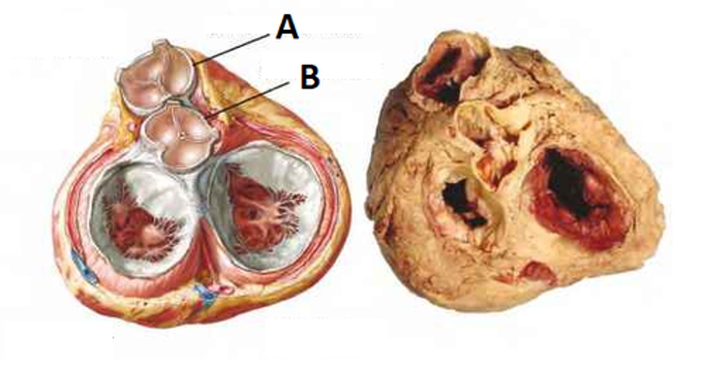
(A) Bicusspid Right AV Valve
(B) Tricuspid Left AV Valve
Closed during Systole
Name A & B
What phase of the heartbeat do the close?
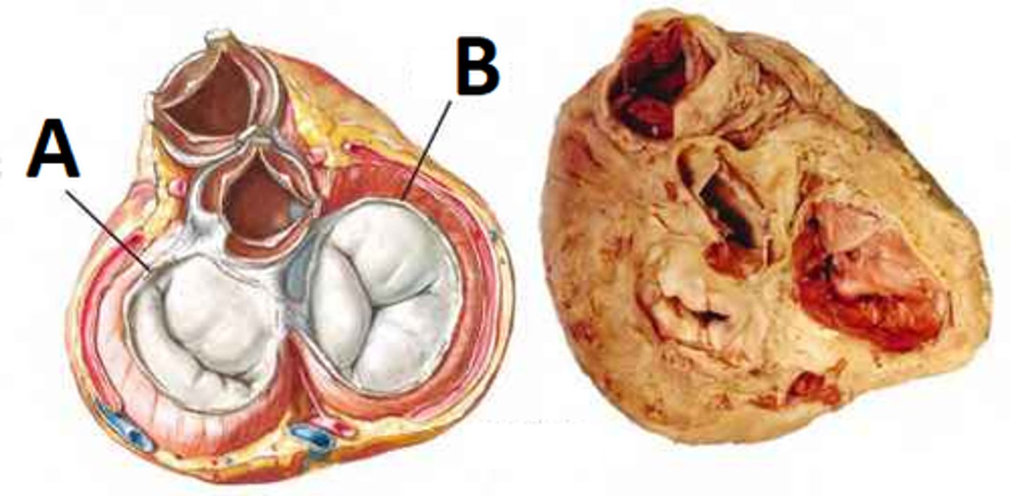
(A) Pulmonary Semiluar Valve
(B) Aortic Semilunar Valve
Closed during diastole
Name A & B
What phase of the heartbeat do the close?
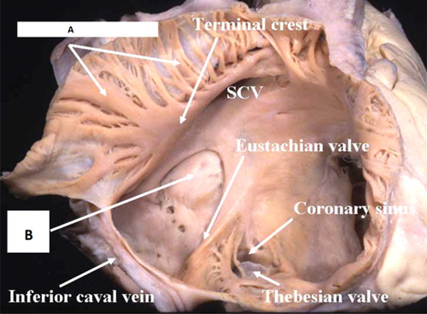
(A) Pectinate Muscle
(B) Fossa Ovale (remnant of the foramen ovale from fetal life)
Name A & B
What is significant about B?
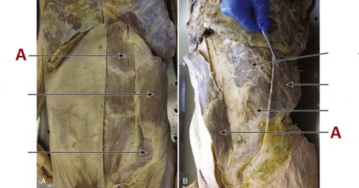
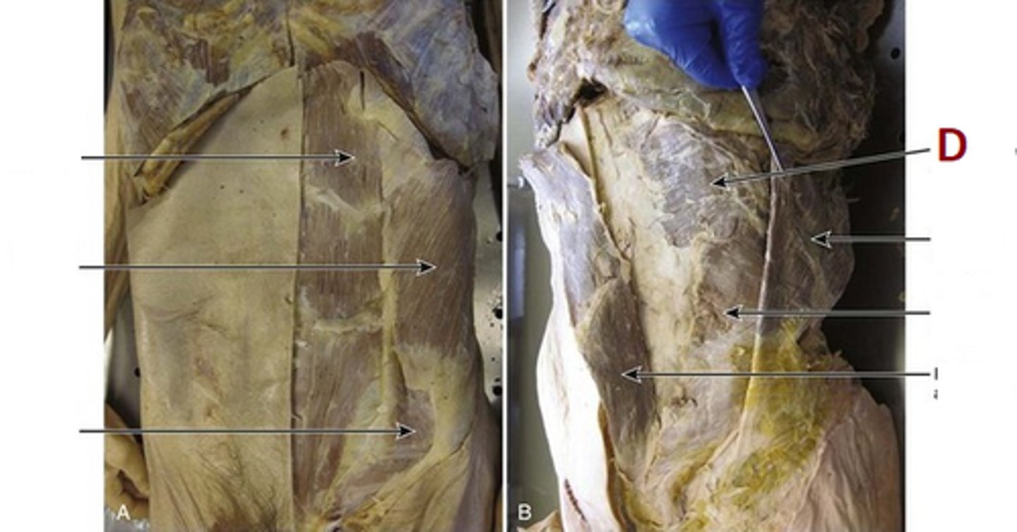
Rectus Abdominis Muscle
Name A
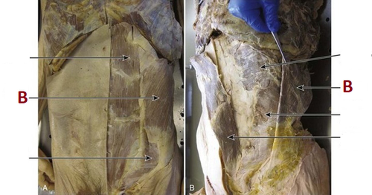
External Oblique Muscle
Name B
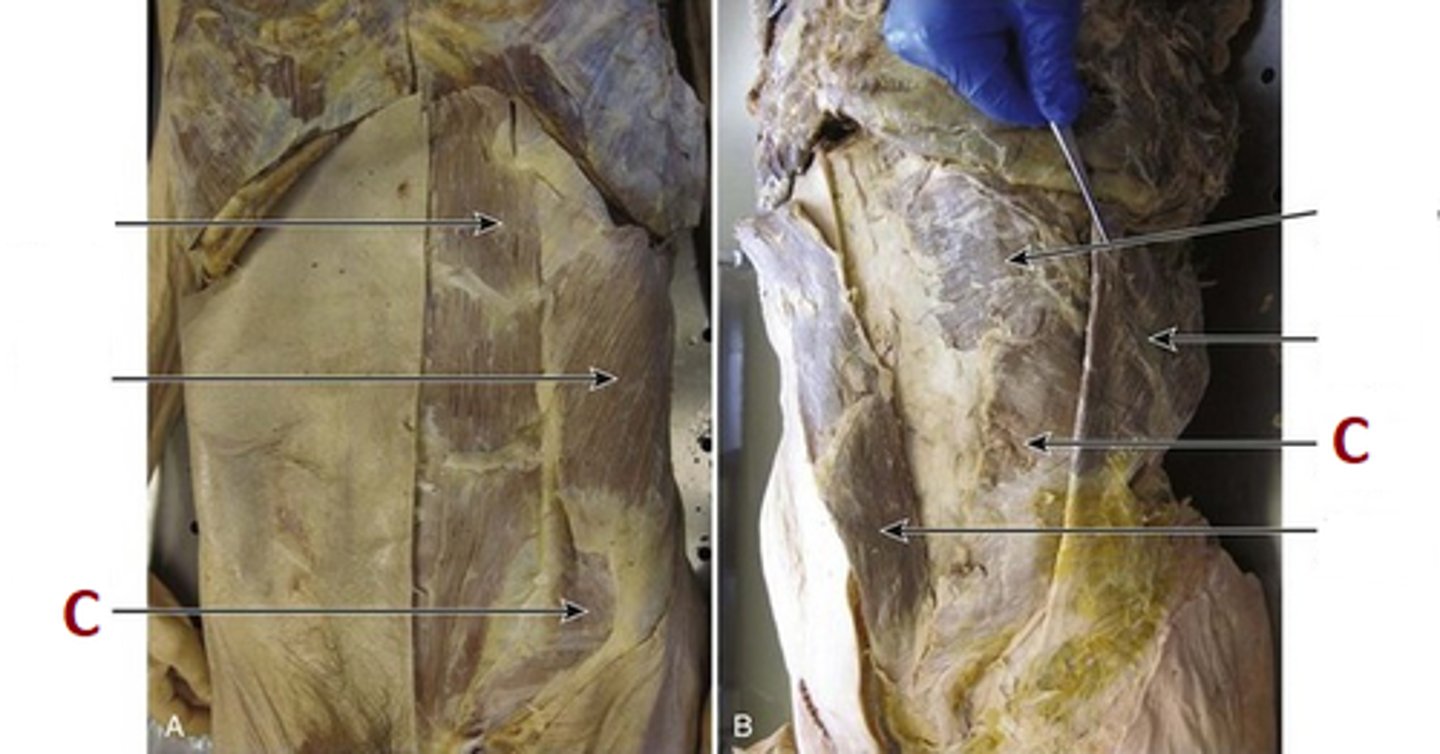
Internal ObliqueMuscle
Name C
Tranverse Abdominis Muscle
Name D
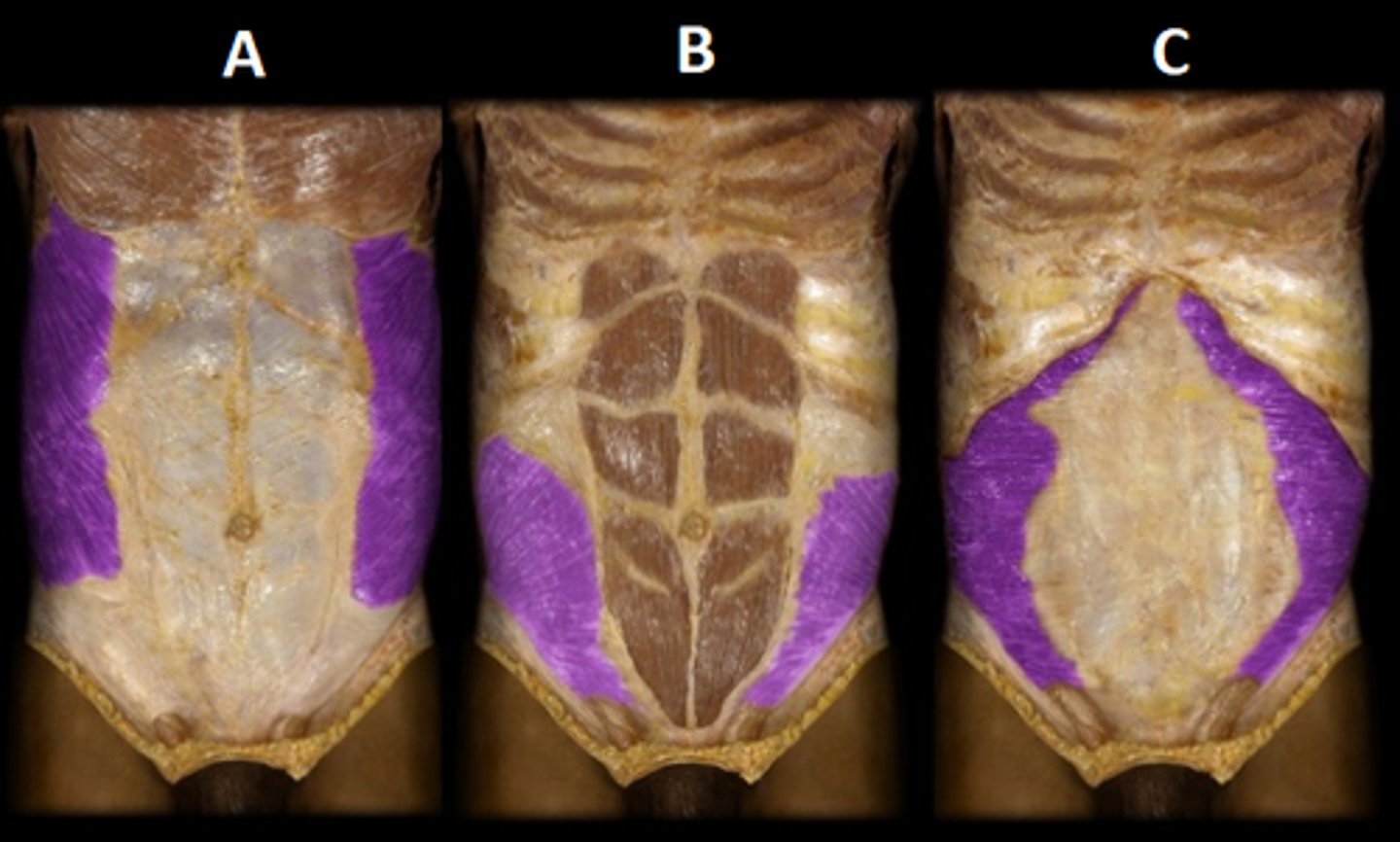
(A) External Abdominal
(B) Internal Abdominal
(C) Transverse Abdominis
Name A.B.C.
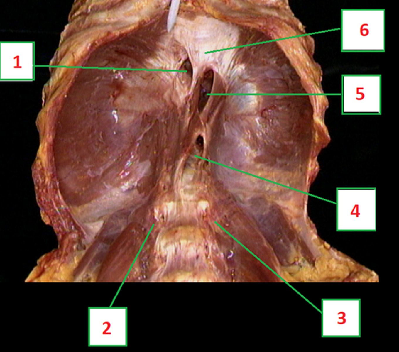
(1) Vena Caval Foramen (opening for Inferior Vena Cava)
(2) Left Crus
(3) Right Crus
(4) Aortic Hiatus
(5) Esophogeal Hiatus
(6) Centeral Tendon
Diaphragm:
Name 1 - 6
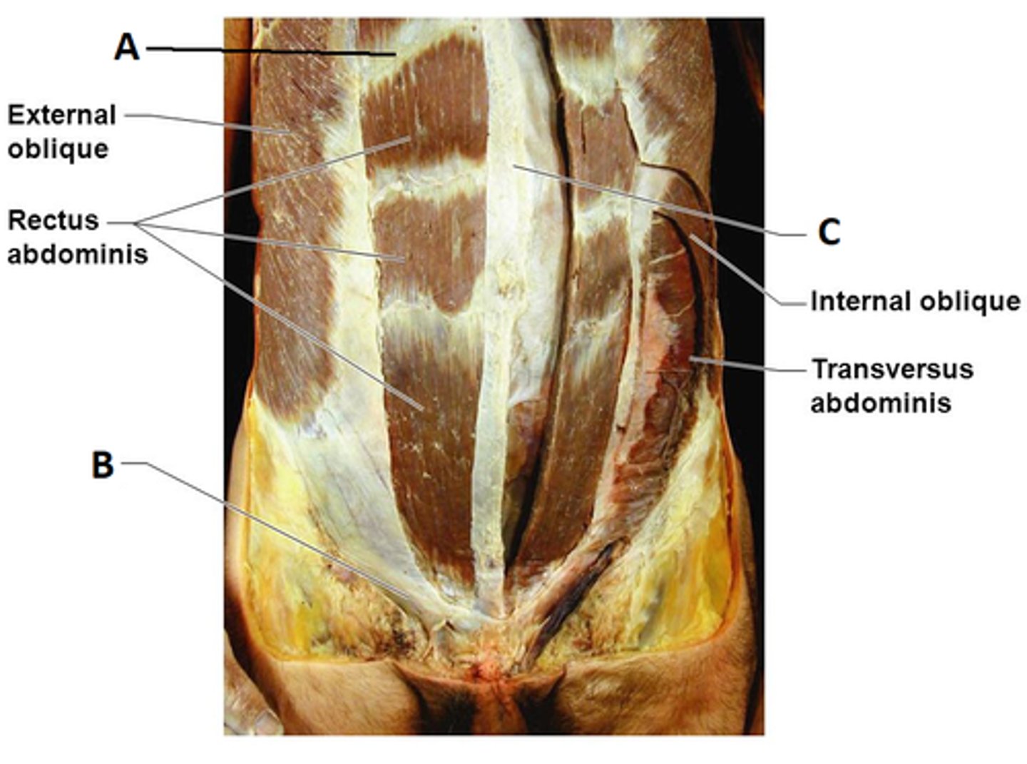
(A) Tendinsous Inscriptions (intersections)
(B) Inguinal Ligament
(C) Linea Alba
Connective tissue Structures:
Name A,B & C
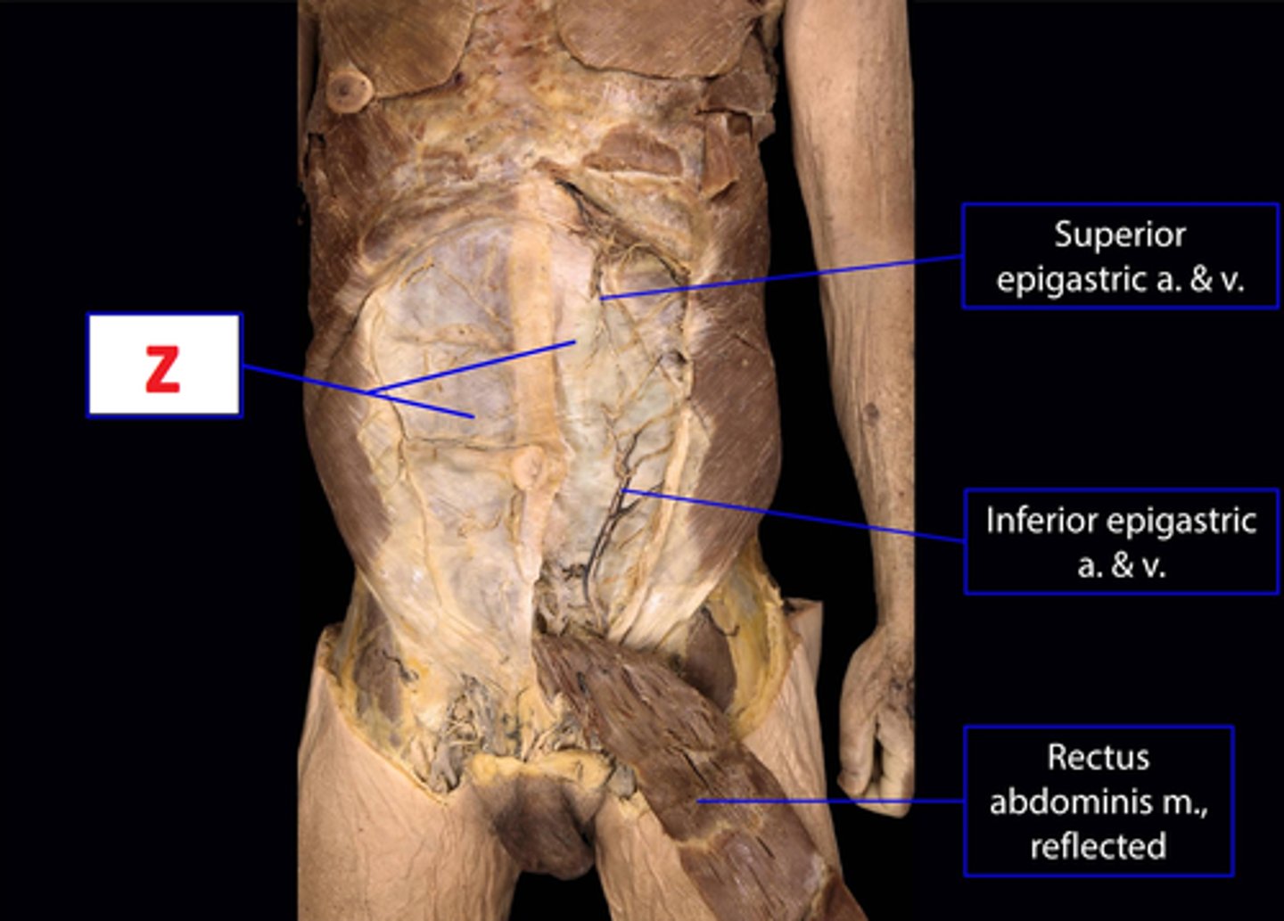
Rectus Sheath
Connective tissue:
Name Z
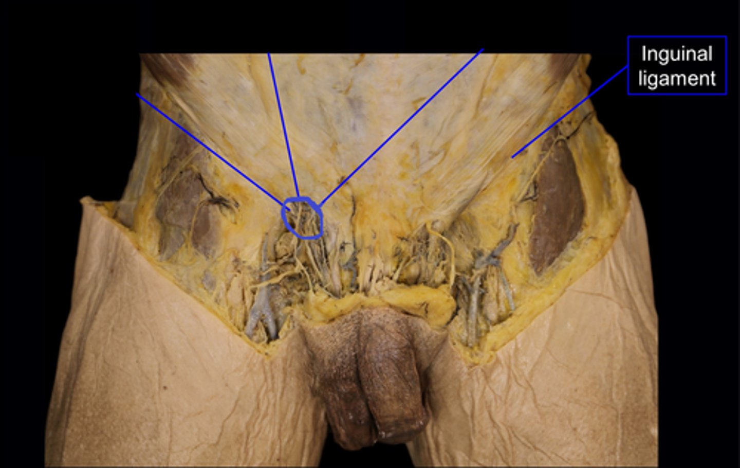
Superficial Inguinal Canal
Name the area circled in purple
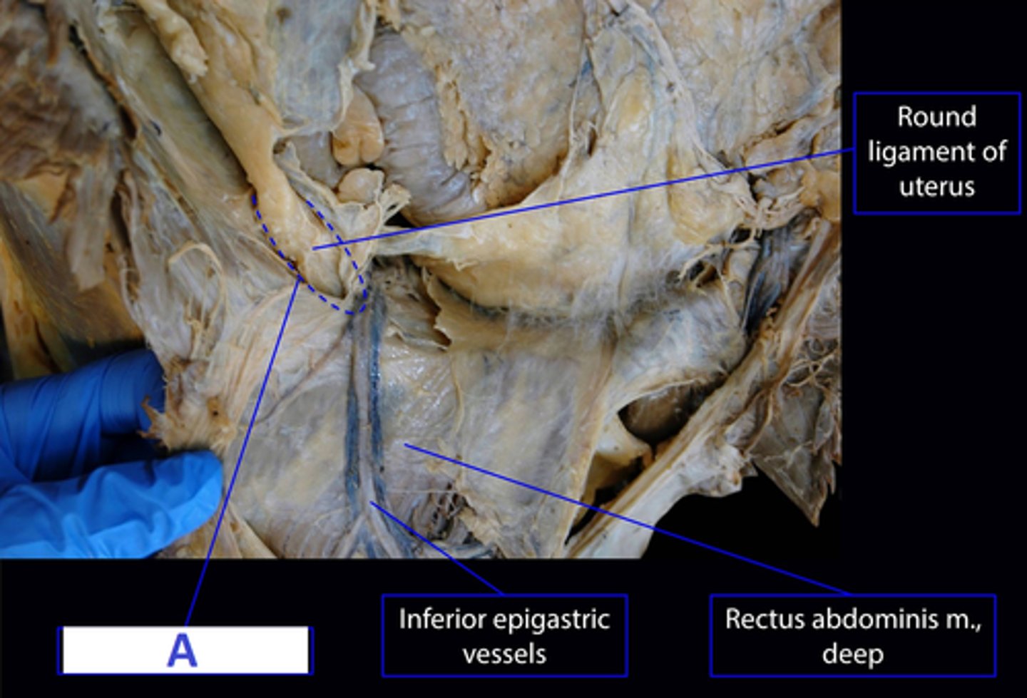
Deep Inguinal Canal
Name A
Lateral Umbilical (epigastic) Fold
Formed by Inferior Epigastric Vessels
Name A
What is its significance?
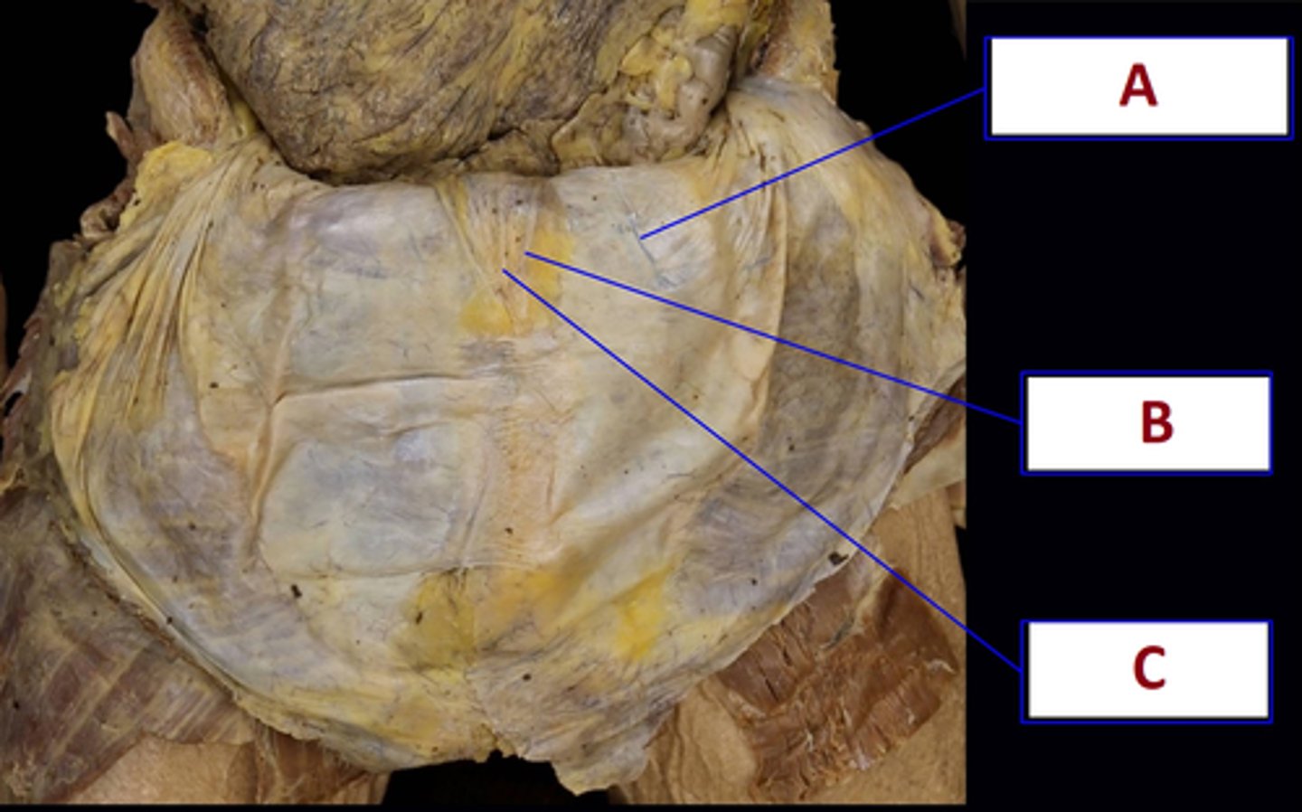
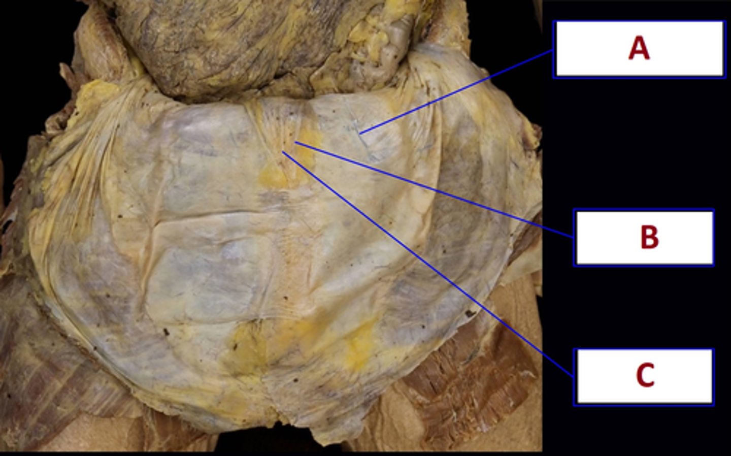
Medial Umbilical Fold
Remnant of Umbilical Arteries
Name B
What is its significance?
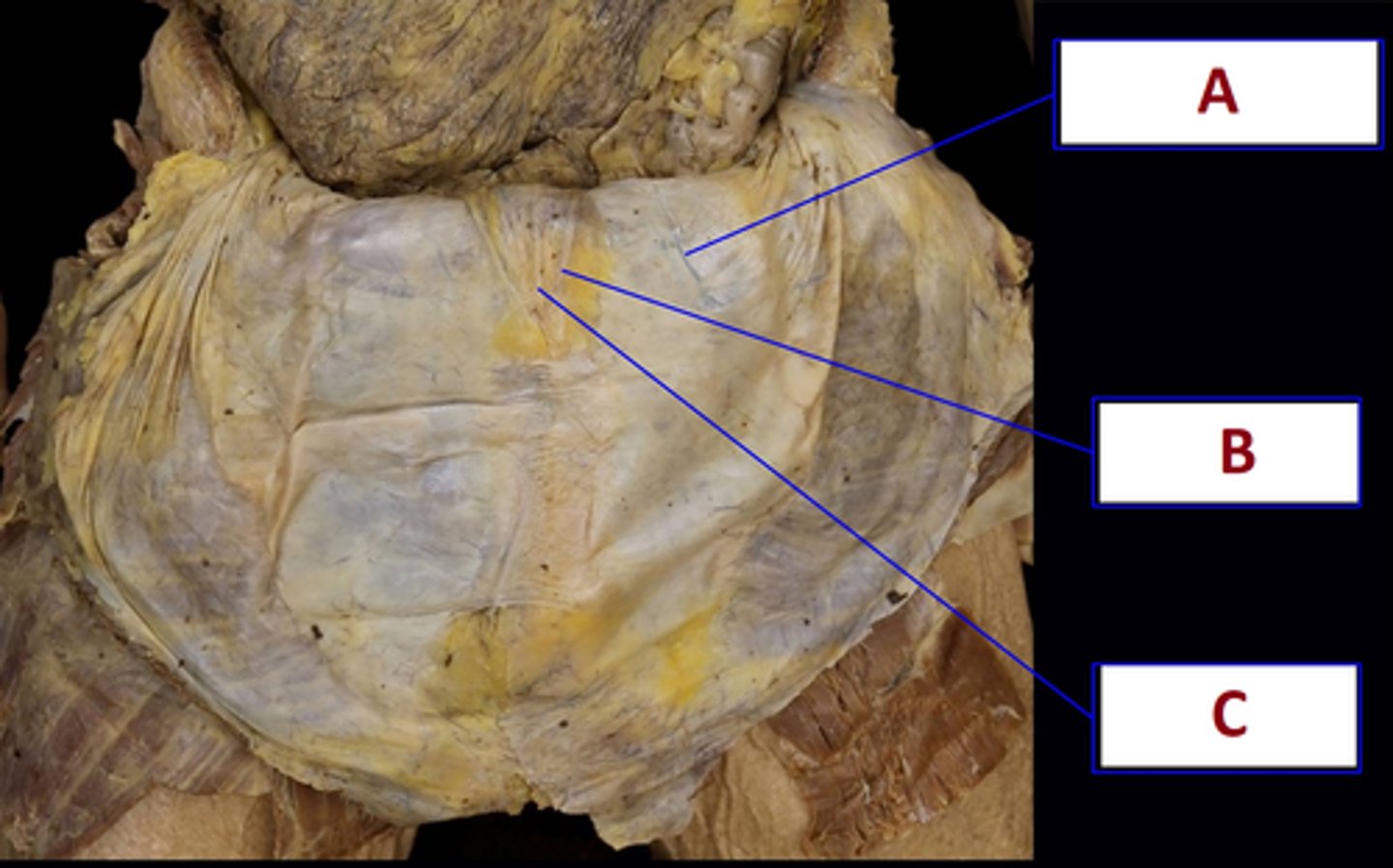
Median Umbilical Fold
Remnant of the Urachus
Name C
What is its significance?
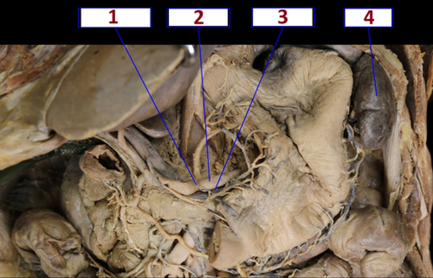
(1) Celiac Trunk
(2) Splenic Artery
(3) Splenic Vein
(4) Spleen
Name 1 - 4
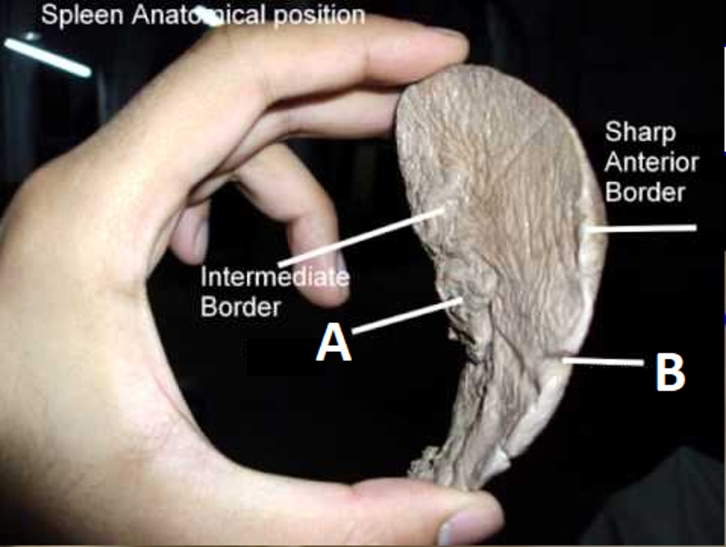
(A) Spleen Hilum
(B) Spleen Notches
name A & B
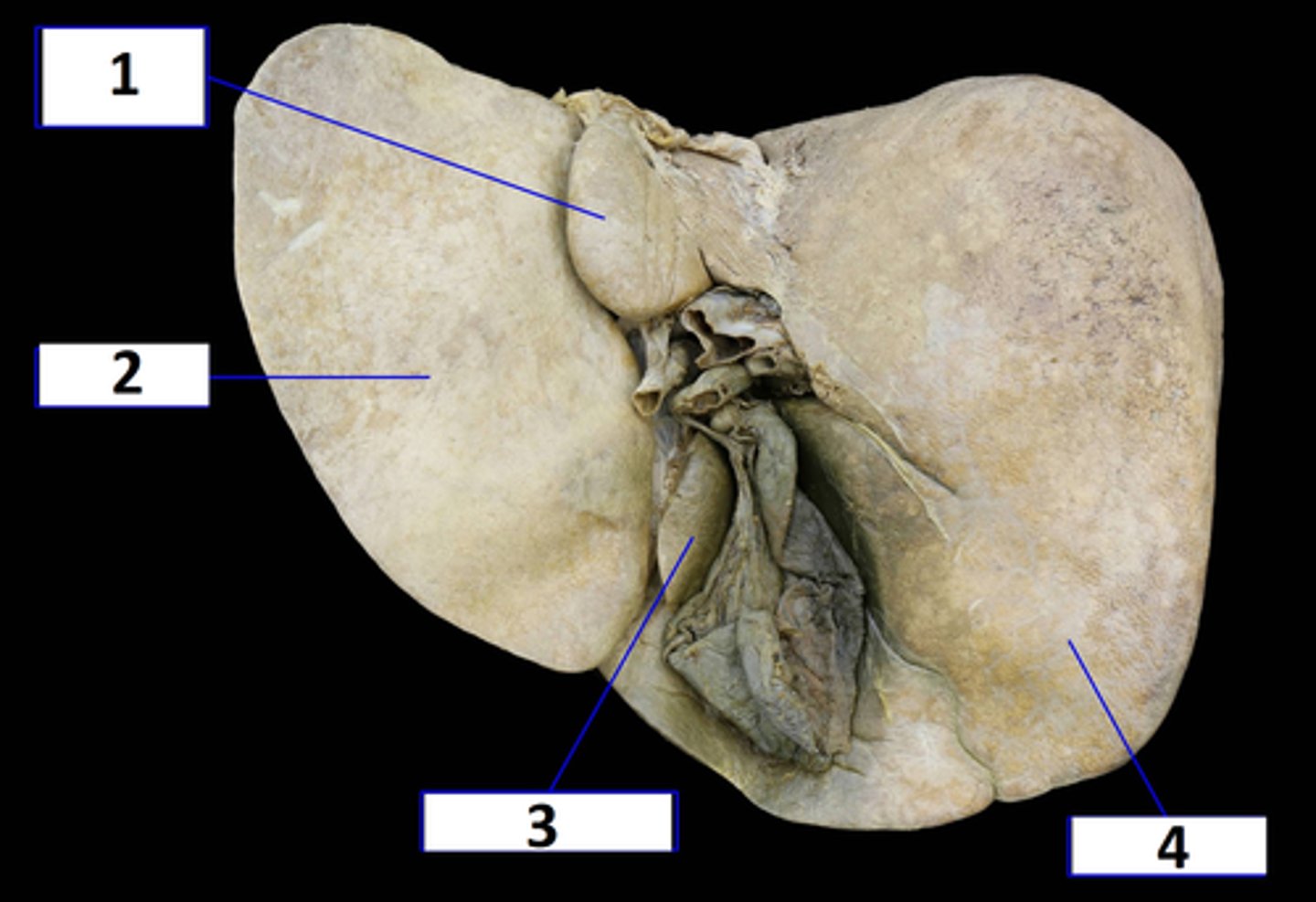
(1) Caudate Lobe
(2) Left Lobe
(3) Quadrate Lobe
(4) Right Lobe
Liver:
Name 1 - 4
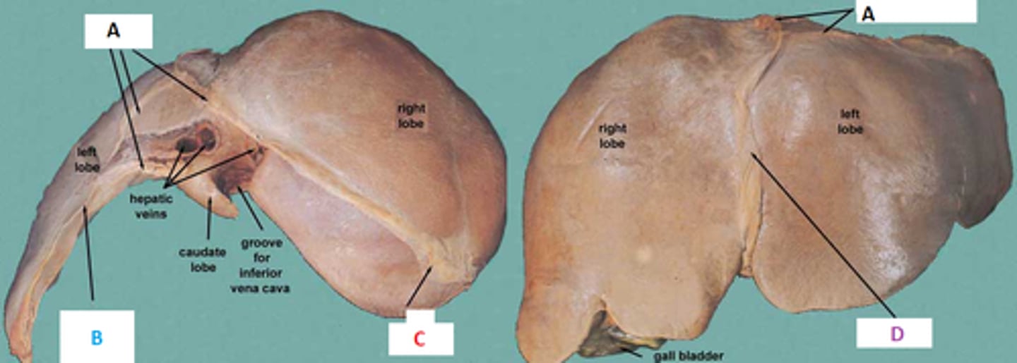
(A) Coronal/ Coronary Ligament
(B) Left Triangular Ligament
(C) Right Triangular Ligament
(D) Falciform Ligament
Liver:
Name A - D
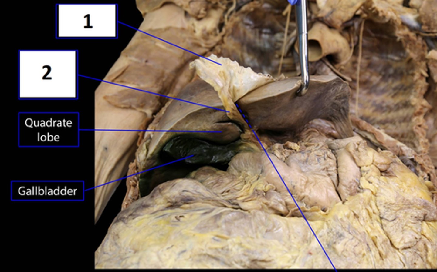
(1) Falciform Ligament
(2) Ligamentum Teres Hepatis (round ligament of liver)
#2: remnant of the umbilical v.
Liver:
Name 1 & 2
What is Significant about 2?
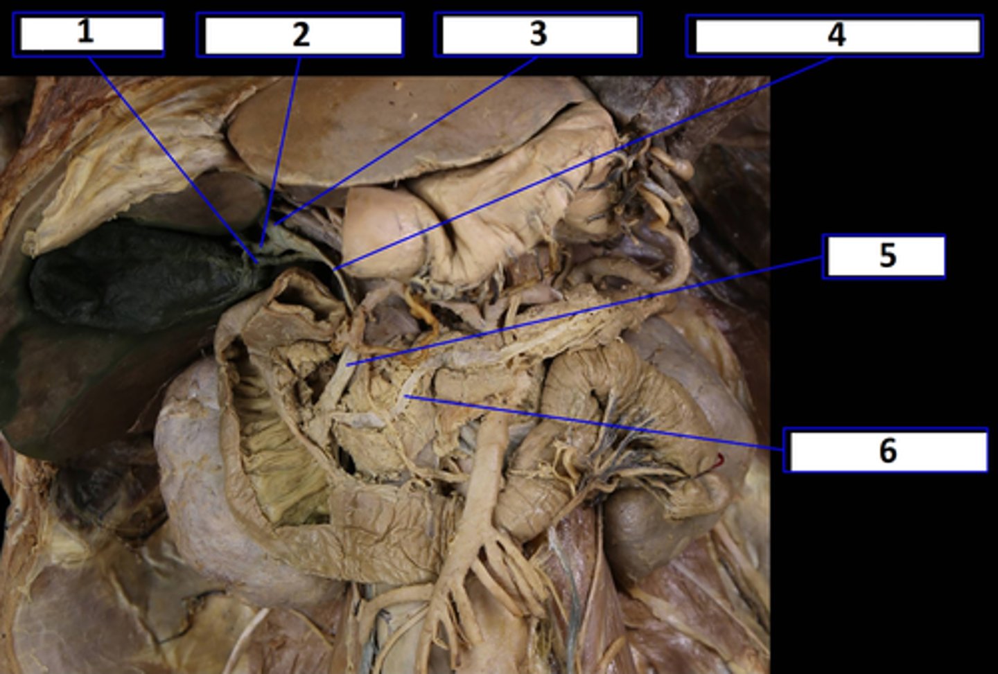
(1) Cyst Duct
(2) Right Hepatic Duct
(3) Left Hepatic Duct
(4) Common Hepatic Duct
(5) Bile Duct
(6) Pancreatic Duct
Hepatic, ducts Ducts:
name 1-6
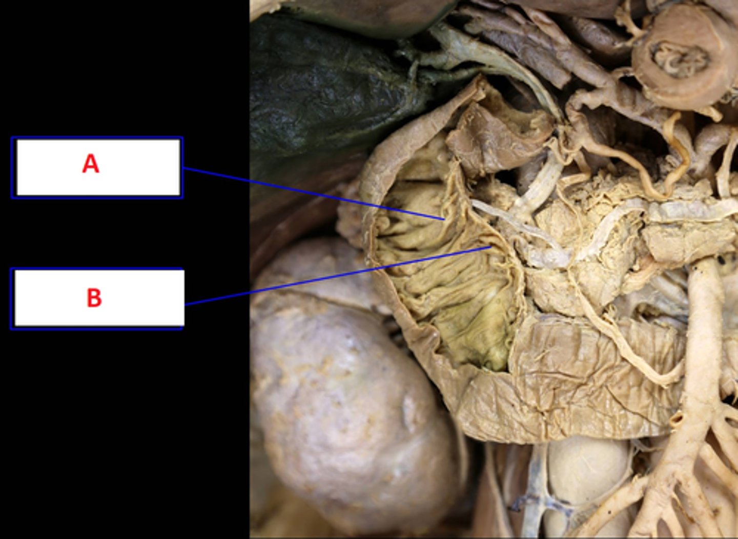
(A) Duodenal Papilla (Minor)
(B) Duodenal Papilla (Major)
Name A & B
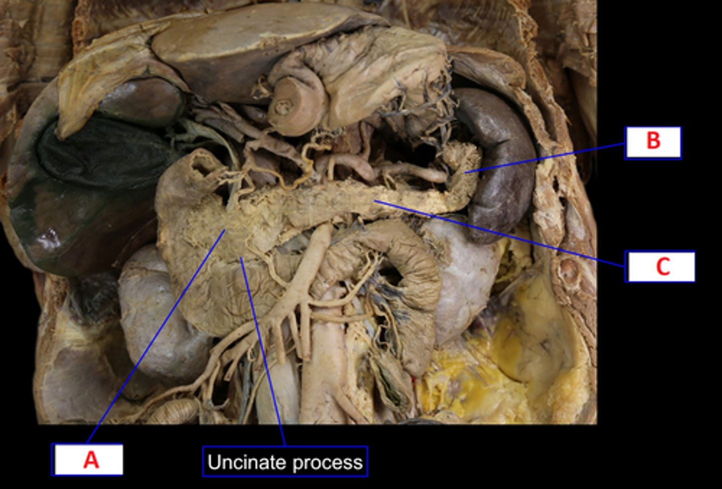
Pancreas
(A) Head
(B) Tail
(C) Body
What body part is this?
Name A, B & C
For open lab tutoring contact:
Sforbes@college.lifewest.edu
For open lab tutoring contact:
Sforbes@college.lifewest.edu