Looks like no one added any tags here yet for you.
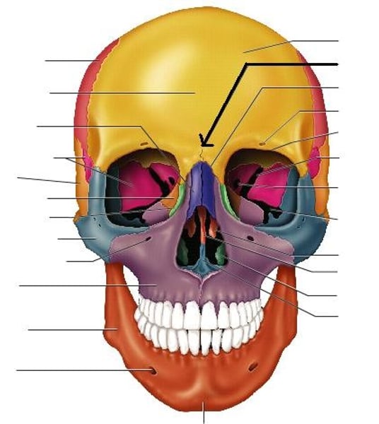
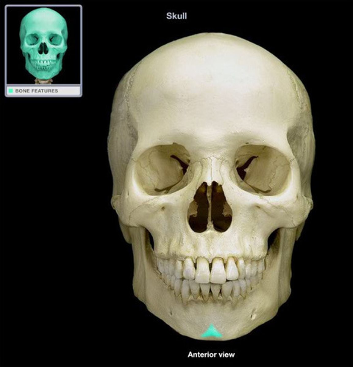
Glabella
Middle concha (of Ethmoid Bone)
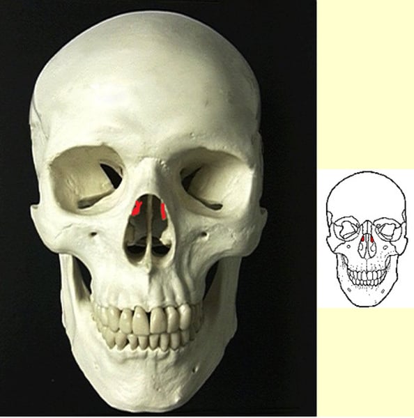
Perpendicular plate of Ethmoid; top part of nasal septum
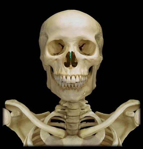
Vomer; inferior part of nasal septum
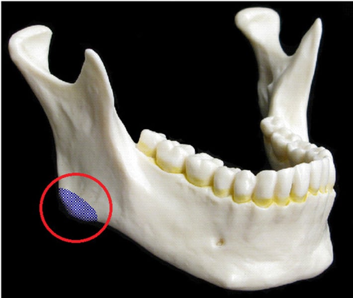
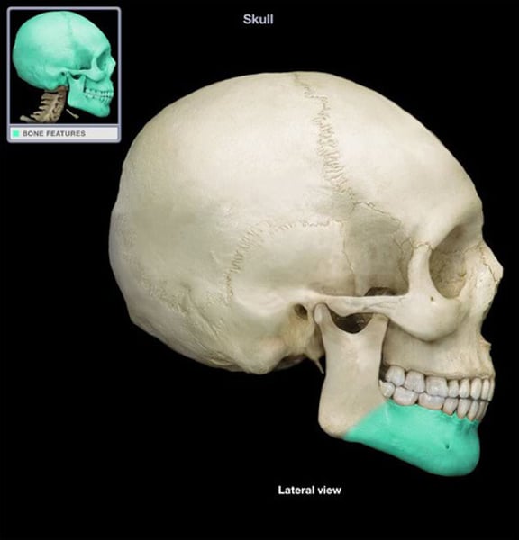
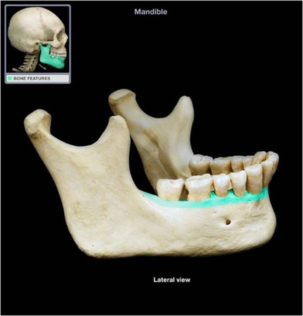
Angle of mandible; lower posterior of ramus
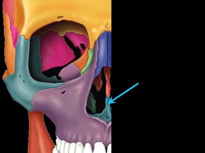
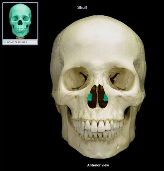
inferior conchae; paired
Note: this is a separate bone from the ethmoid (ethmoid has a superior and middle nasal conchae)
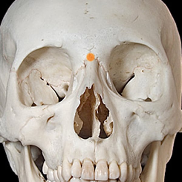
Nasion
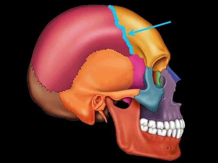
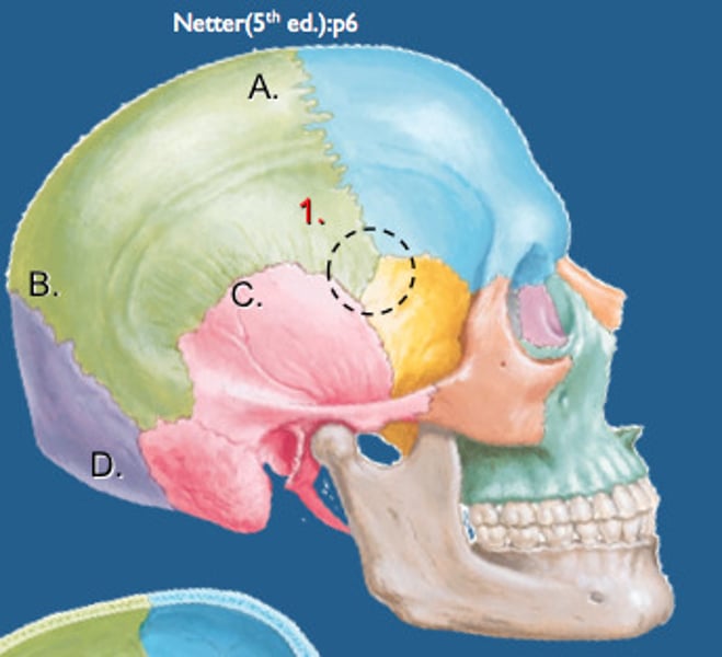
Coronal suture
-Between frontal bone and parietal bones
Pterion
-H-shaped junction between the frontal, parietal, temporal, and sphenoid
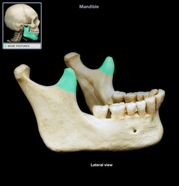
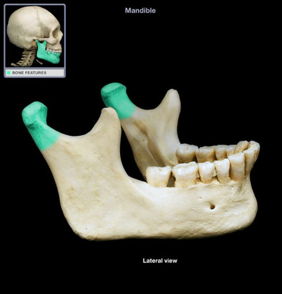
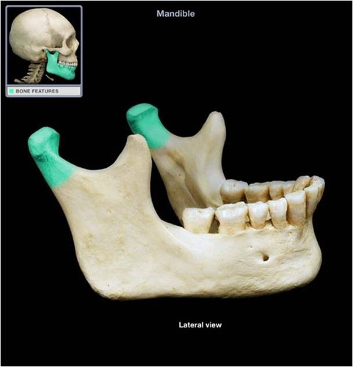
Coronoid process
Head of mandible/Condylar process
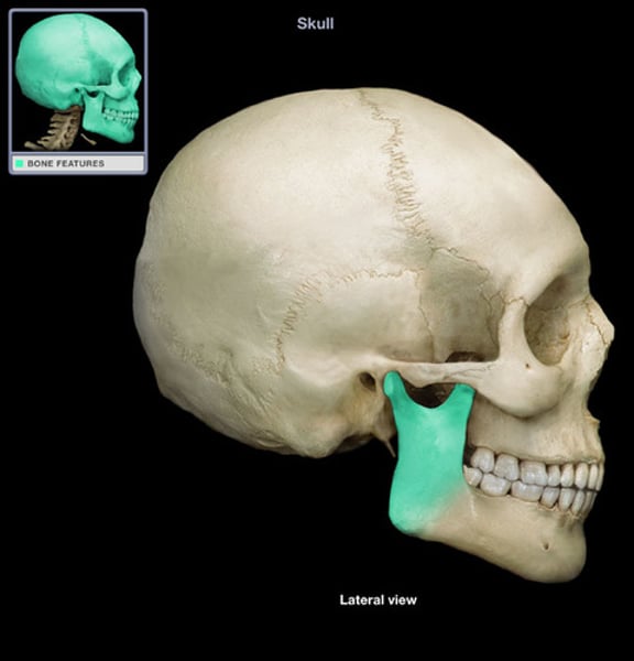
Ramus of mandible
Body of mandible
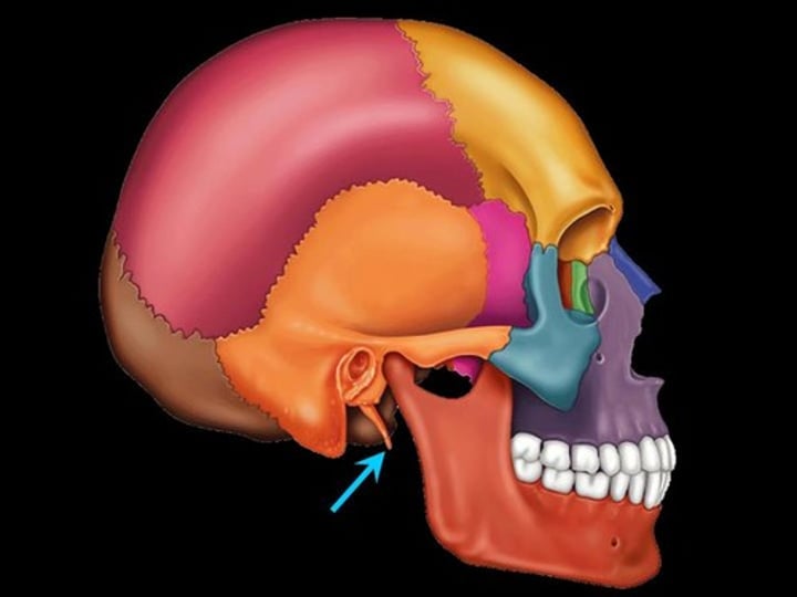
Styloid process
-part of temporal bone
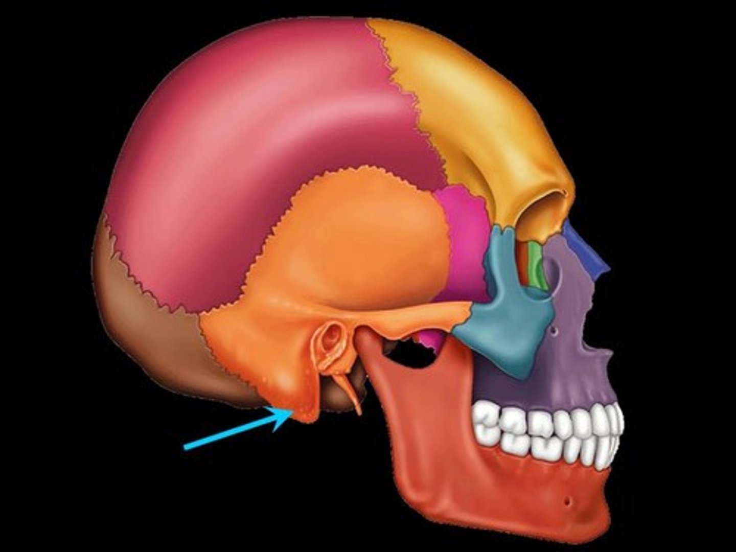
Mastoid process
-round projection on temporal bone behind ear; palpable
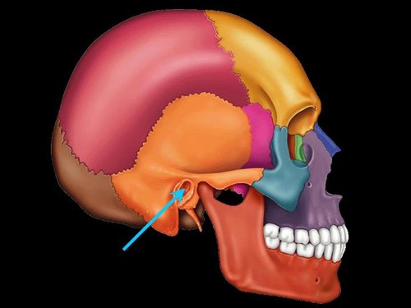
External acoustic meatus
(ear canal)
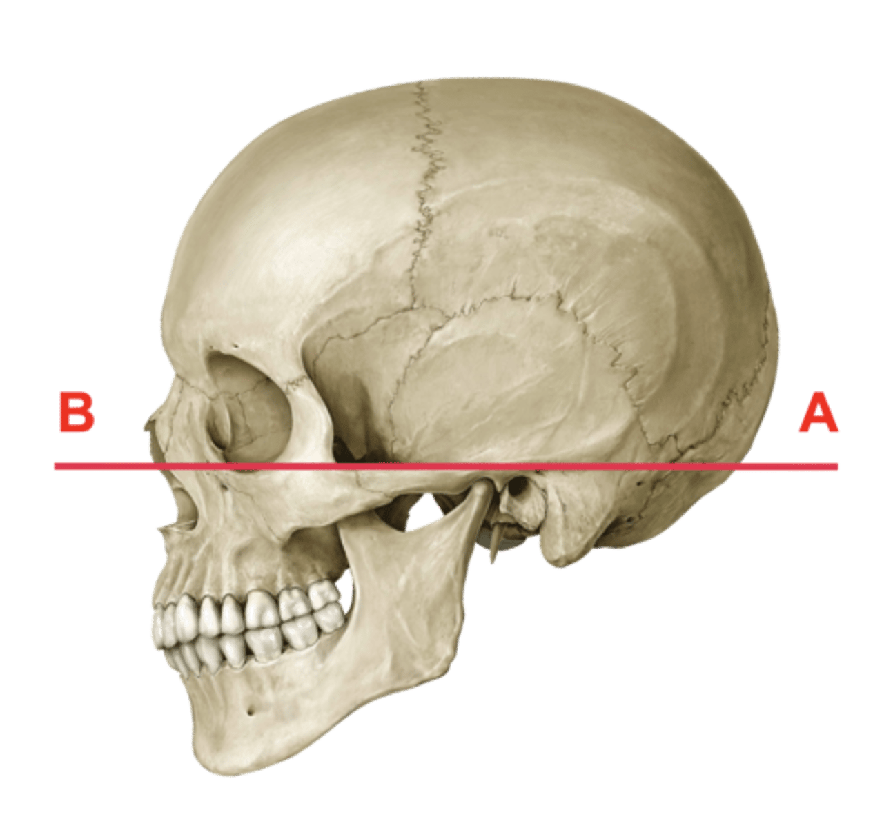
Orbitomeatal plane
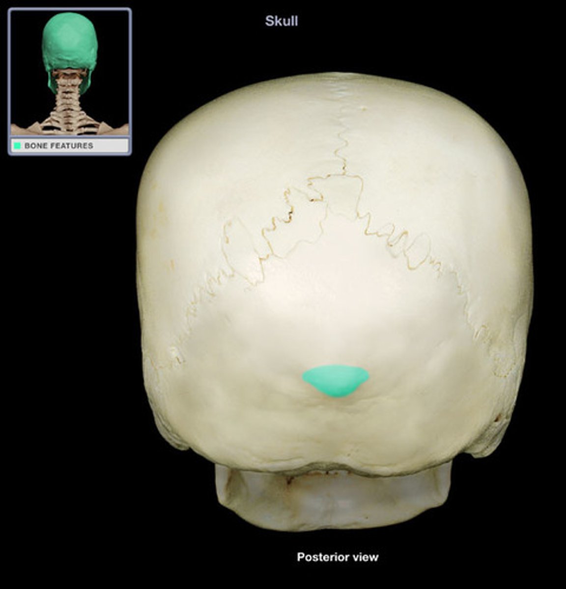
External occipital protuberance
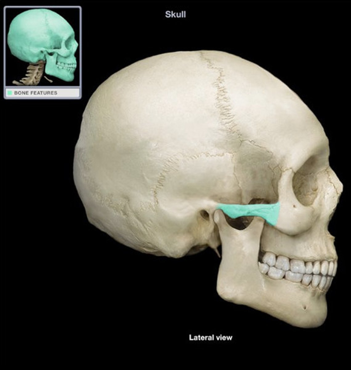
zygomatic arch
-paired
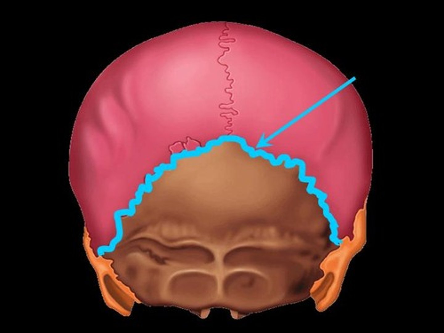
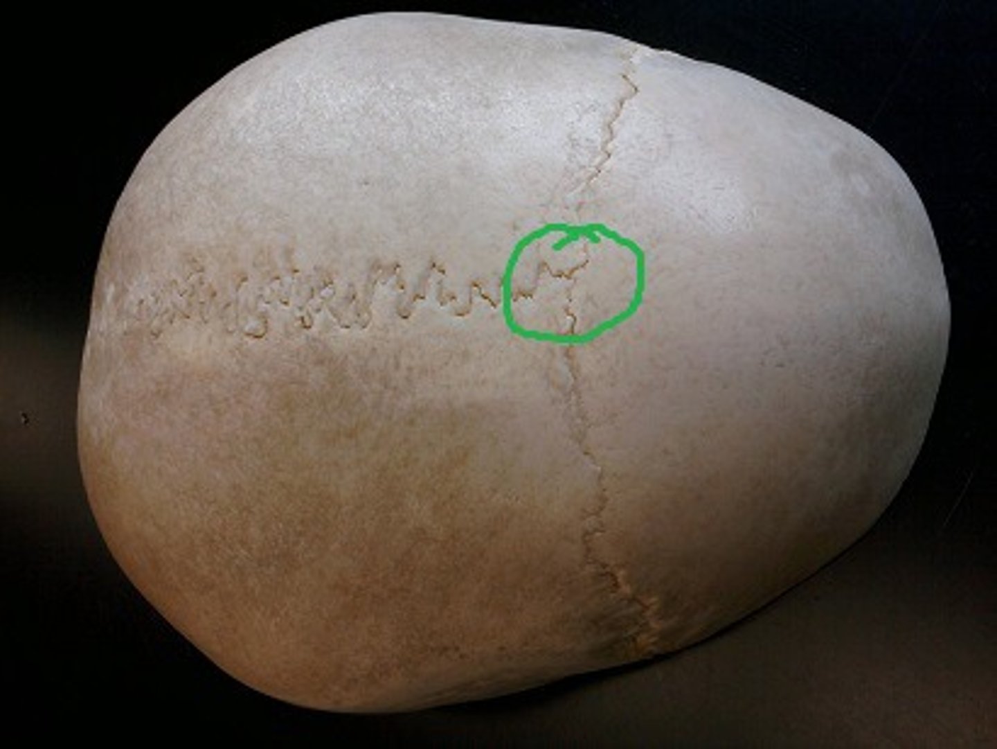
Lambdoid suture
-divides parietal and occipital bones
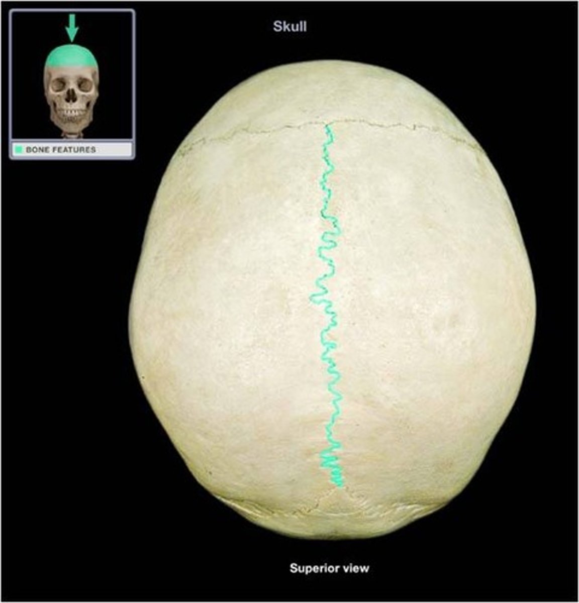
Sagittal suture
-between parietal bones
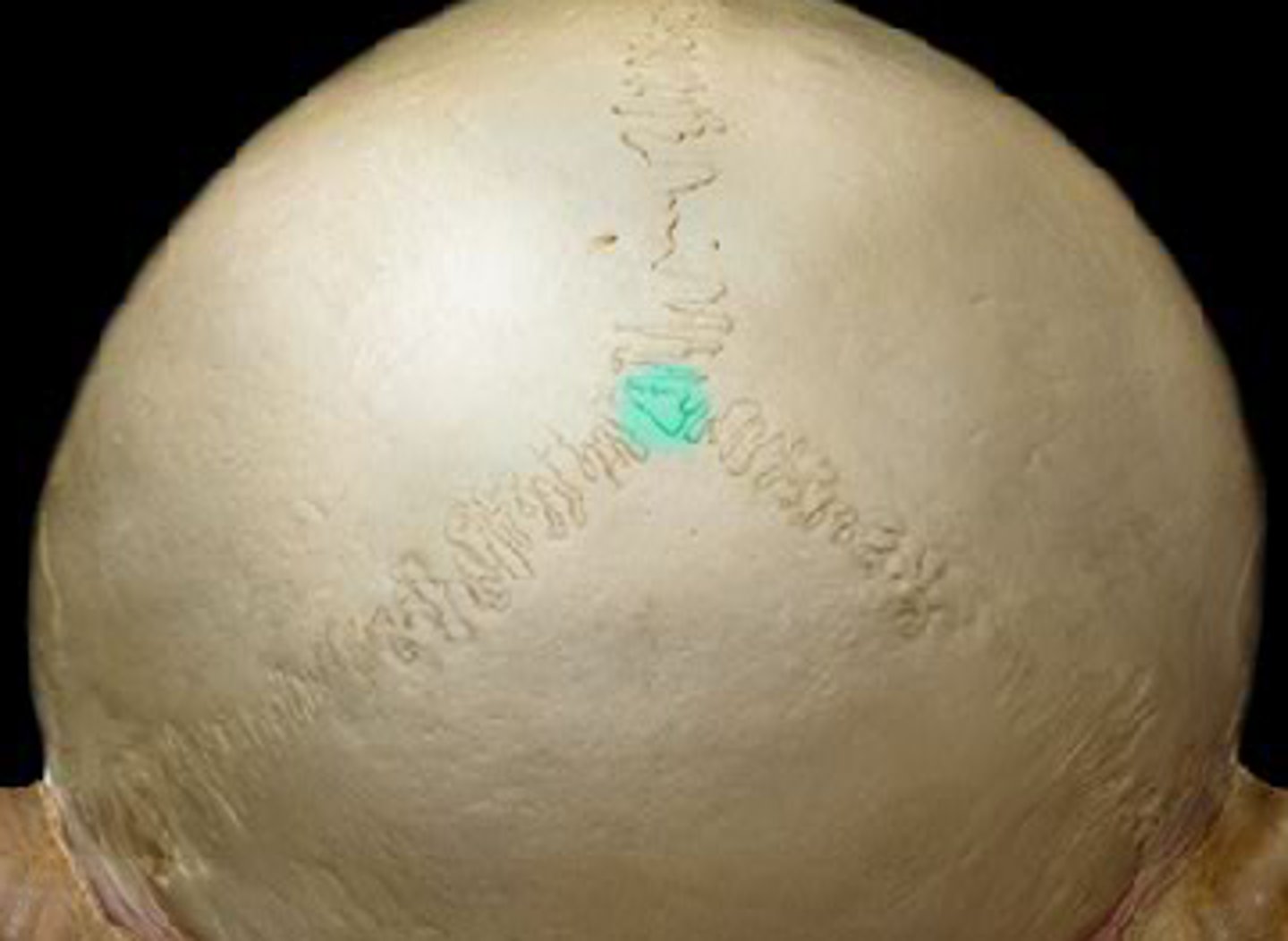
Lambda
-where lambdoid suture and sagittal sutures meet (separating parietal bones and occipital bones)
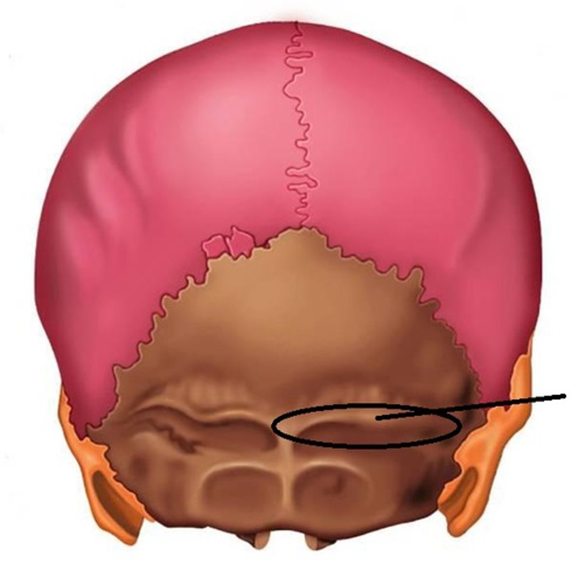
Superior nuchal line
-runs horizontally from external occipital protuberance
Inferior nuchal line
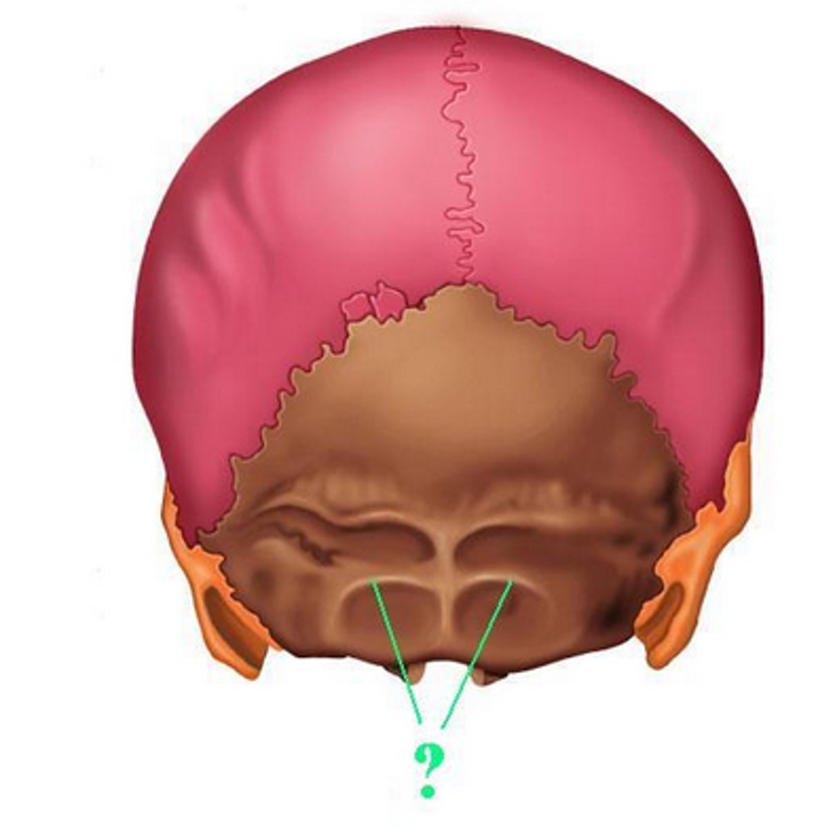
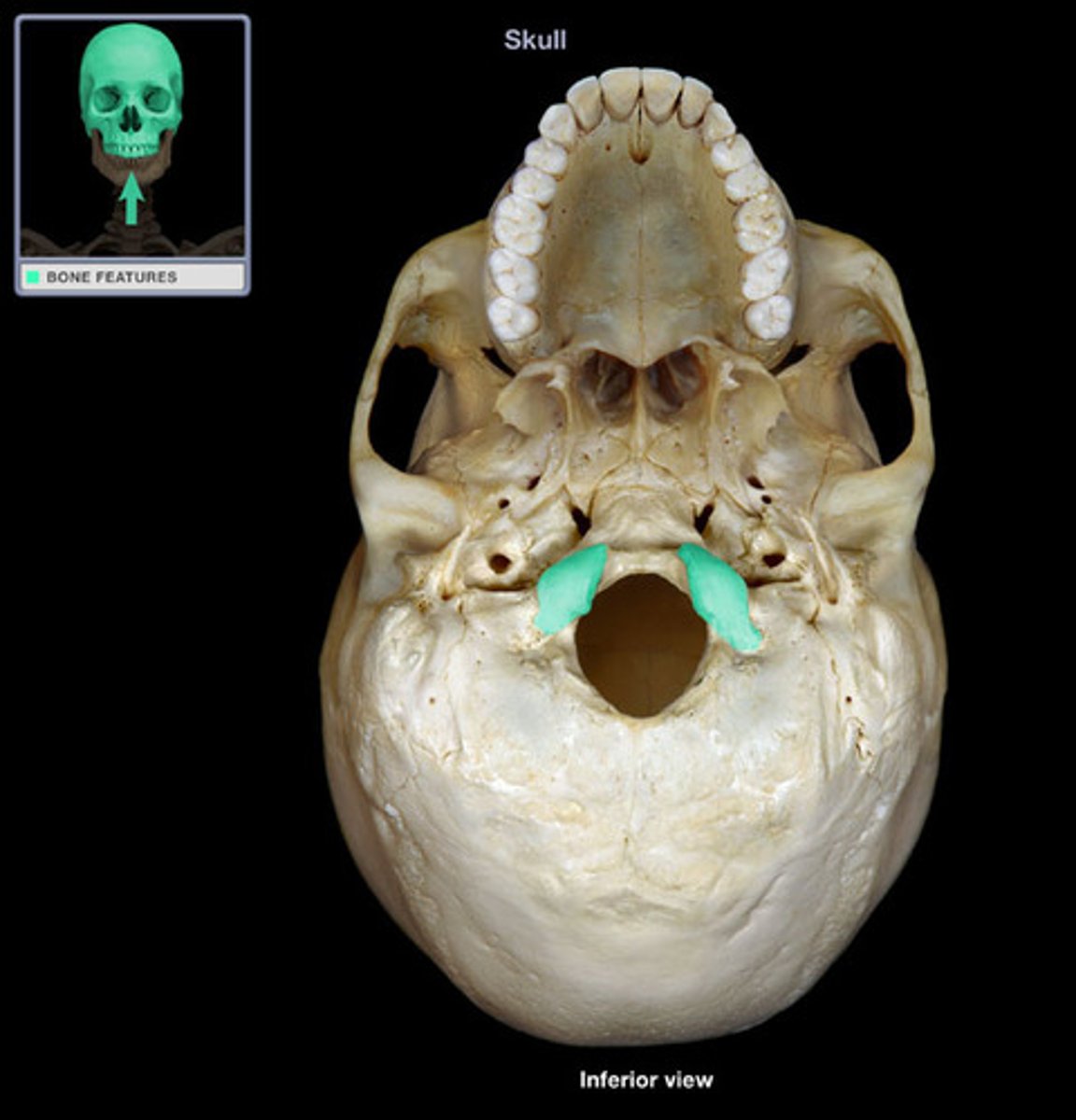
Occipital condyle
-round prominence on right and left of foramen magnum
bregma (junction of coronal and sagittal sutures)
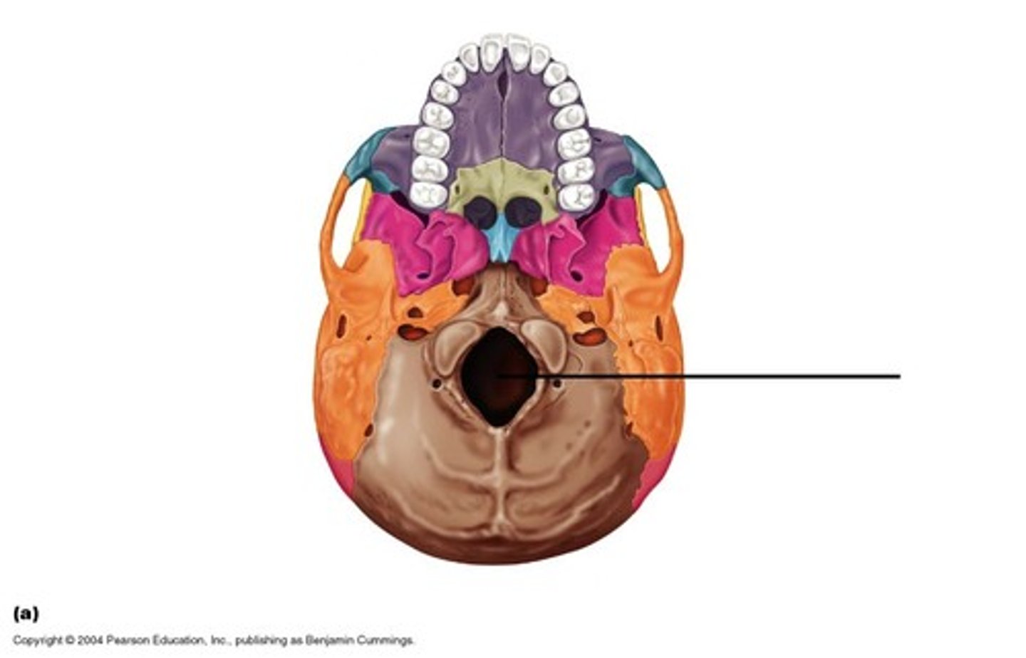
Foramen magnum
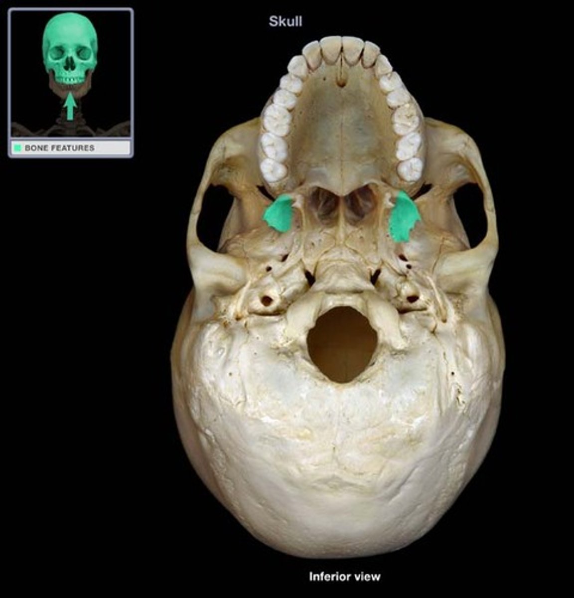
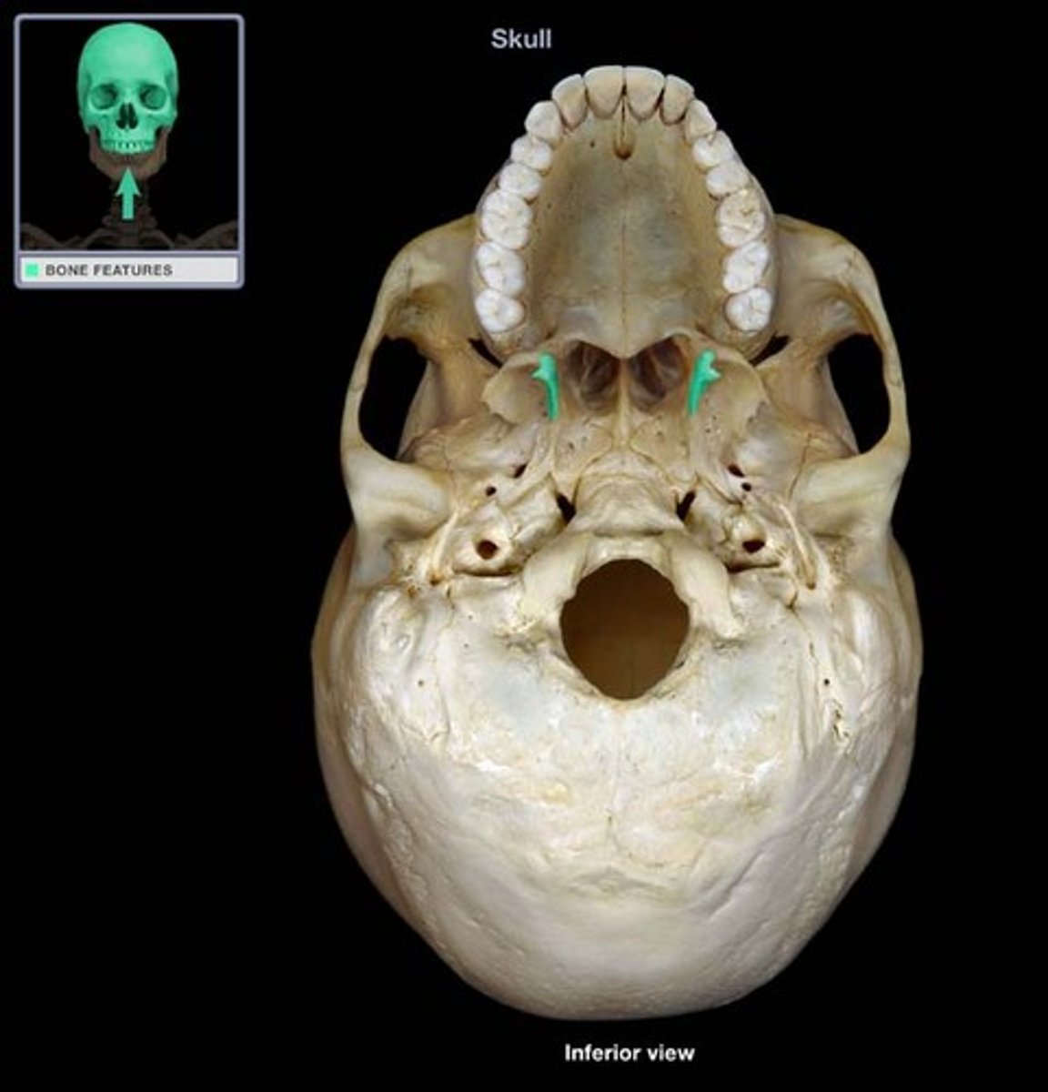
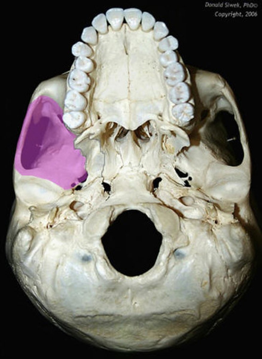
Lateral pterygoid plate
-part of sphenoid bone
Att: Lateral pterygoid muscle, medial pterygoid muscle
N: Mandibular N
What attaches here and their Nerves?
Medial pterygoid plate
-part of sphenoid bone
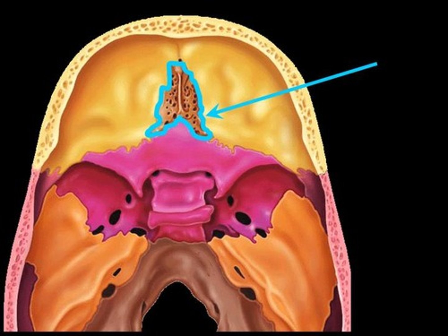
Cribriform plate
-part of ethmoid bone
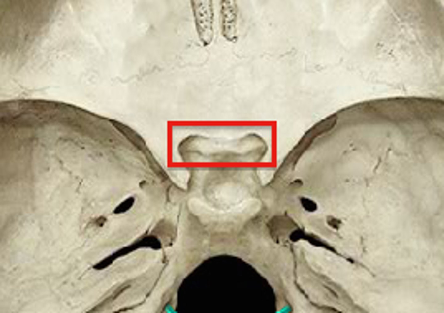
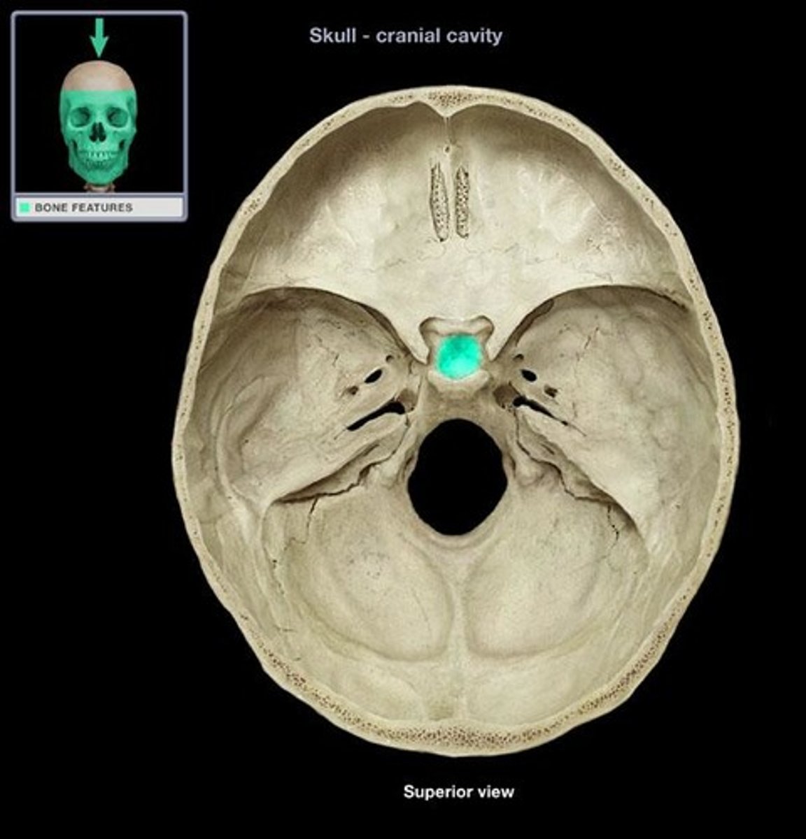
Tuberculum sellae
-part of sphenoid bone; sella turcica
Hypophysial fossa
-depression in sella turcica
temporal fossa
infratemporal fossa
Condylar process
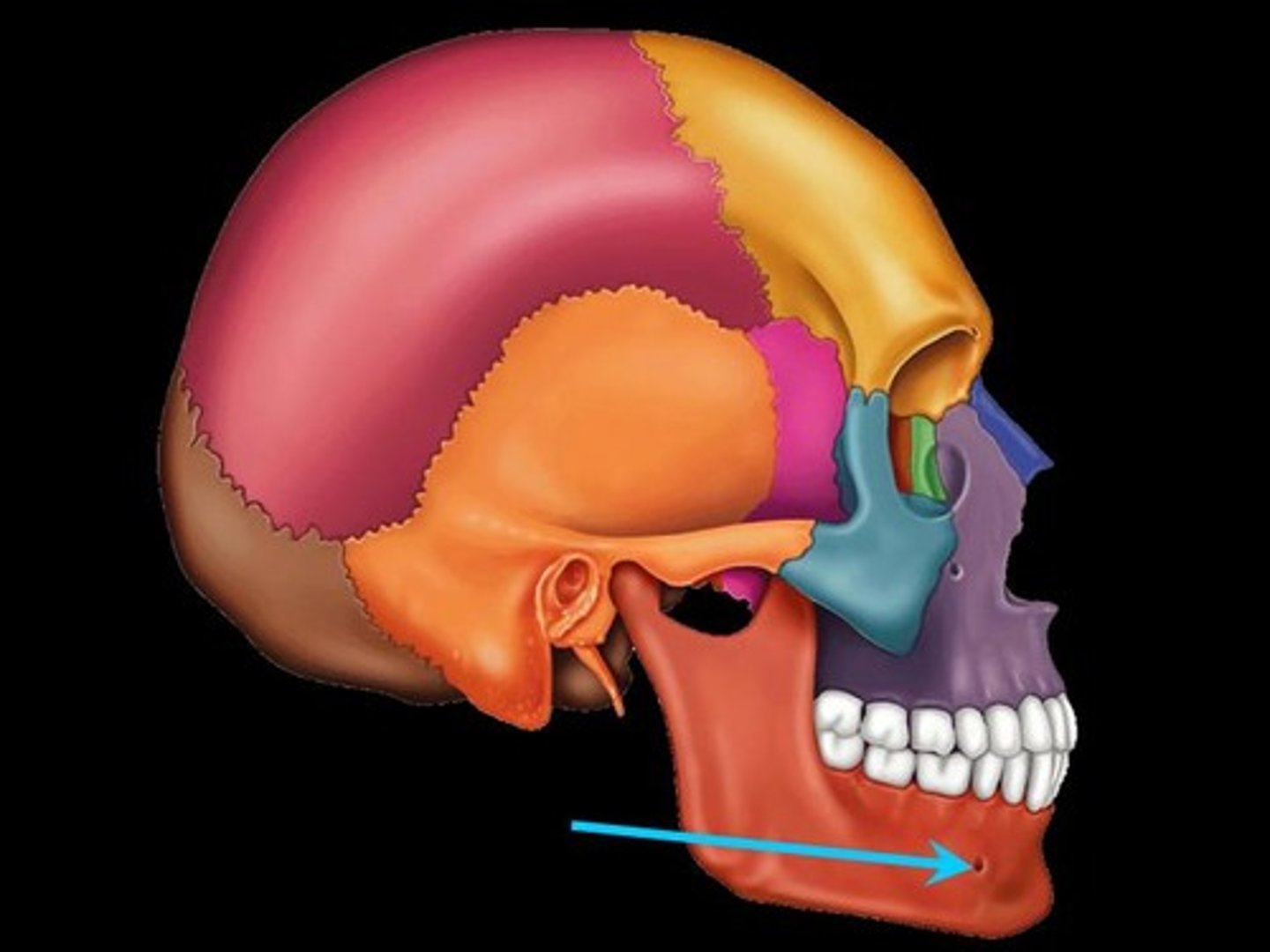
mental foramen
mental protuberance
Alveolar process
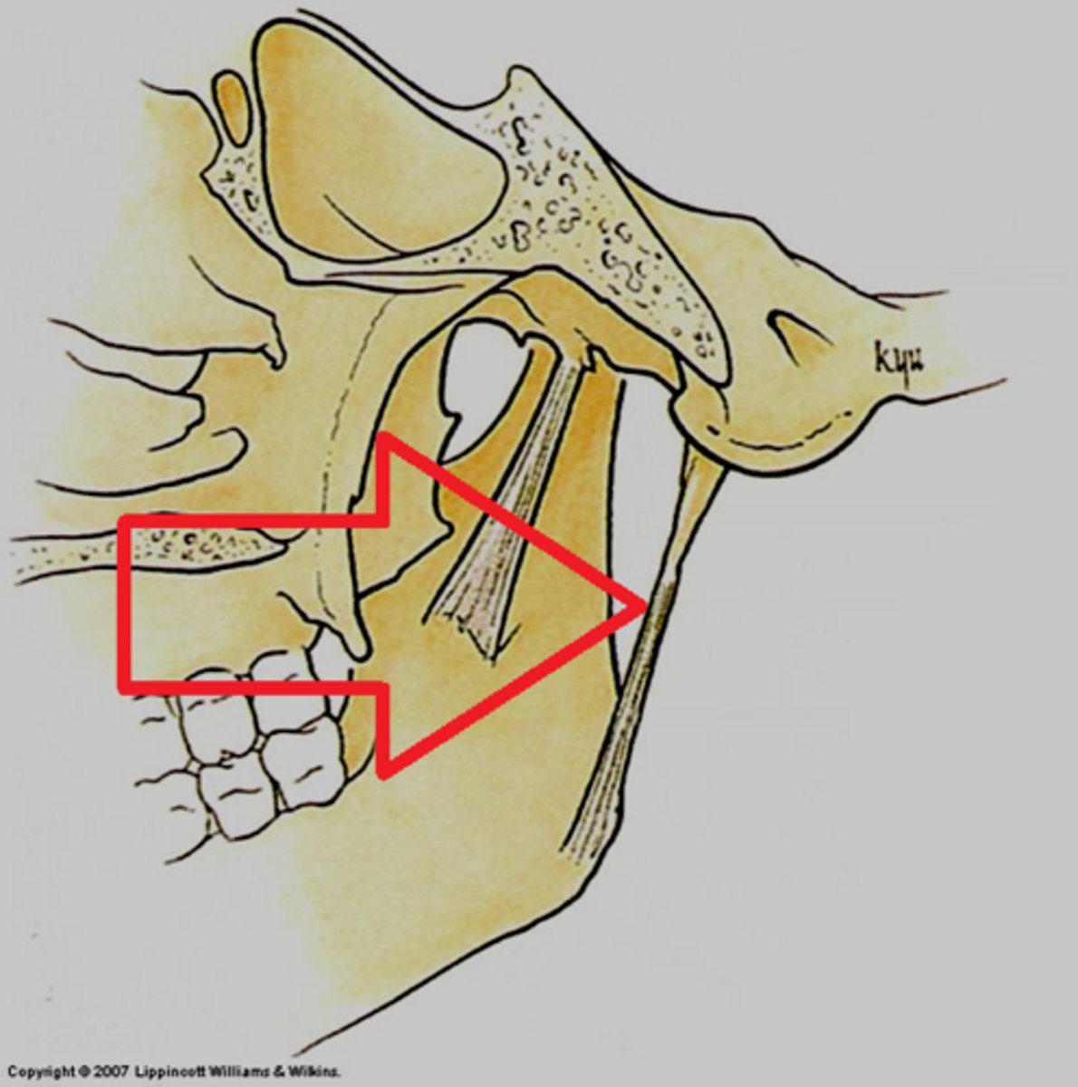
sphenomandibular ligament
-runs from sphenoid bone to ramus of mandible
Function: pivot point; allows the jaw to swing
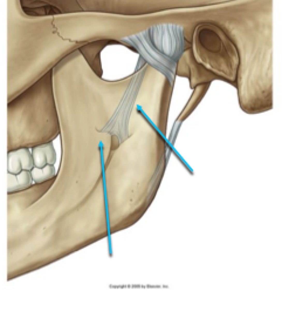
Stylomandibular ligament
-runs from styloid process to angle of mandible
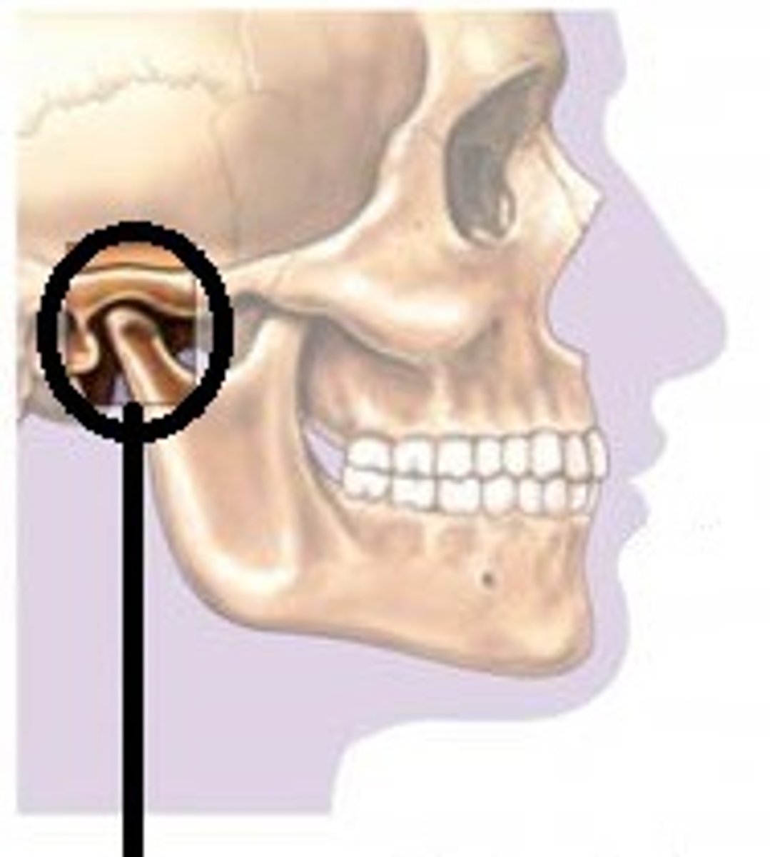
TMJ joint
-modified hinge joint; allows for movement in 3 planes
-Articular surfaces: Head of mandible, articular tubercle (of temporal bone), and mandibular fossa
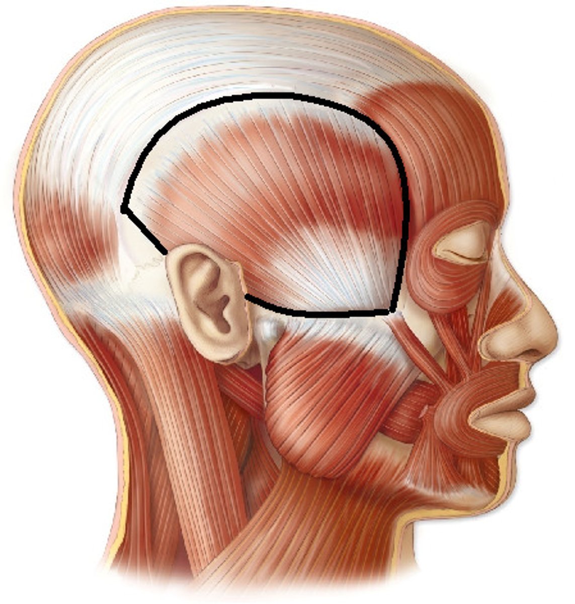
temporalis muscle
Attachments: temporal fossa, coronoid process
-passed behind/deep to zygomatic jaw
Function: elevates mandible, closes jaw, retracts
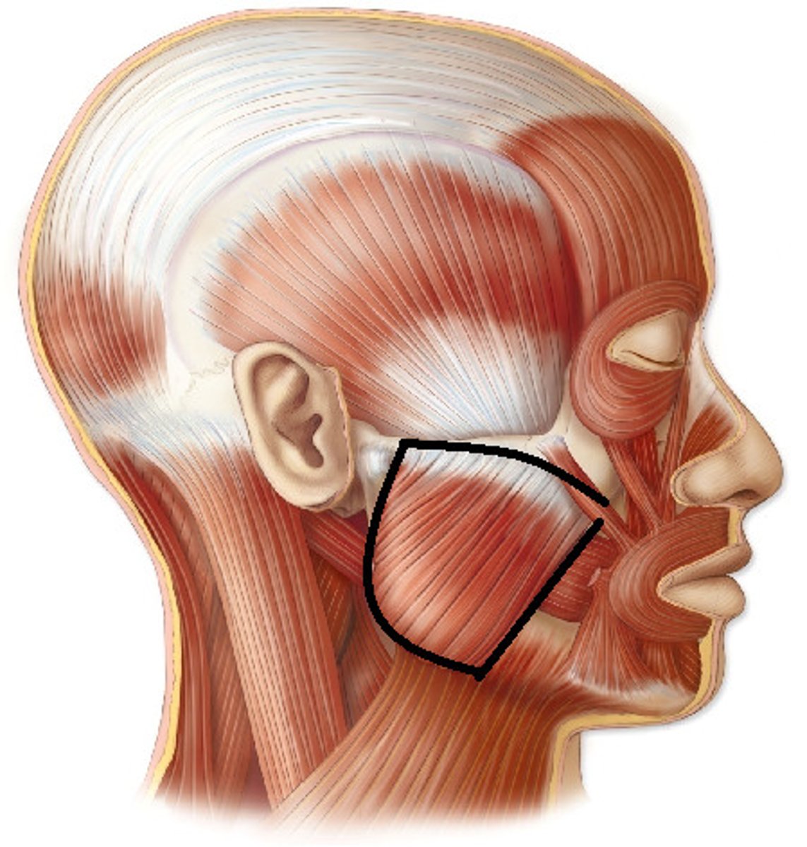
Masseter
Attachments: maxillary process of zygomatic bone and Arch, Ramus of mandible (**Class notes: Zygomatic arch-->Angle of Mandible)
Function: elevates mandible
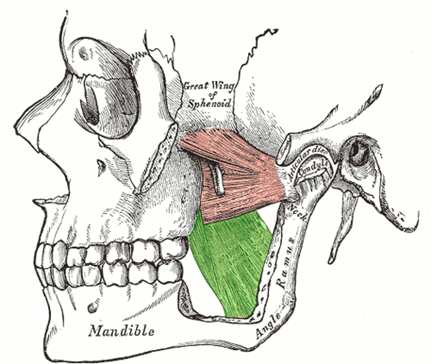
Medial pterygoid
Attachments: Lateral pterygoid plate, tuberosity of maxilla, Ramus of mandible
Function: elevates mandible
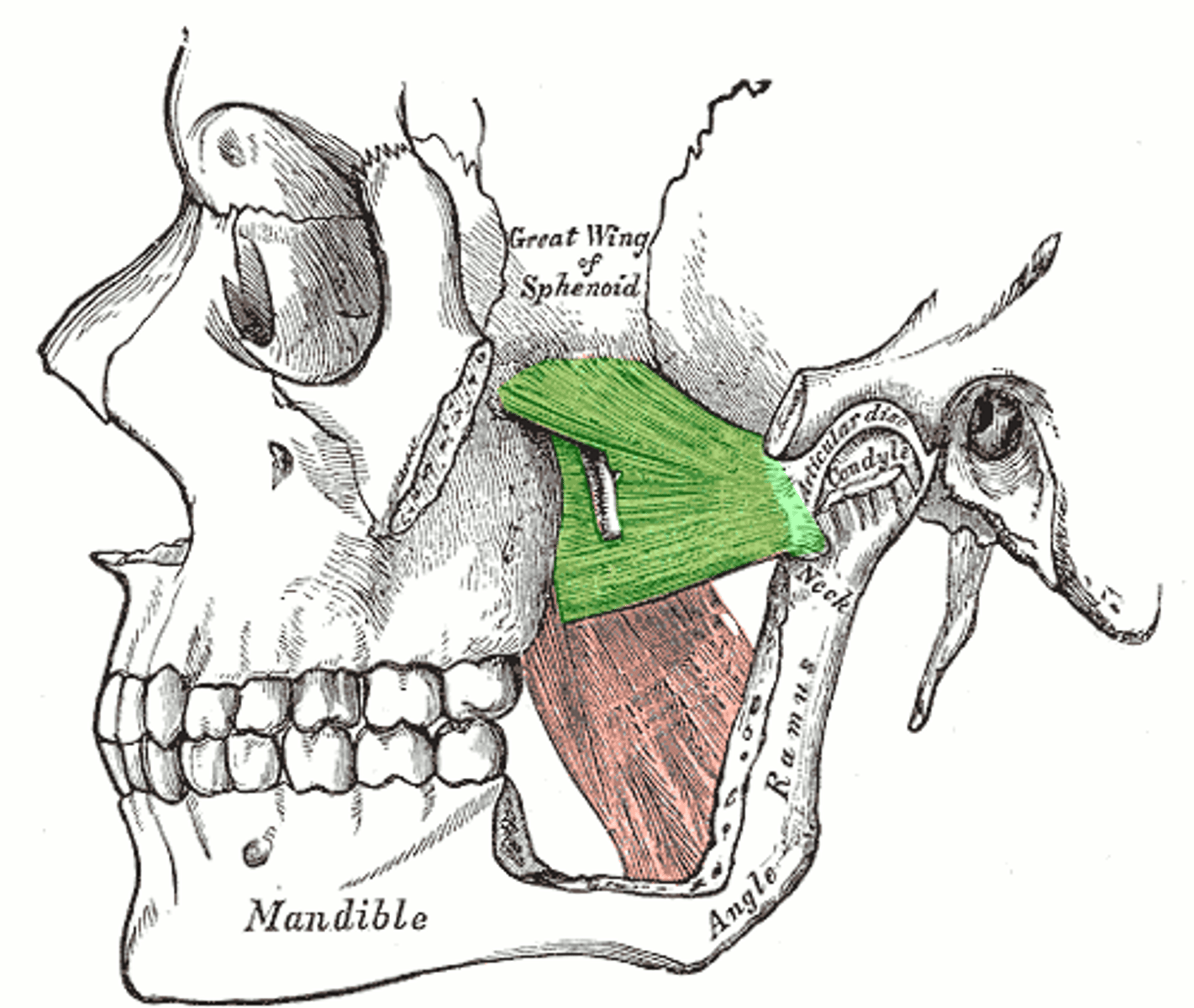
Lateral pterygoid
Attachments: Greater wing of sphenoid and lateral pterygoid plate, Joint capsule and articular disc of TMJ
Function: protracts mandible and swings jaw side to side (contralateral jaw movements, like when a cow eats)
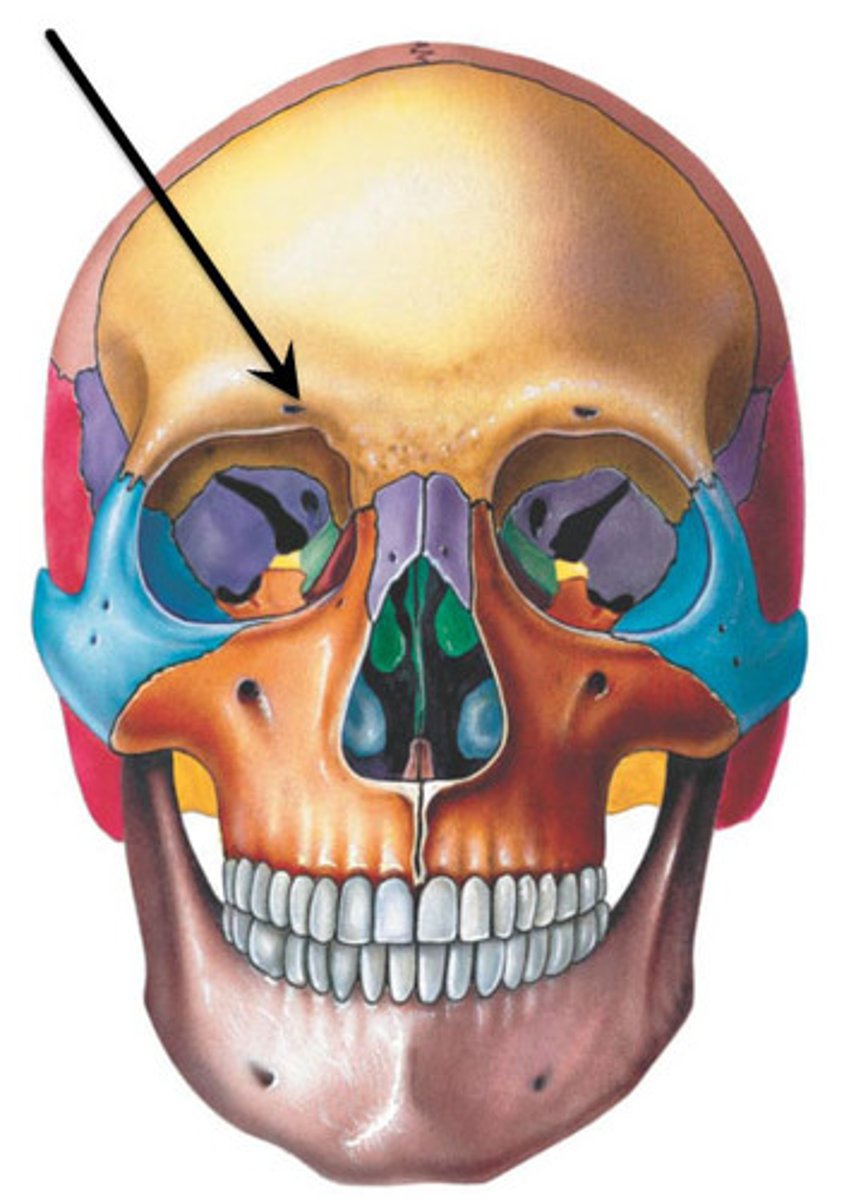
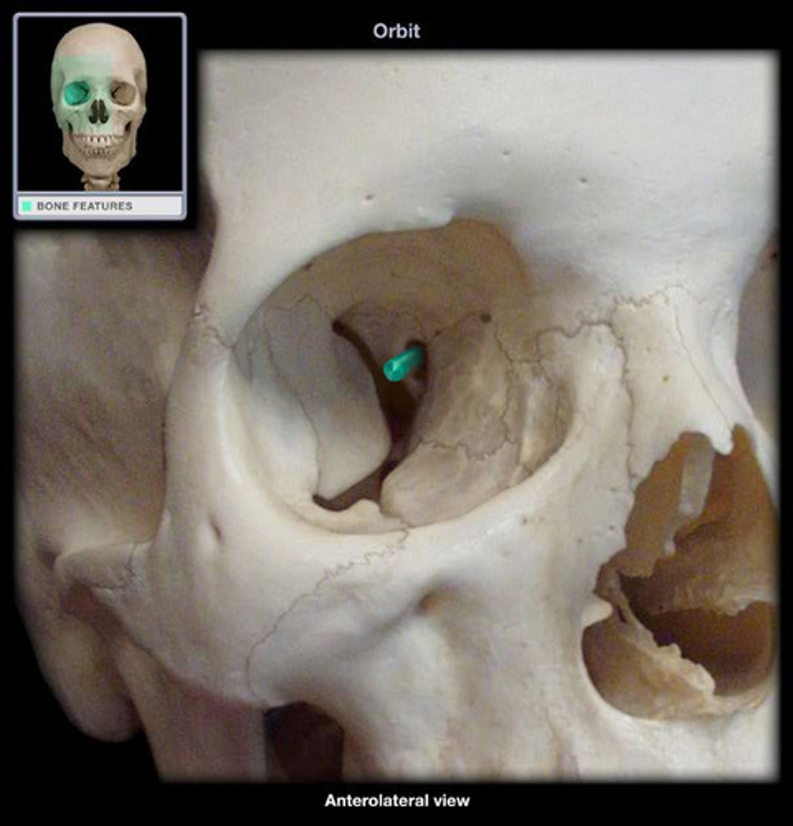
Supra orbital notch
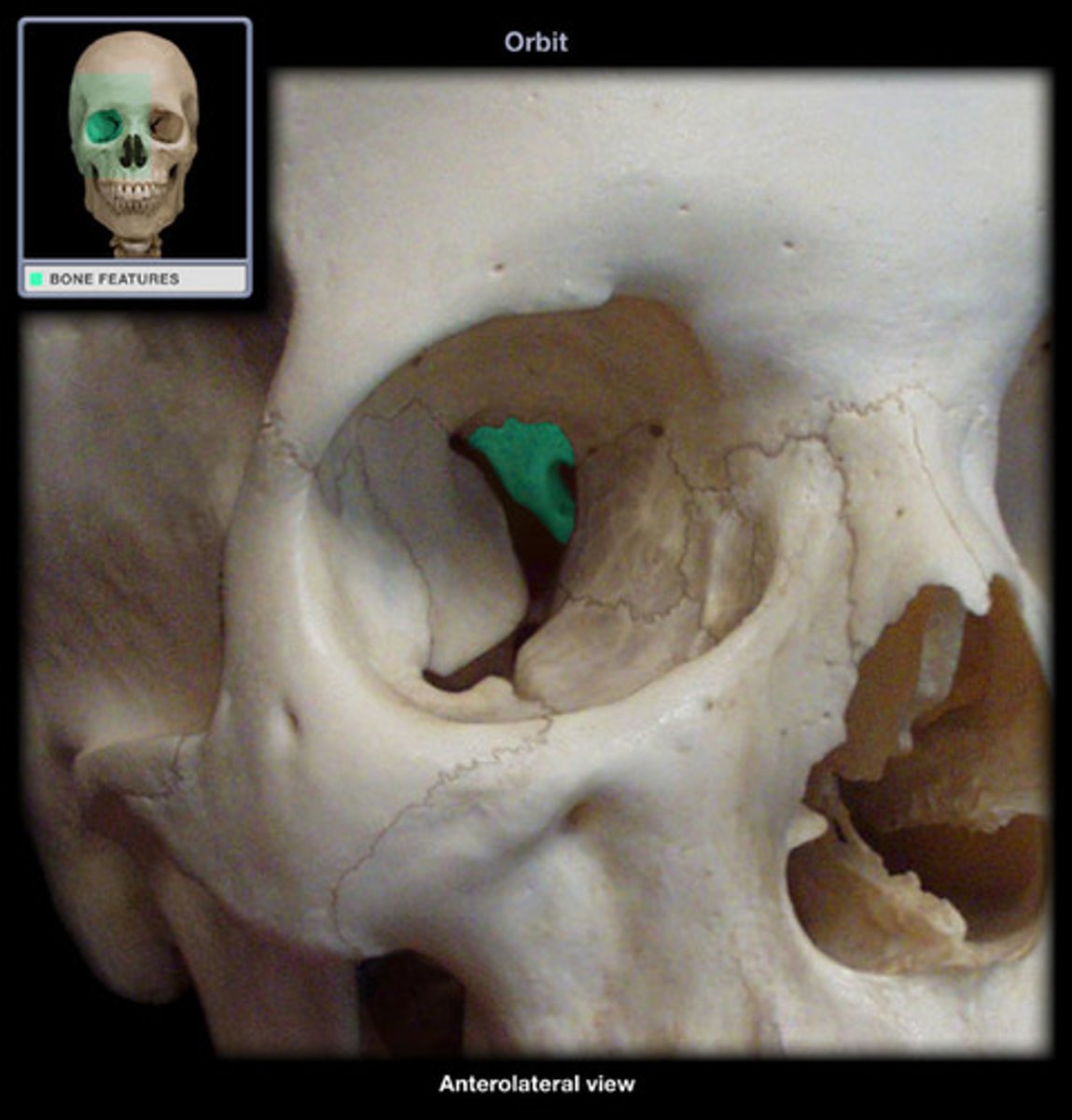
Greater wing of sphenoid
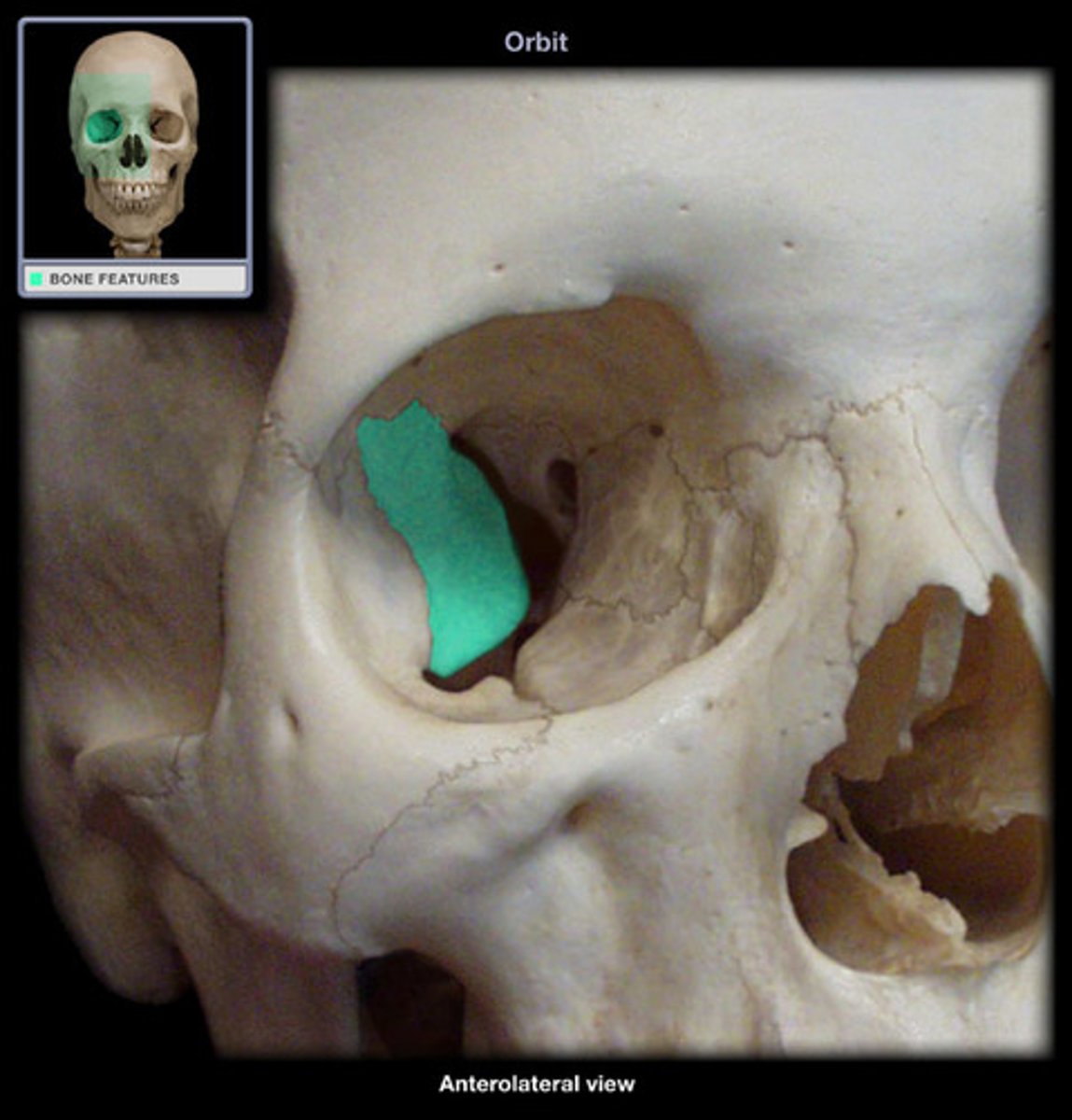
Lesser wing of sphenoid
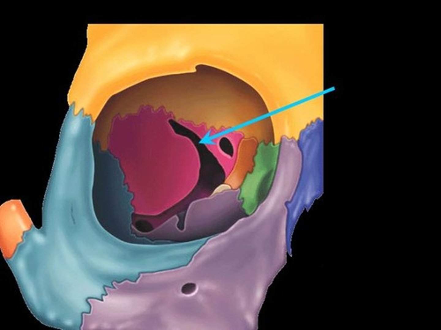
Optical canal
Superior orbital fissure
Inferior orbital fissure
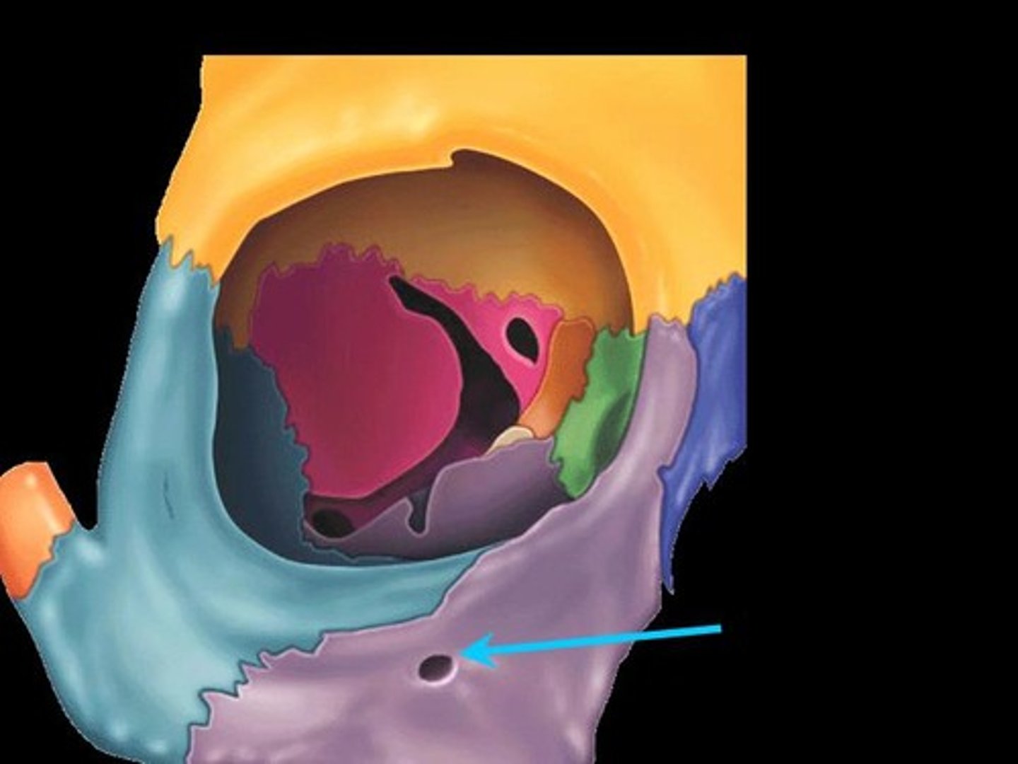
Infraorbital foramen
Mandibular nerve
Nerve supply to the muscles of mastication?
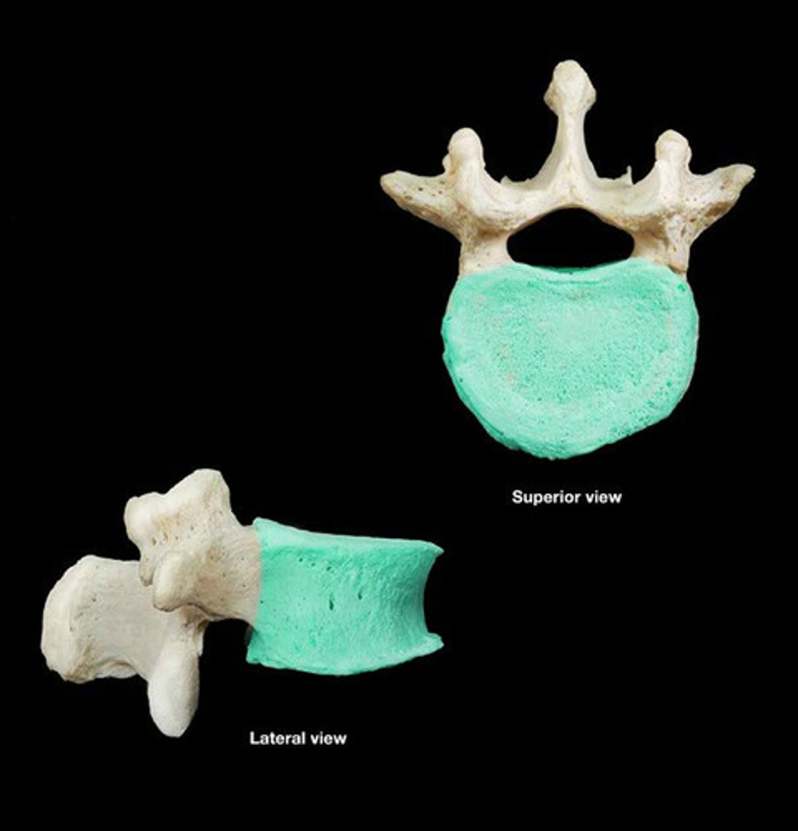
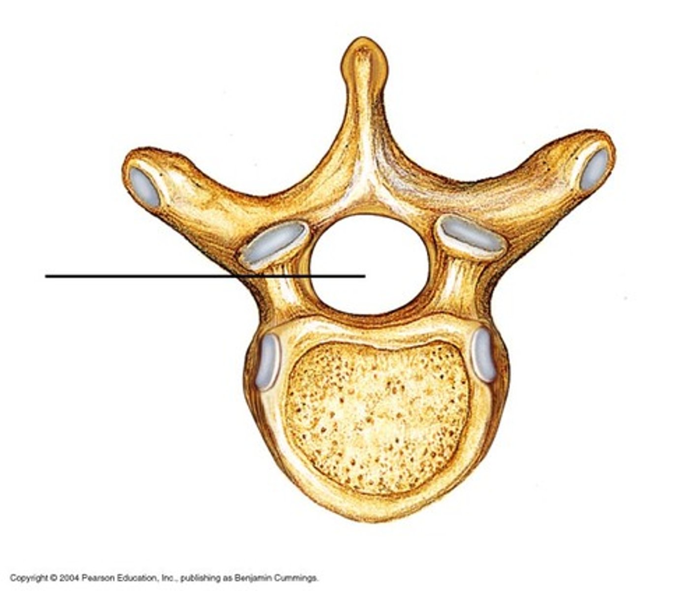
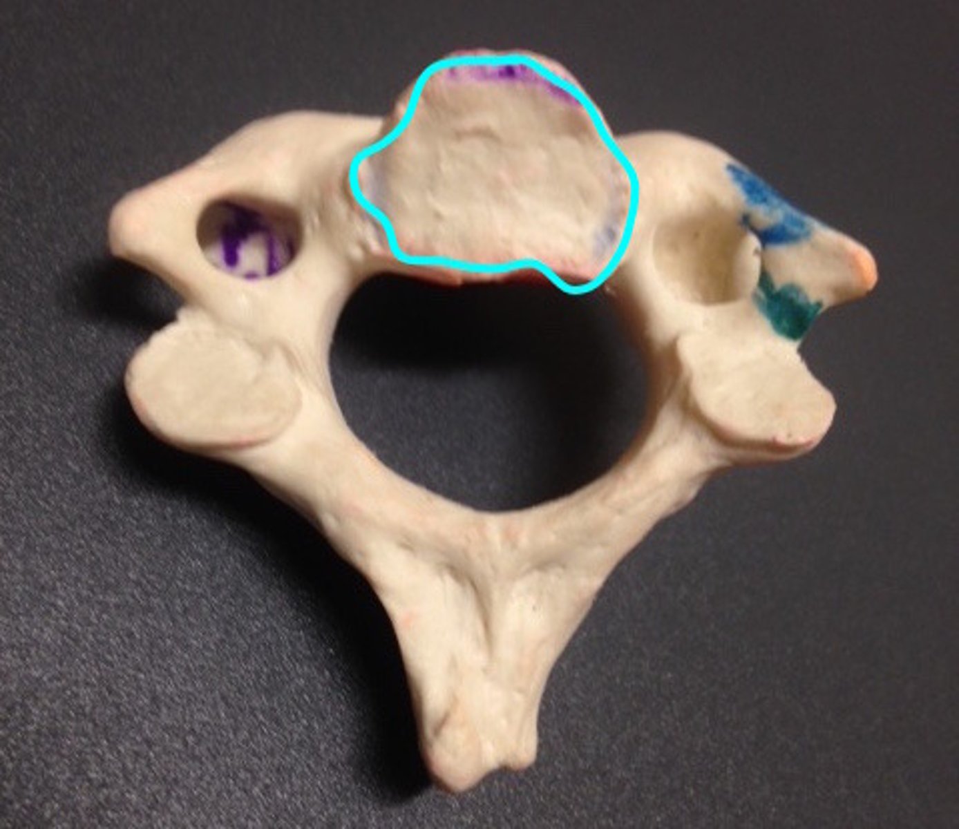
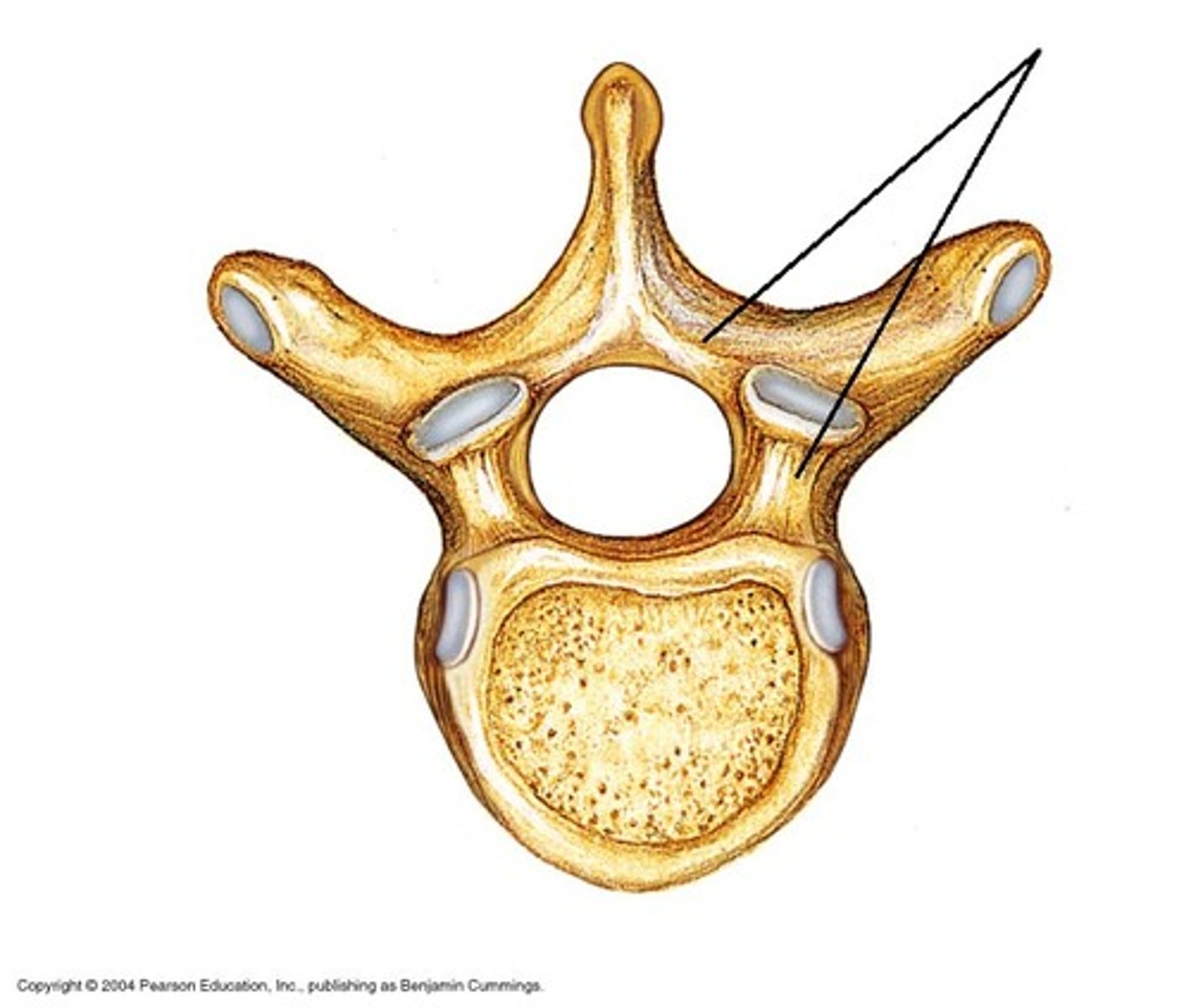
Vertebral body
Function: supports body weight
Vertebral Arch
-Formed by lamina and pedicles
Function: wraps around spinal cord and shields it/PROTECTS
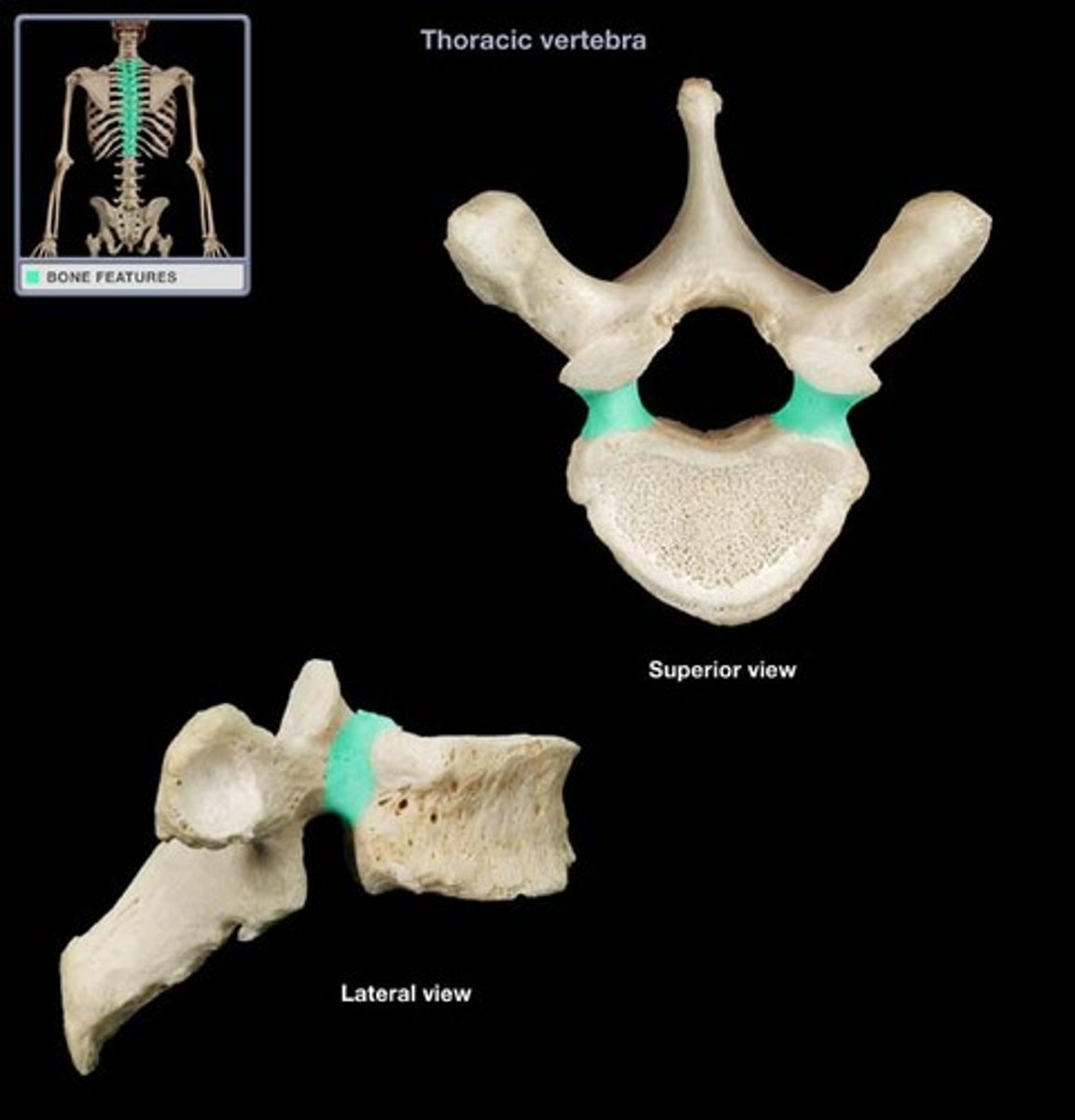
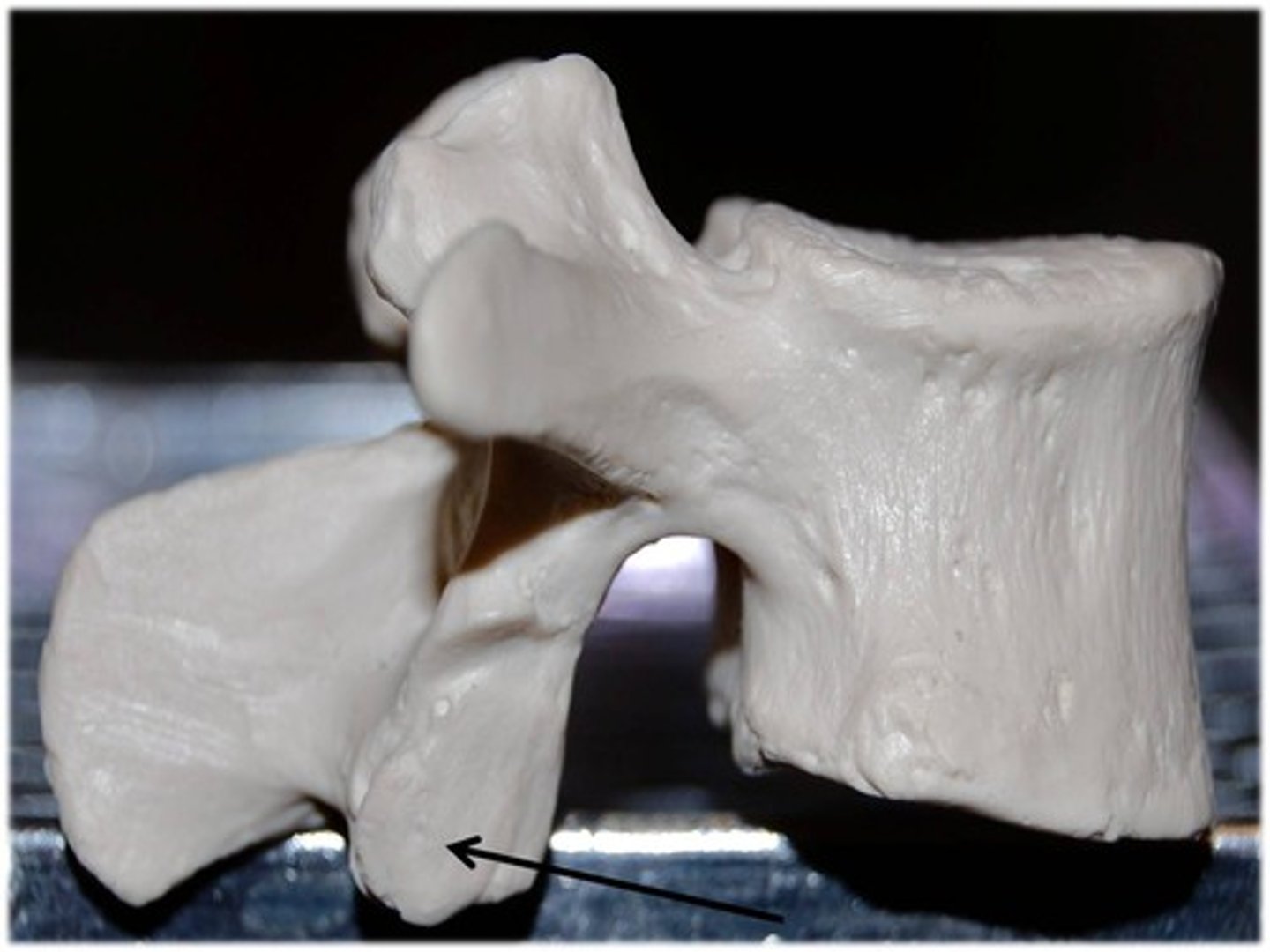
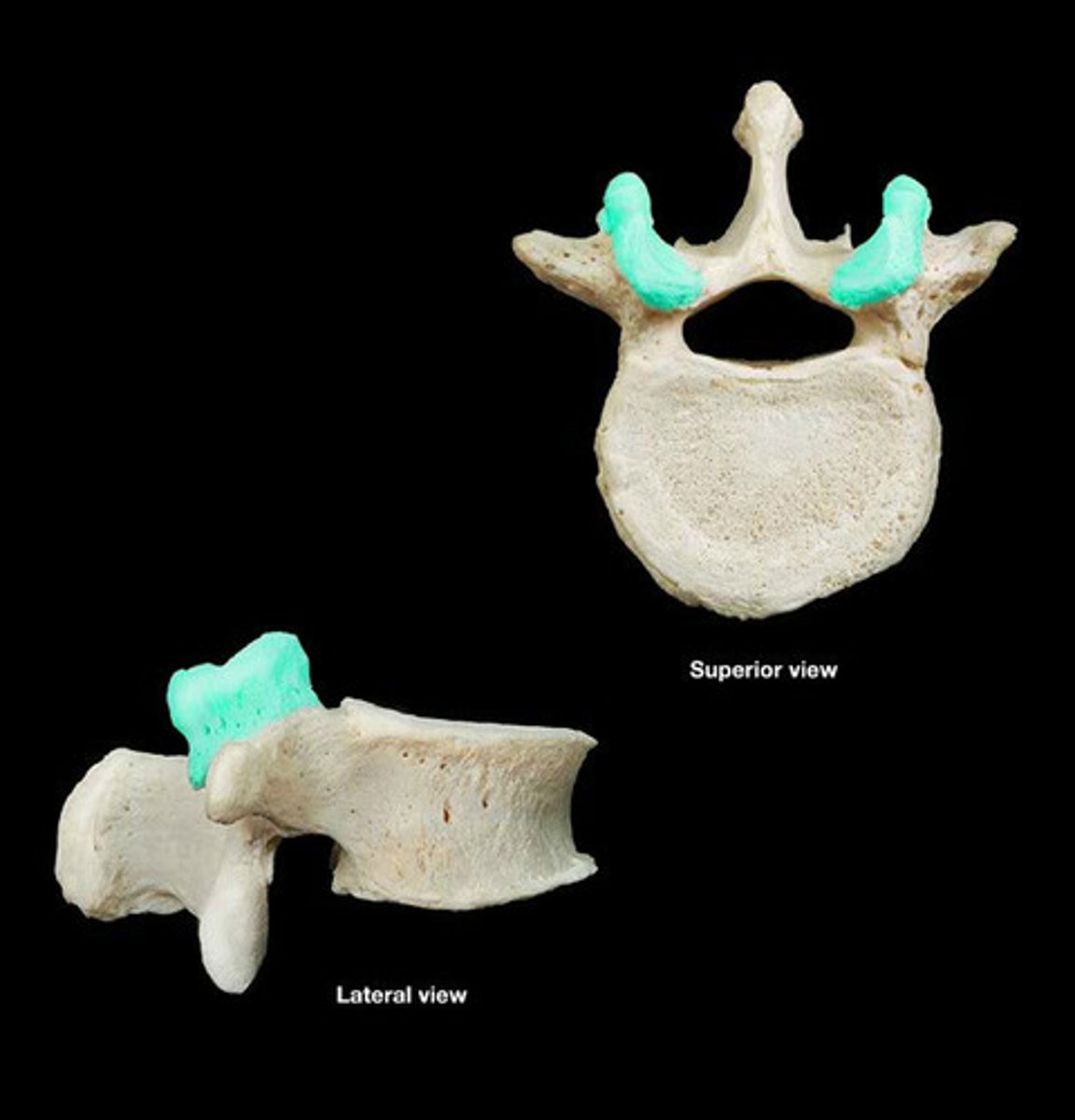
Articular processes
Function: lock vertebra in place. These reach up and down, thus restrict movement
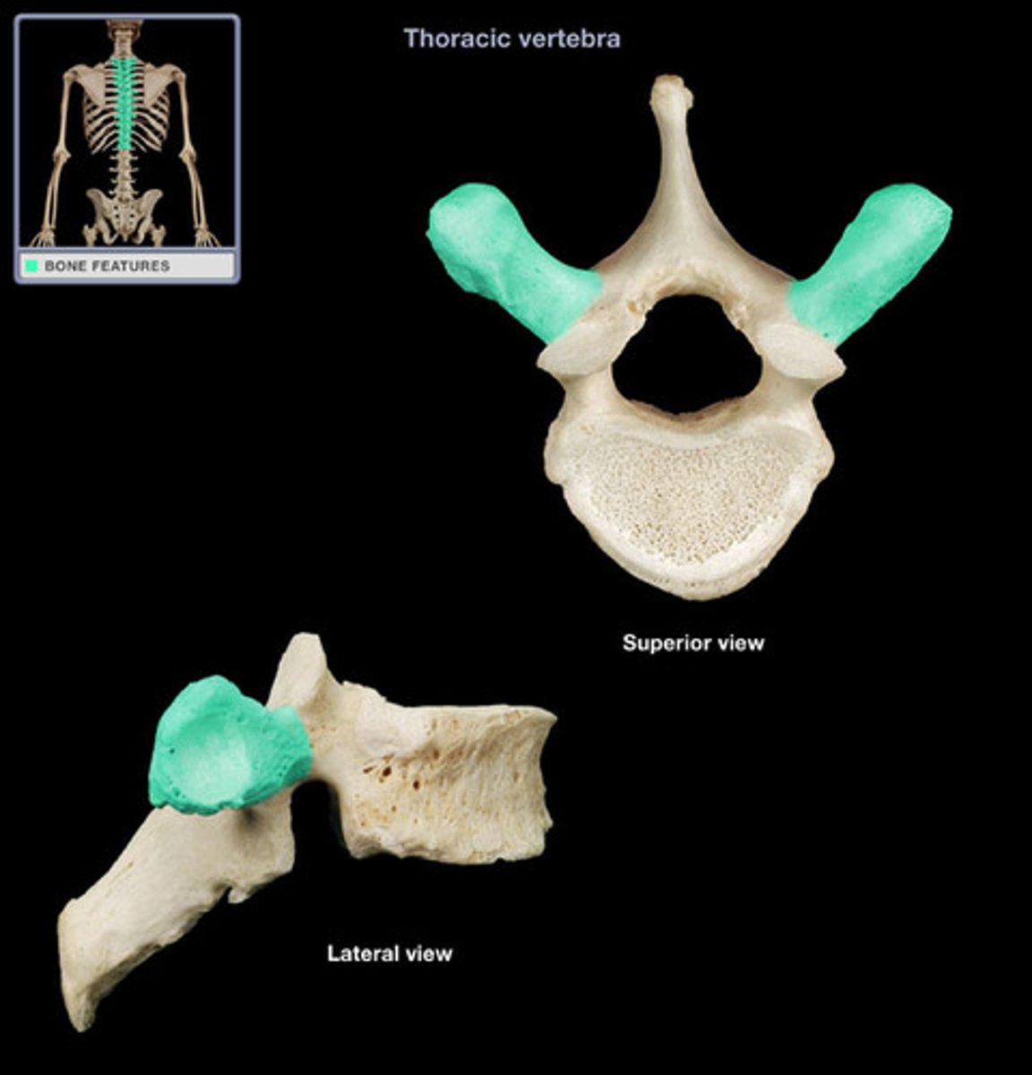
Transverse process
Function: muscle attachments and movement. These stick out a lot so things can attach.
-Allows for increase strength.
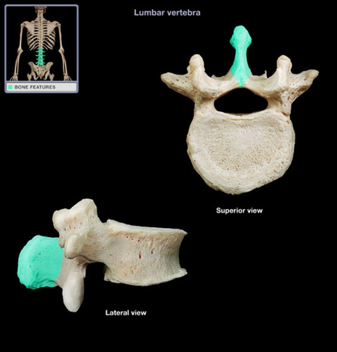
Spinous process
Function: muscle attachments and movement. These stick out a lot so things can attach.
-Allows for increase strength.
Pedicles
Lamina
Vertebral foramen
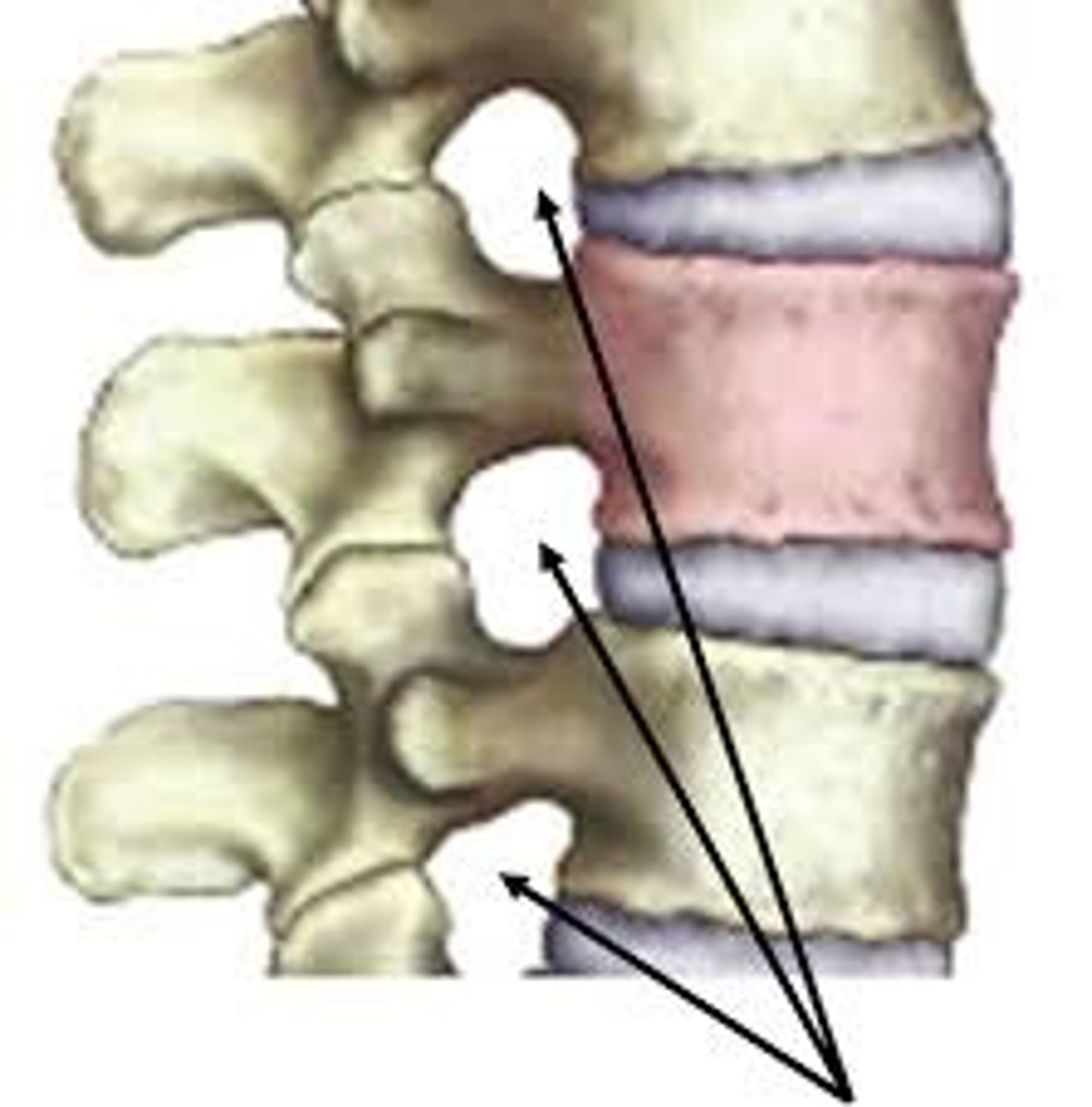
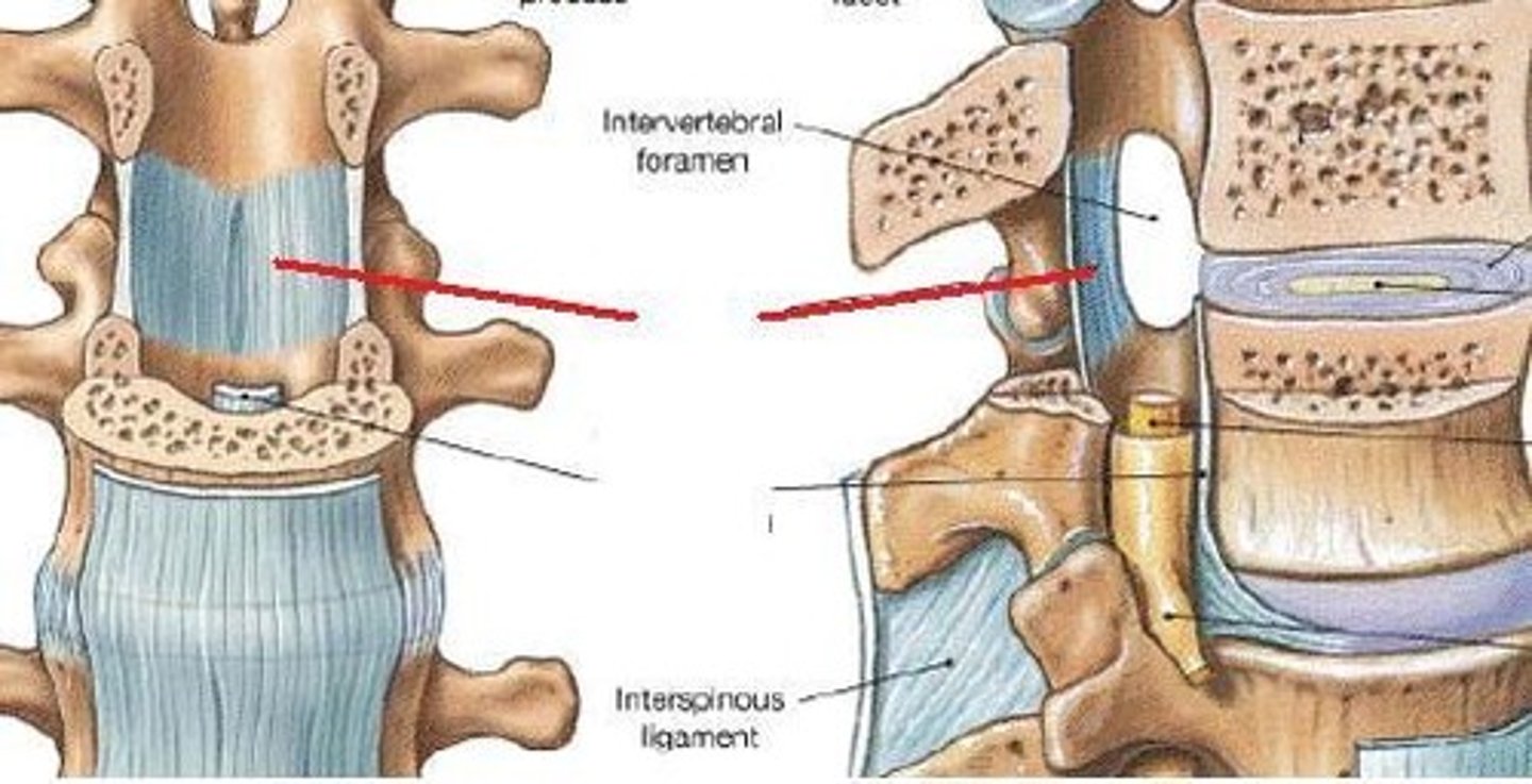
Intervertebral foramina
-doorway between spinal cord and periphery. Nerves and vessels pass through
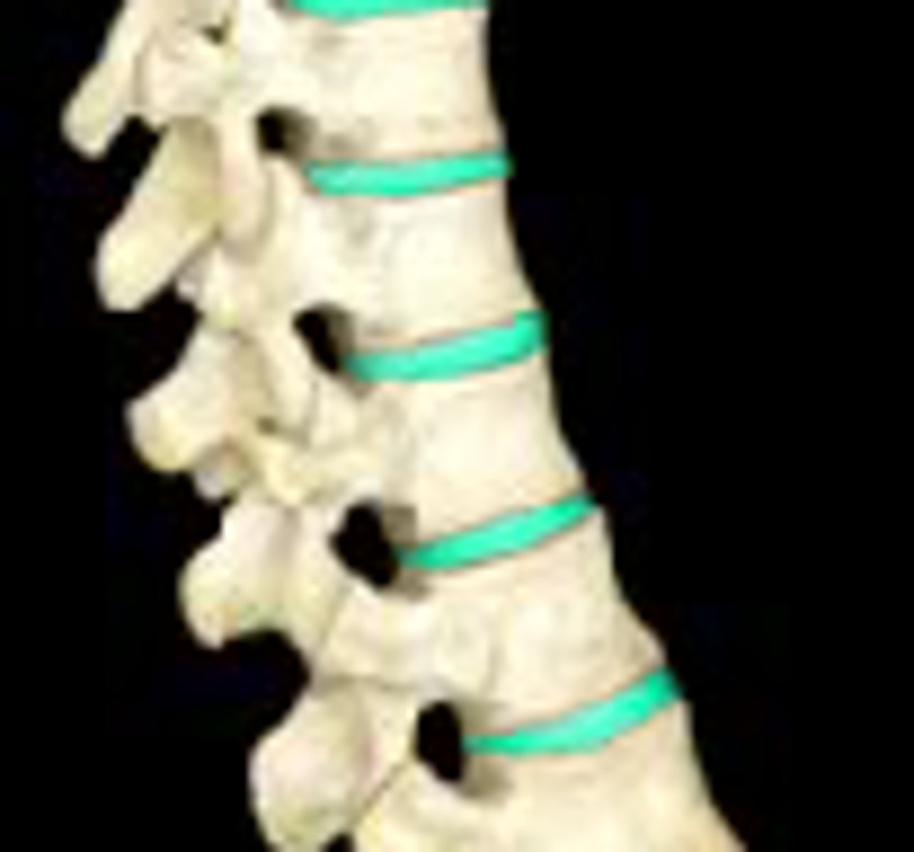
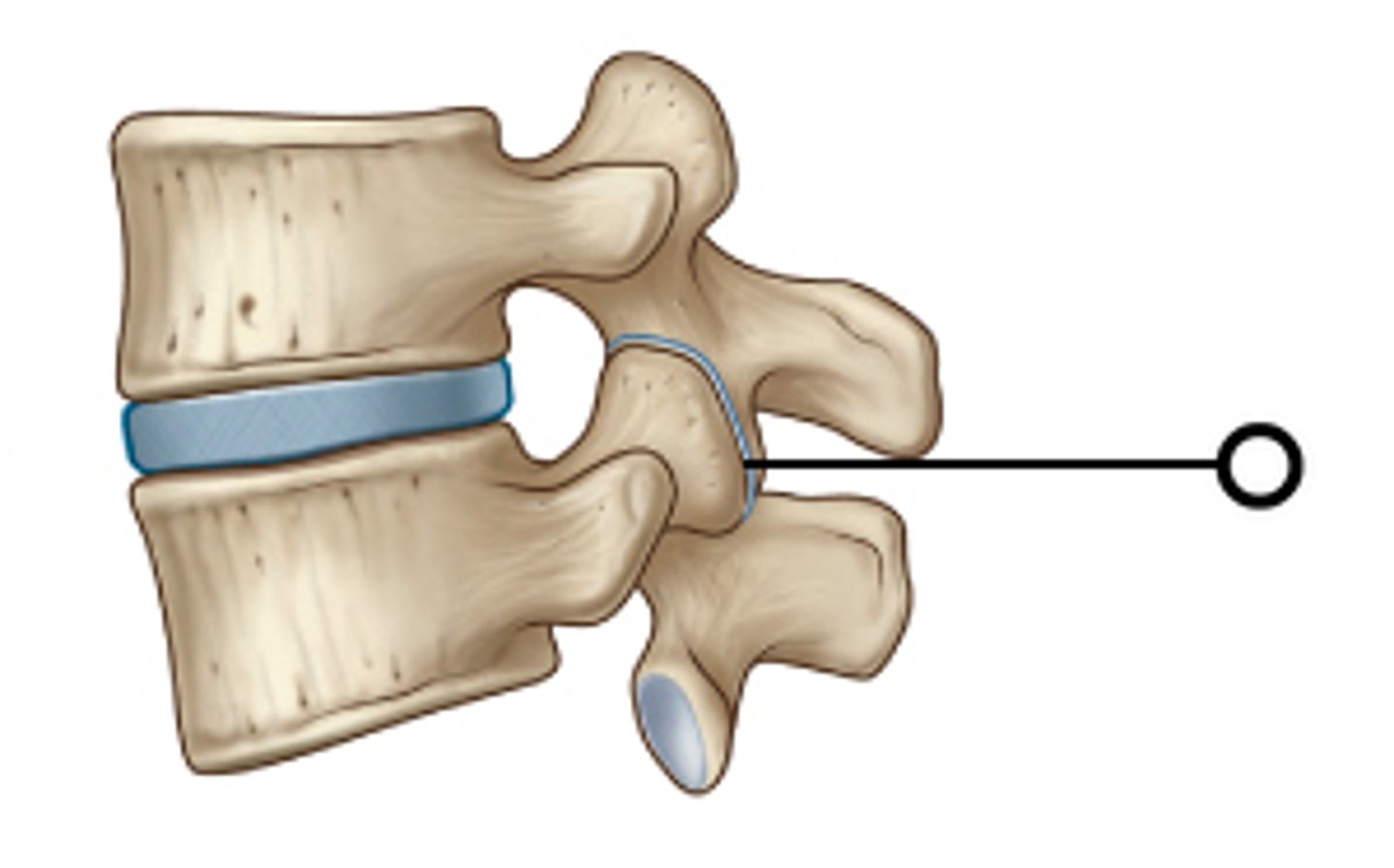
Intervertebral disc
-fibrocartilage between vertebral bodies; Acts as ligament and shock absorber
-permits movement between adjacent vertebrae
-provides strong attachments between vertebral bodies
Zygapophysial joint
(facet joint)
Formed by: superior and inferior articular processes
Function: 1. facilitate and control vertebral column flexibility
2. provides structural stability to vertebral column as a whole
3. guides and constrains motions in spine due to their geometry
Inferior articular process
Superior articular process
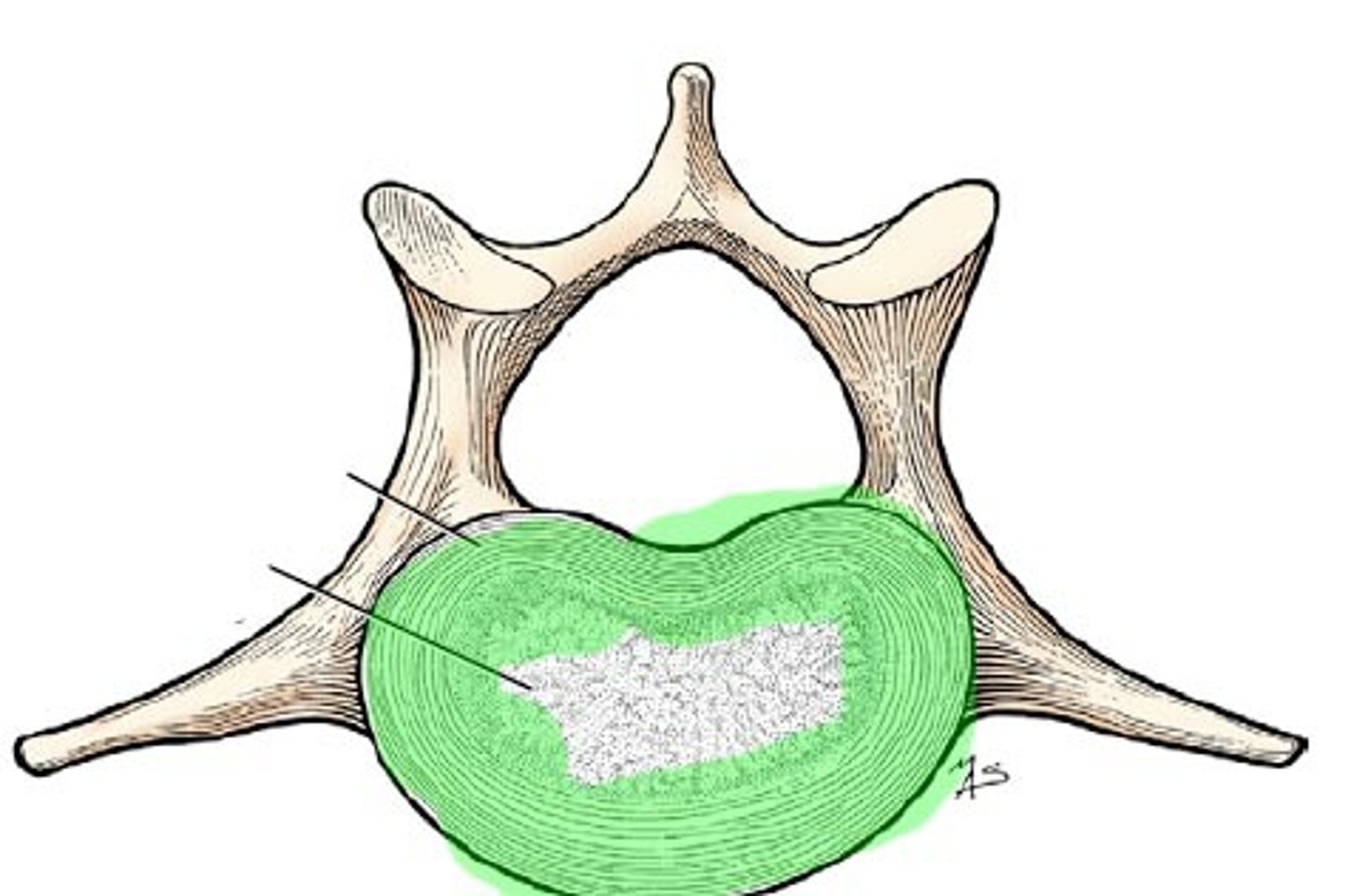
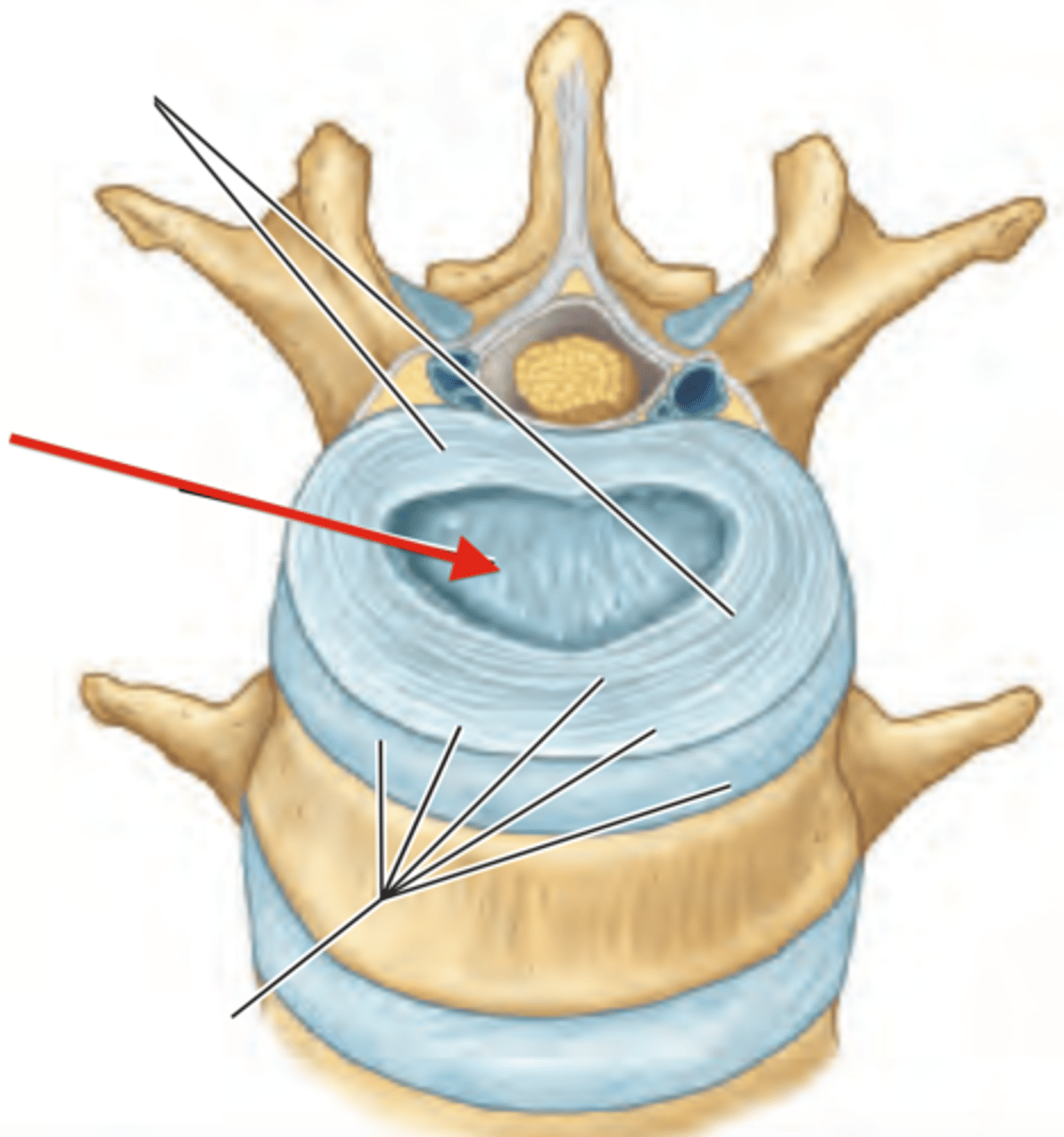
Annulus fibrosus
-fibrocartilage outer portion of intervertebral disc
Nucleus pulposus
-center of intervertebral disc
*closer to the back/canal thus prone to herniating/moving backward
-85% water at birth, dehydrate as we age - then IV discs lose strength and become stiffer, more resistant to deformation
Ligamentum flavum
Runs between lamina - from lamina above to lamina below. Yellowish
FUNCTION: prevents flexion (resists separation of lamina)
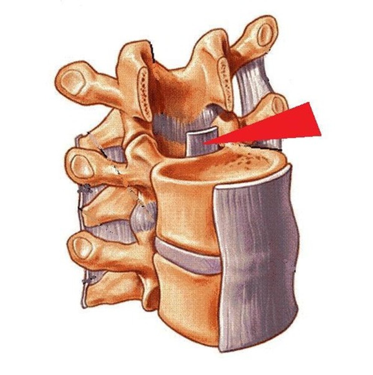
Posterior longitudinal ligament
-inside canal, mainly attached to IV discs.
Runs C-spine --> sacrum; Runs along posterior aspect of vertebral bodies
-not very wide
FUNCTION: 1. prevents hyperflexion of vertebral column
2. prevents herniation of IV discs
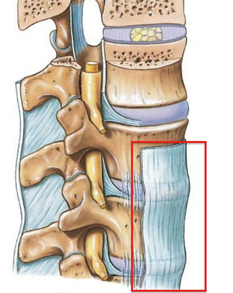
Anterior longitudinal ligament
-Wide, strong, broad
-connects anterolateral aspects of vertebral bodies and IV discs
-Runs from sacrum to C-1 and occipital bone
FUNCTION: limit extension of vertebral column; maintain stability of IV joints
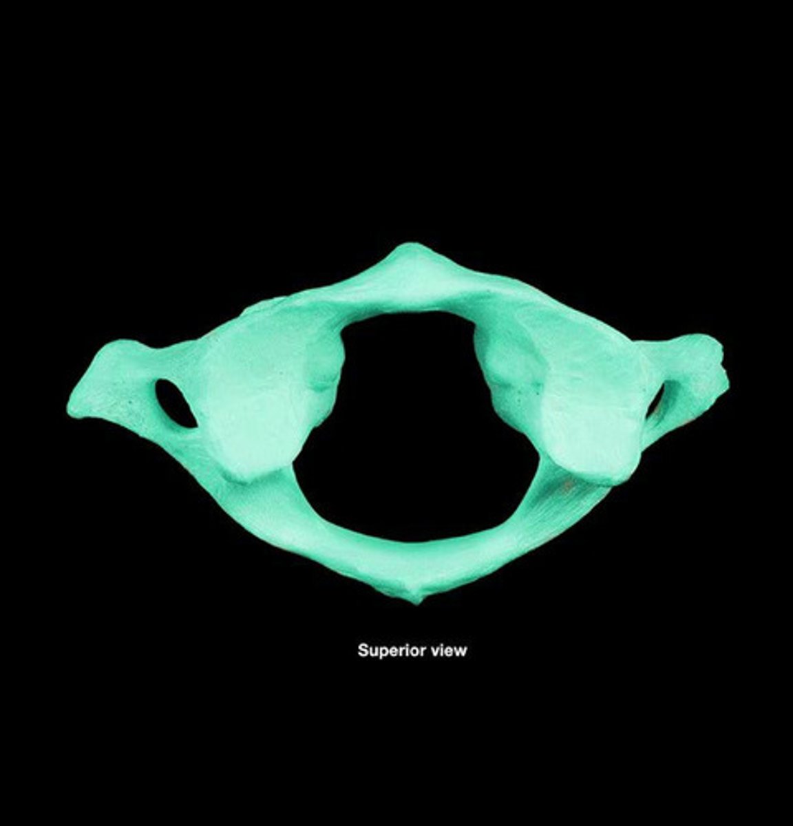
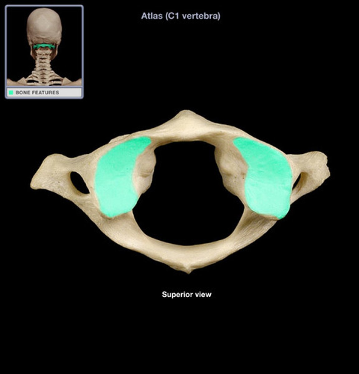
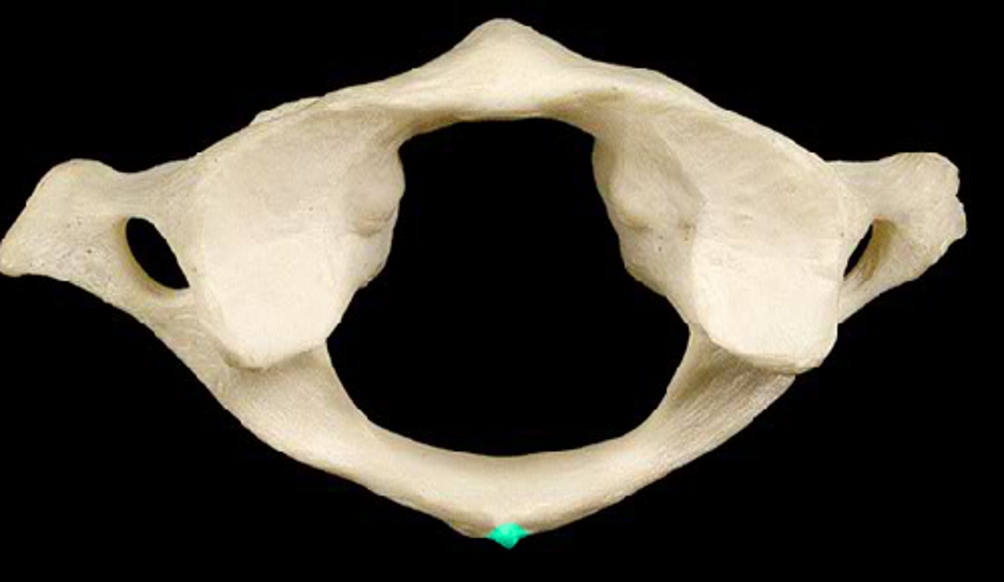
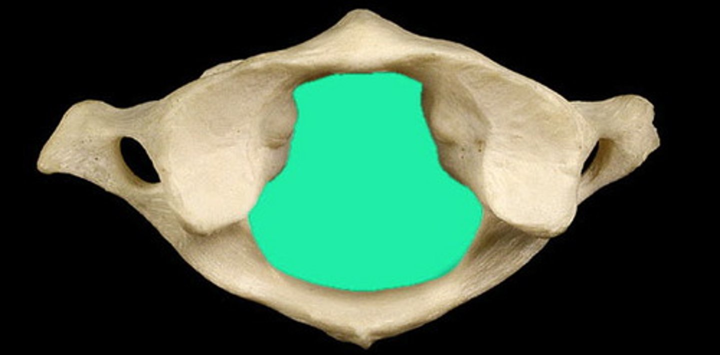
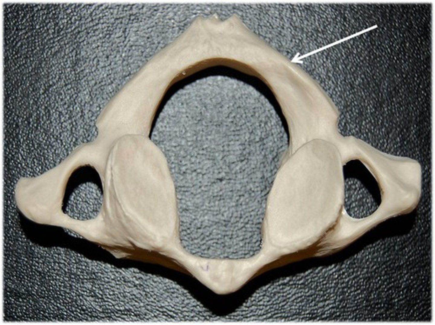
C-1 Atlas
Function: support the skull
-No body
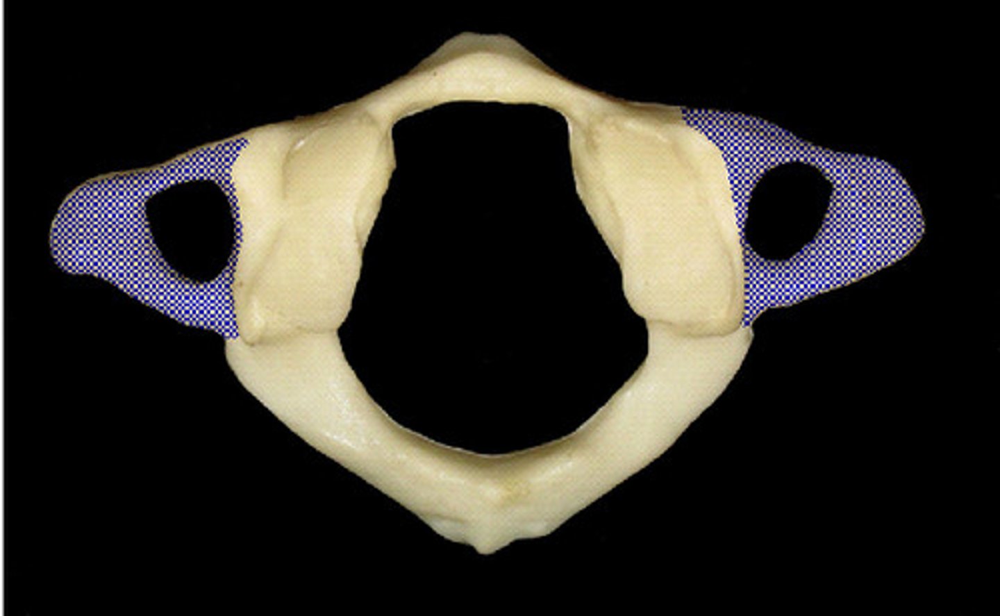
-wide transverse process
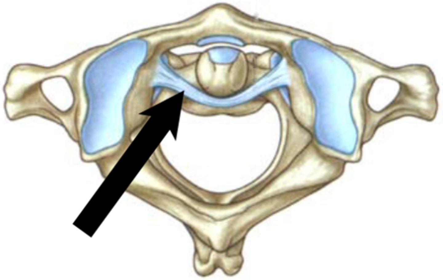
-transverse ligament of atlas
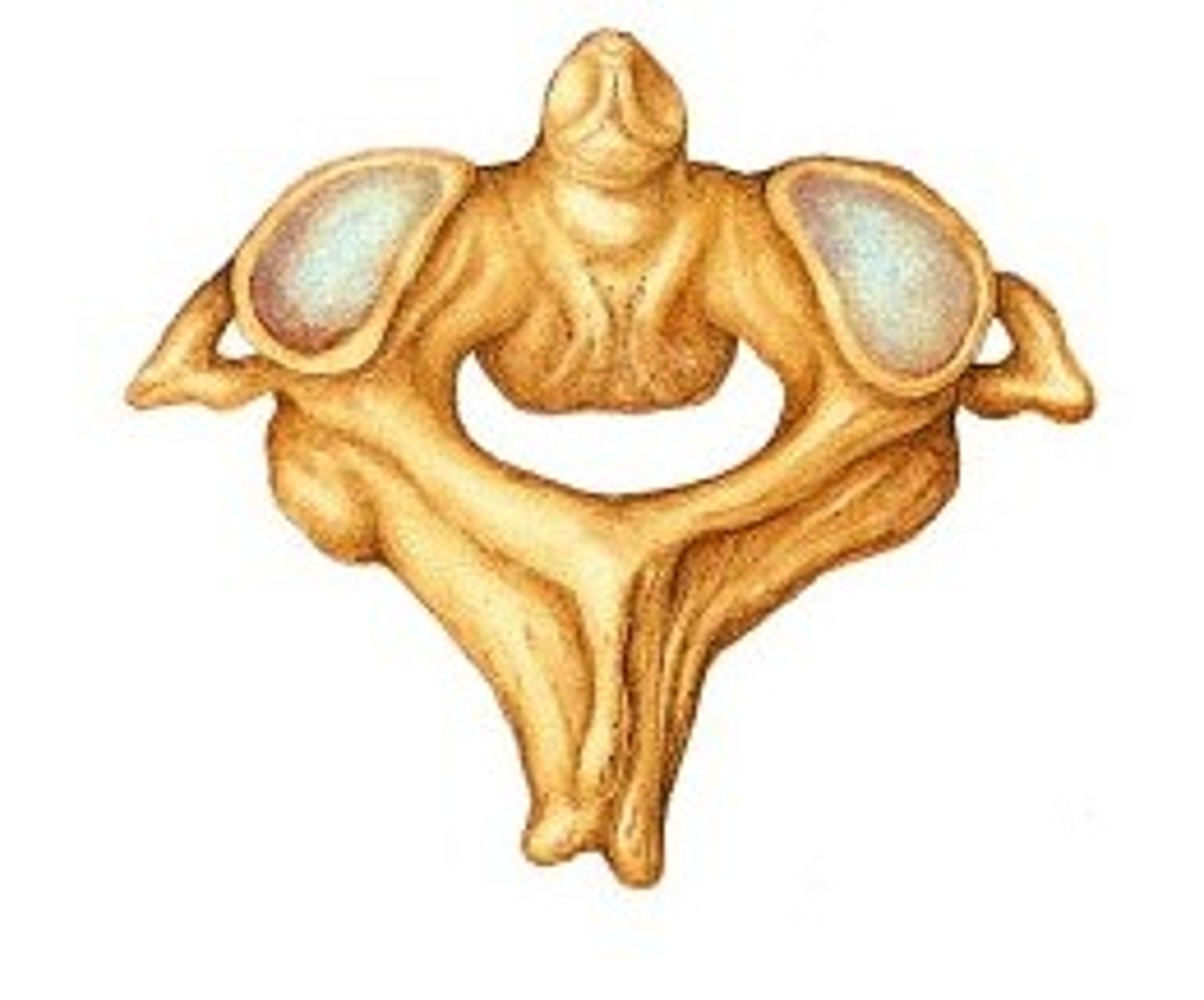
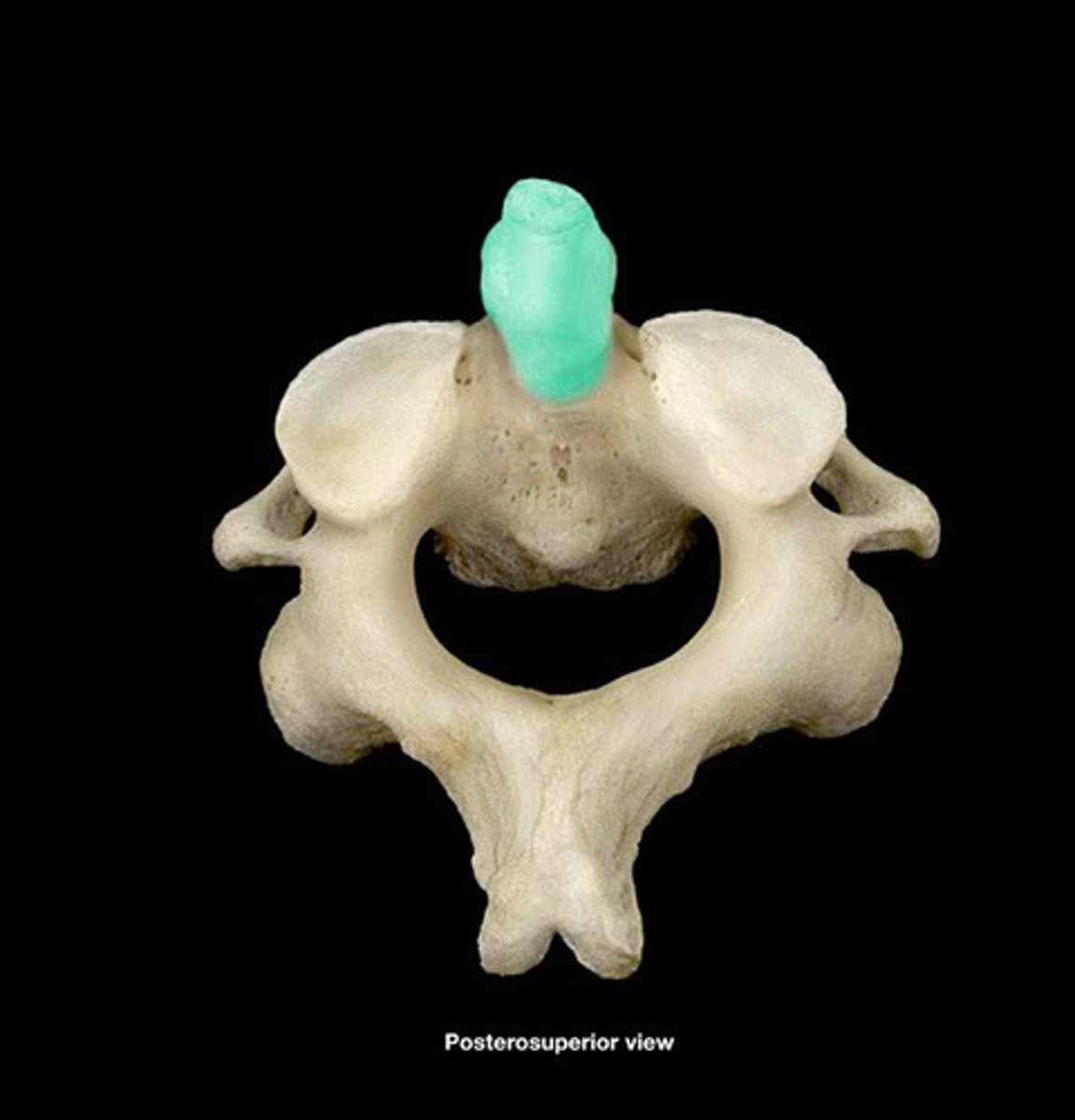
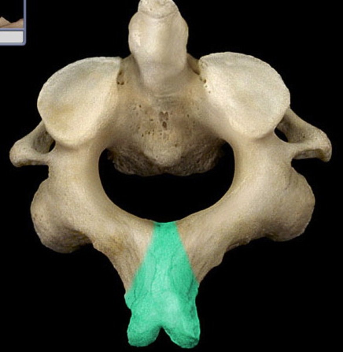
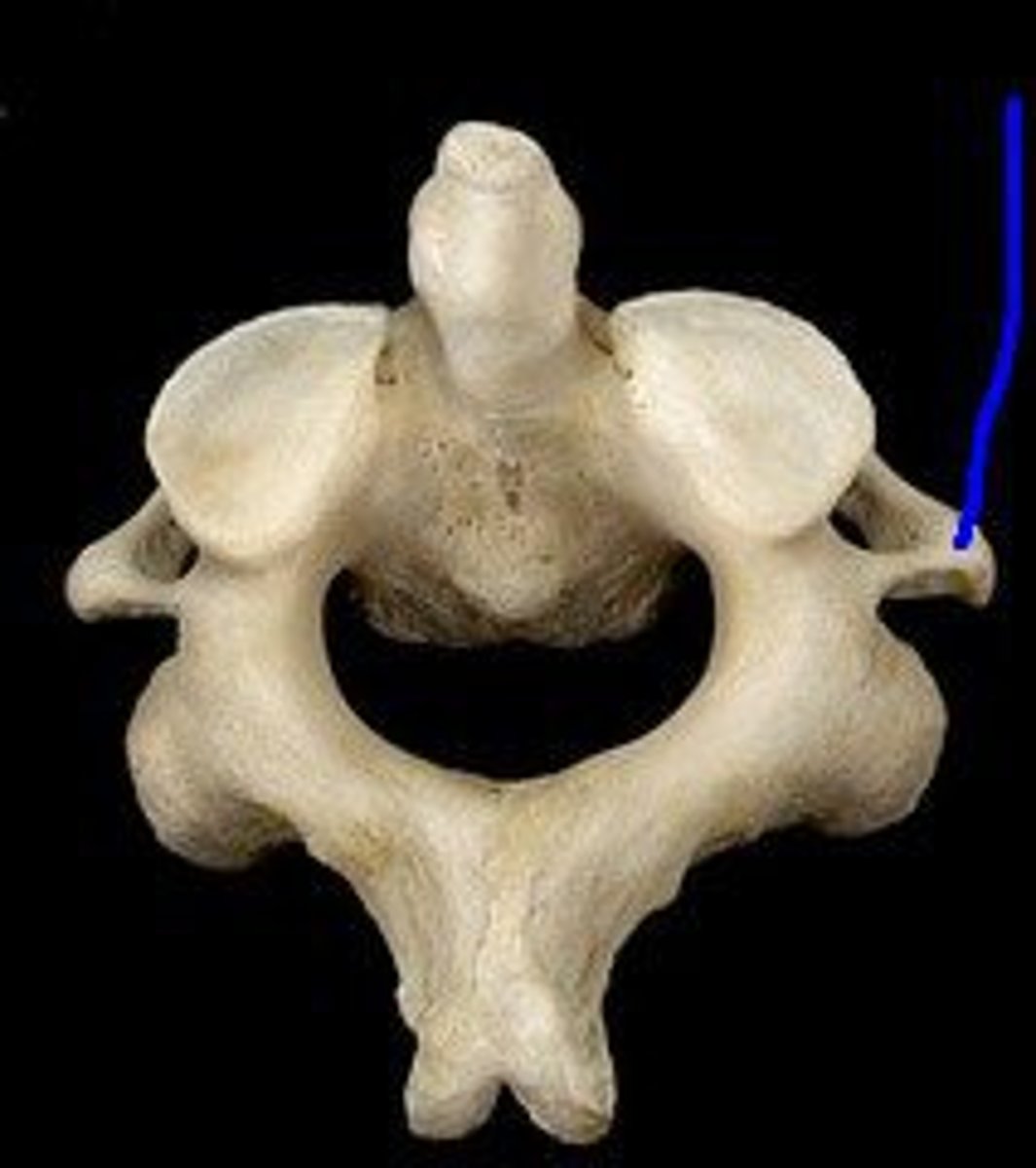
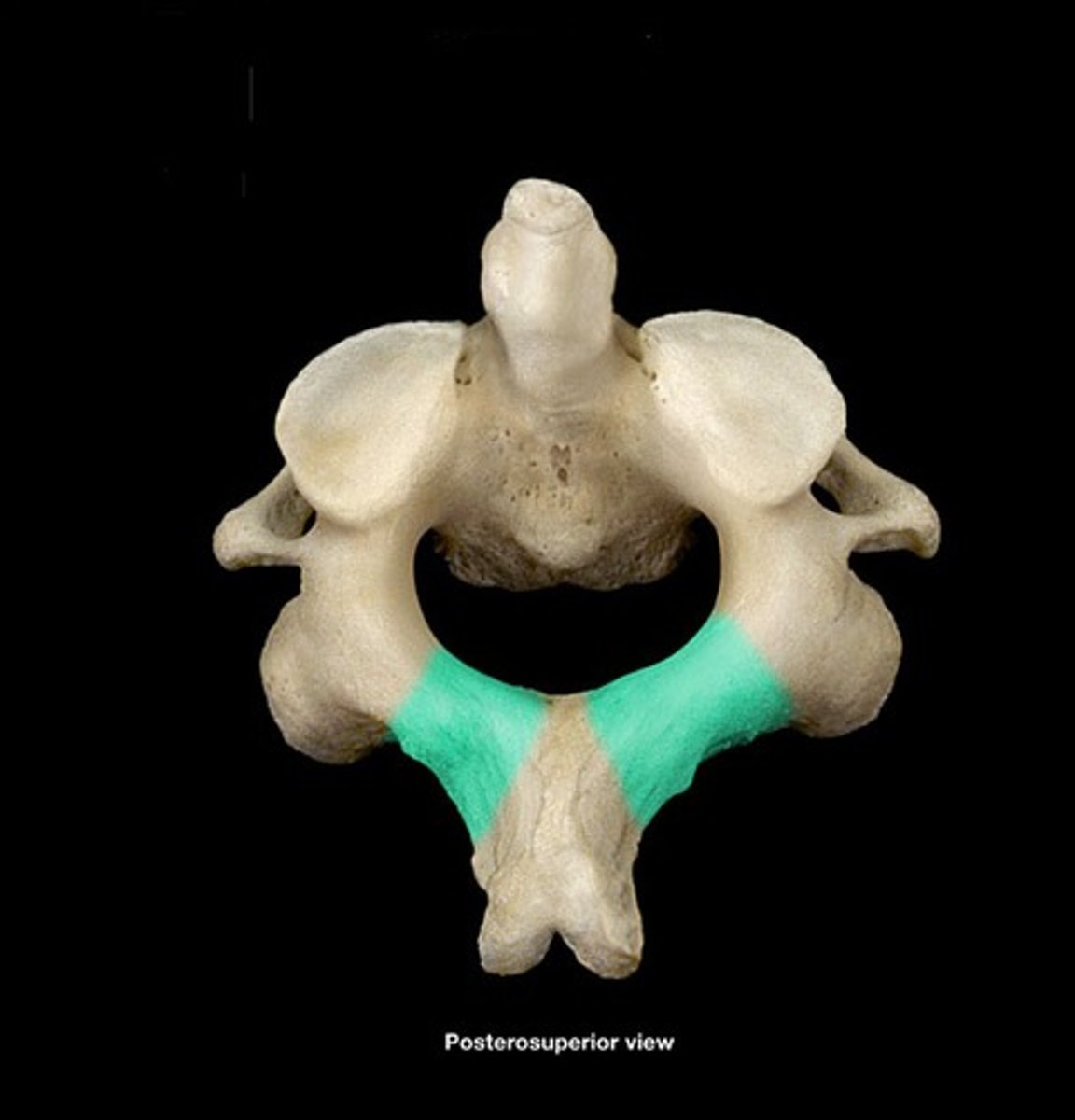
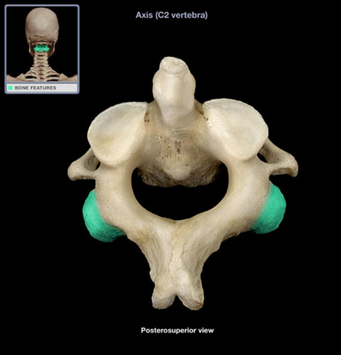
C-2 Axis
Function: support articulating facet; act as base of C-1
-Dens*: body of C-1. Sticks up and goes in between anterior arch and transverse ligament
Dens
-Acts as C-1 body. Sticks up and goes in between Anterior arch and transverse ligament ("foramen for dens")
Facet of atlas
-facets glide across each other
Transverse ligament of atlas
-holds dens of C2 against C1
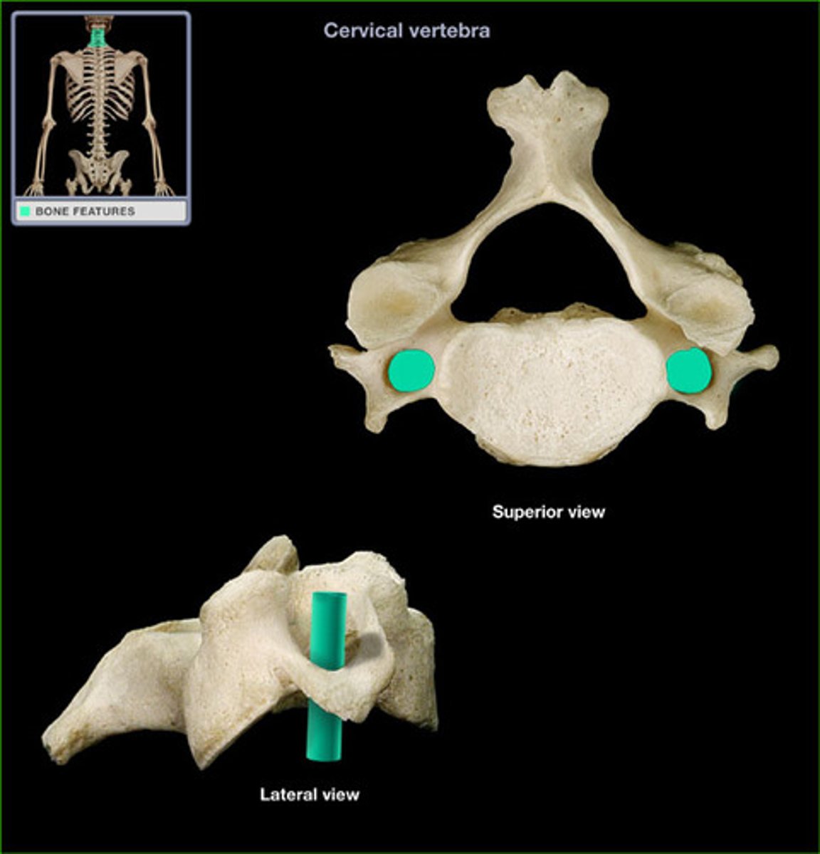
Foramen transversarium/Transverse foramina
In Cervical vertebrae ONLY
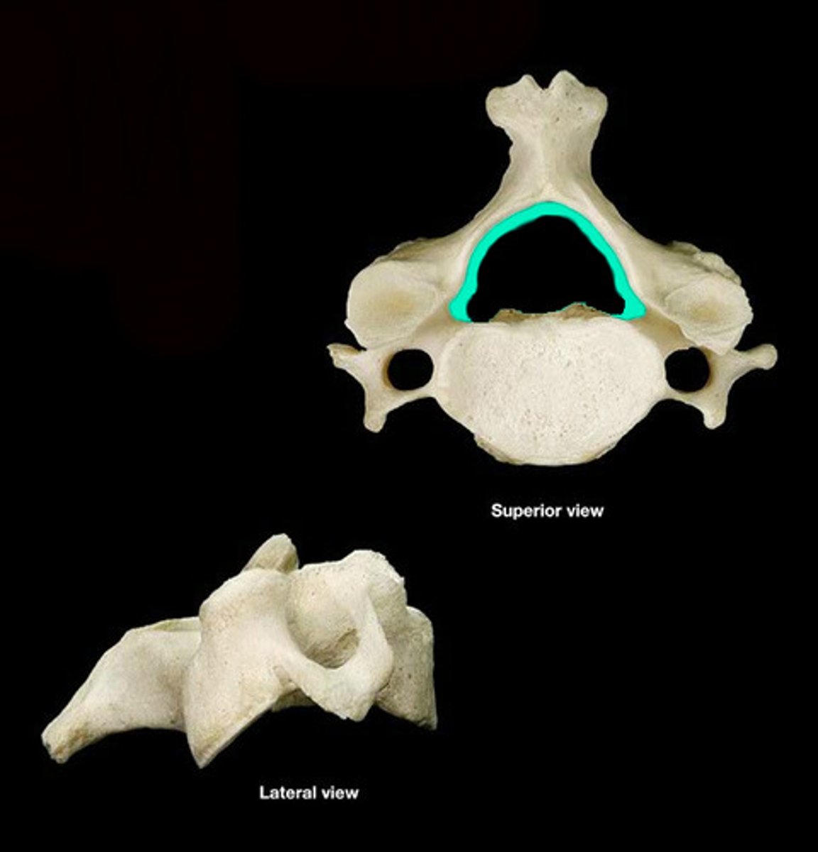
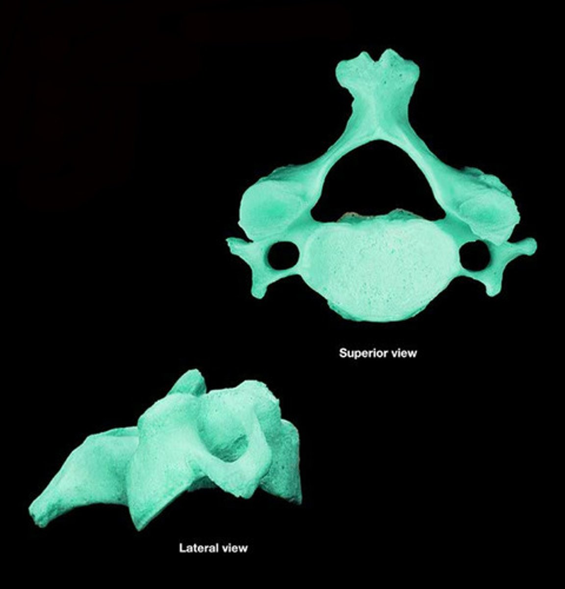
Cervical vertebrae
-spinous processes= bi-footed (bifid)
-Big foramen (for vertebral artery) - protects artery. Distinct. Big and triangular
*Have transverse foramen (foramen in transverse process)
-Facet surface: point superiorly and anteriorly
Epiphysial rim
-Discs directly attach here. Type of bone, is covered in hyaline cartilage.
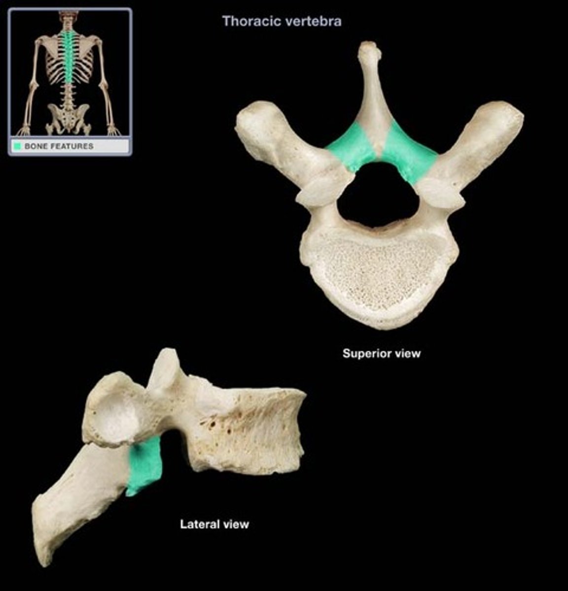
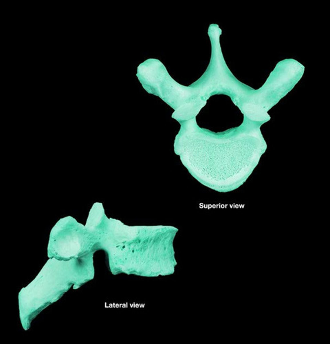
thoracic vertebrae
-Heart shaped body
pronounced transverse processes
-smaller foramen
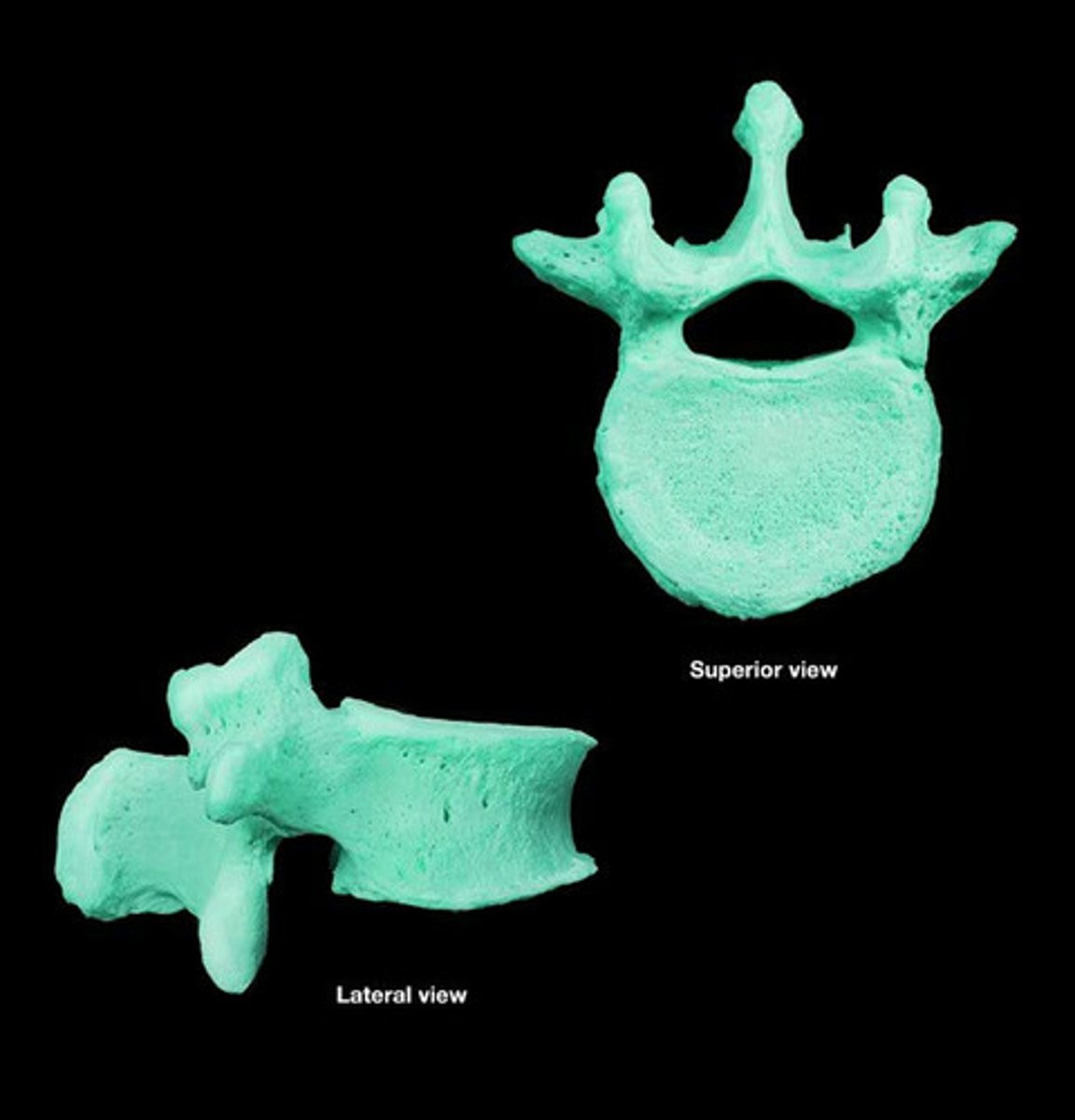
Lumbar vertebrae
-triangular foramen
-big body
-flat spinous processes-> for attachments
-Large IV discs relative to body --> good lateral flexion
-interlocking articular processes--> prevent rotation
-Zygapophysial joints: allow for flexion and extension (Extension>flexion)
Atlas - posterior tubercle
Atlas - transverse process
Atlas vertebral foramen
Atlas posterior arch
Axis spinous process
**bifid
Axis transverse process
Axis lamina
Axis inferior articular process
Vertebral arch=Pedicles+Lamina
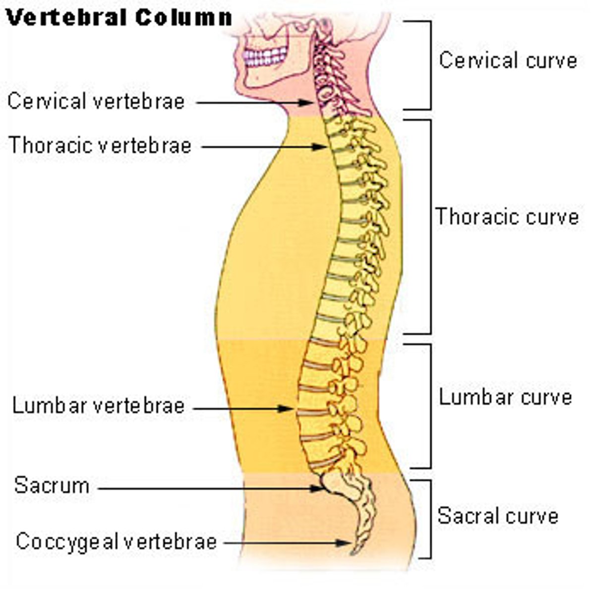
Vertebral column:
-Protects spinal cord and nerves
-supports weight of body superior to the pelvis
-provides partially rigid and flexible axis for body and pivot for head
-plays important role in posture and locomotion
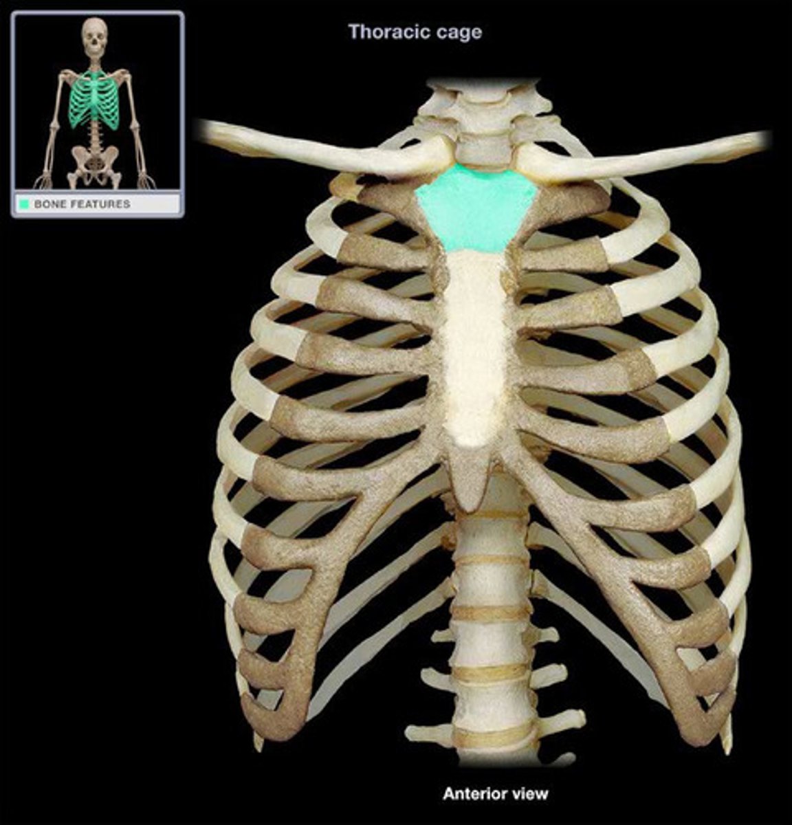
Jugular notch
-top of sternum
Manubrium
Lies at level of T3-4
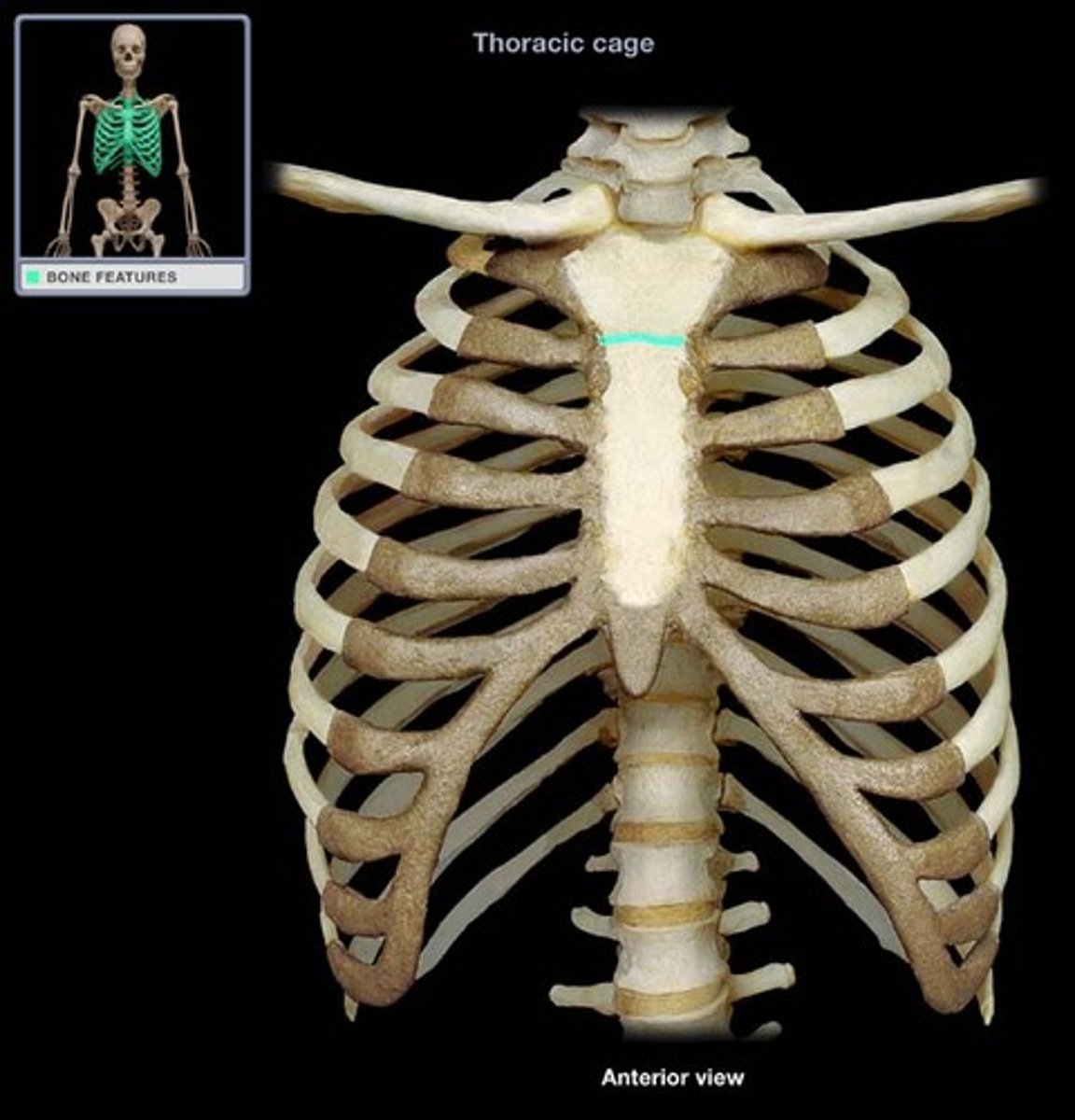
Manubriosternal joint (sternal angle)
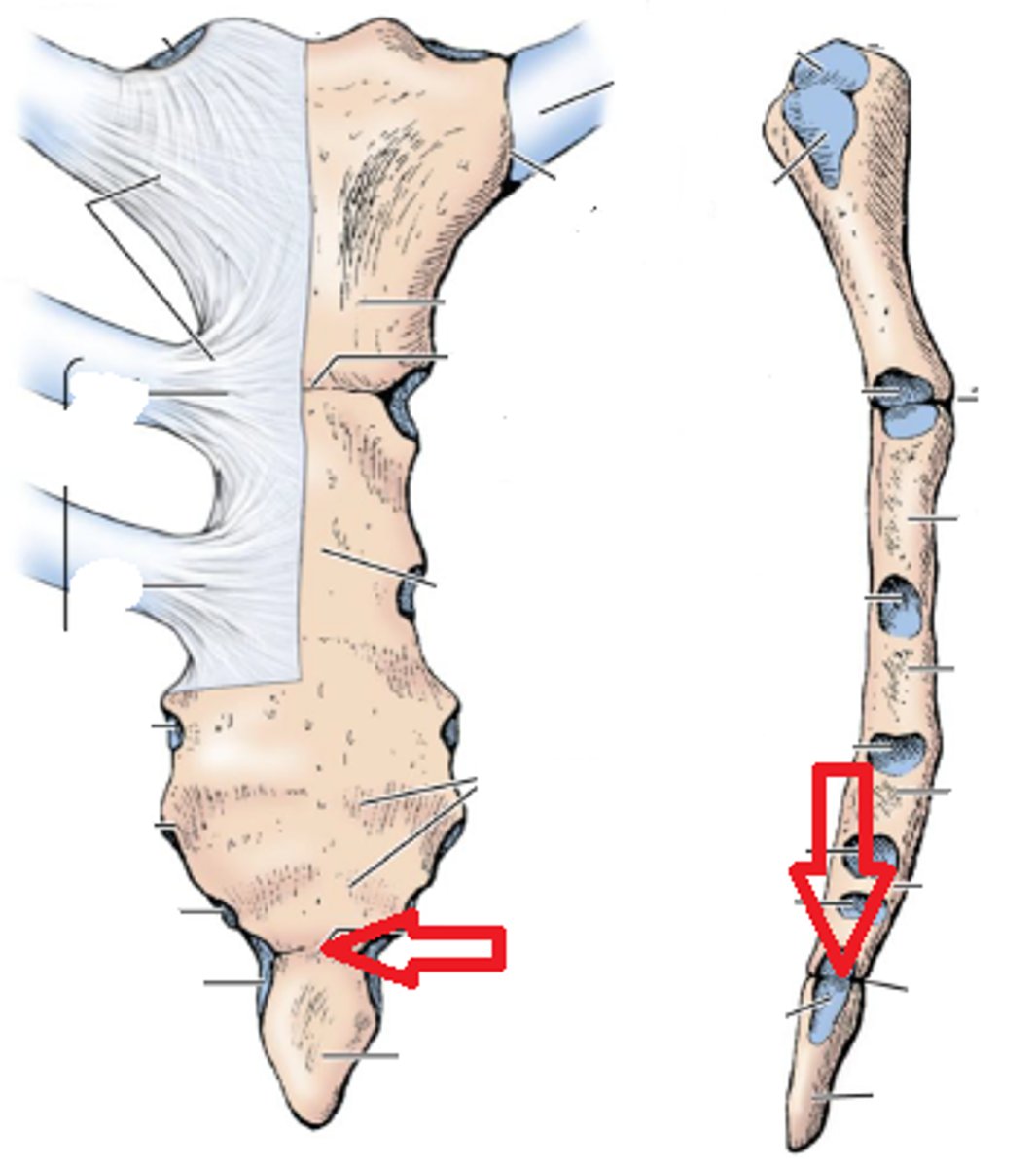
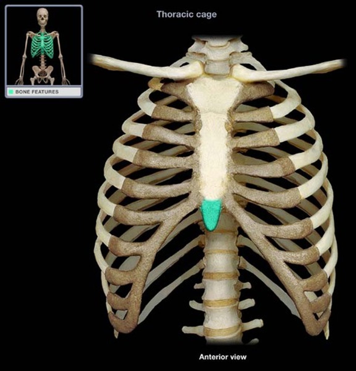
Xiphisternal joint
Xiphoid process
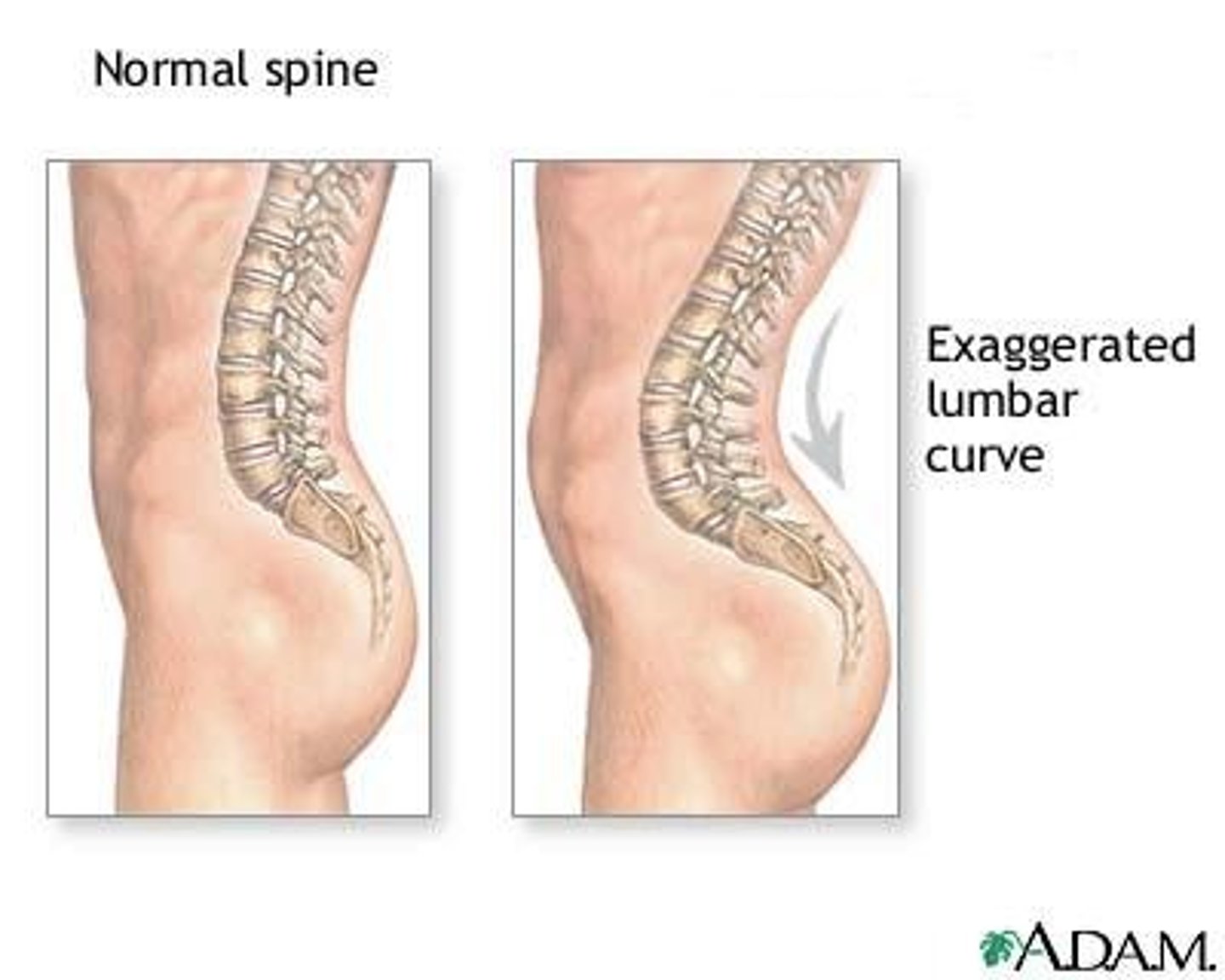
Lordosis
-Anterior rotation of pelvis
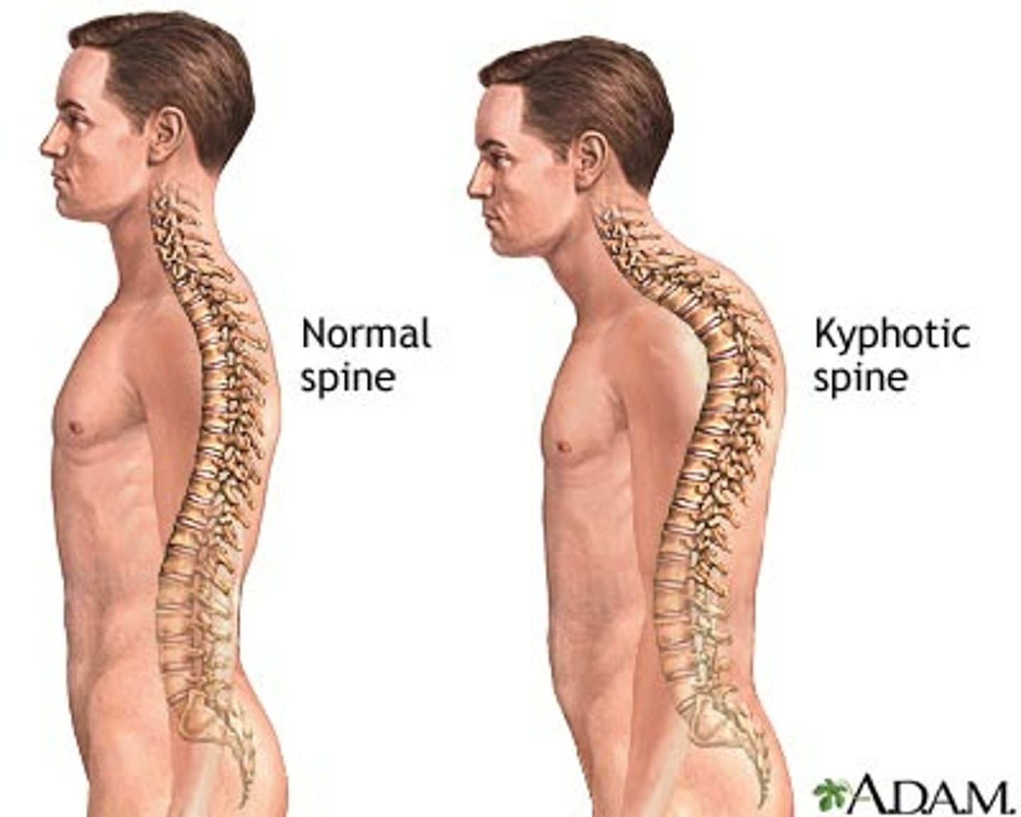
Kyphosis
-abnormal increase in thoracic curvature
-vertebral column curves posteriorly
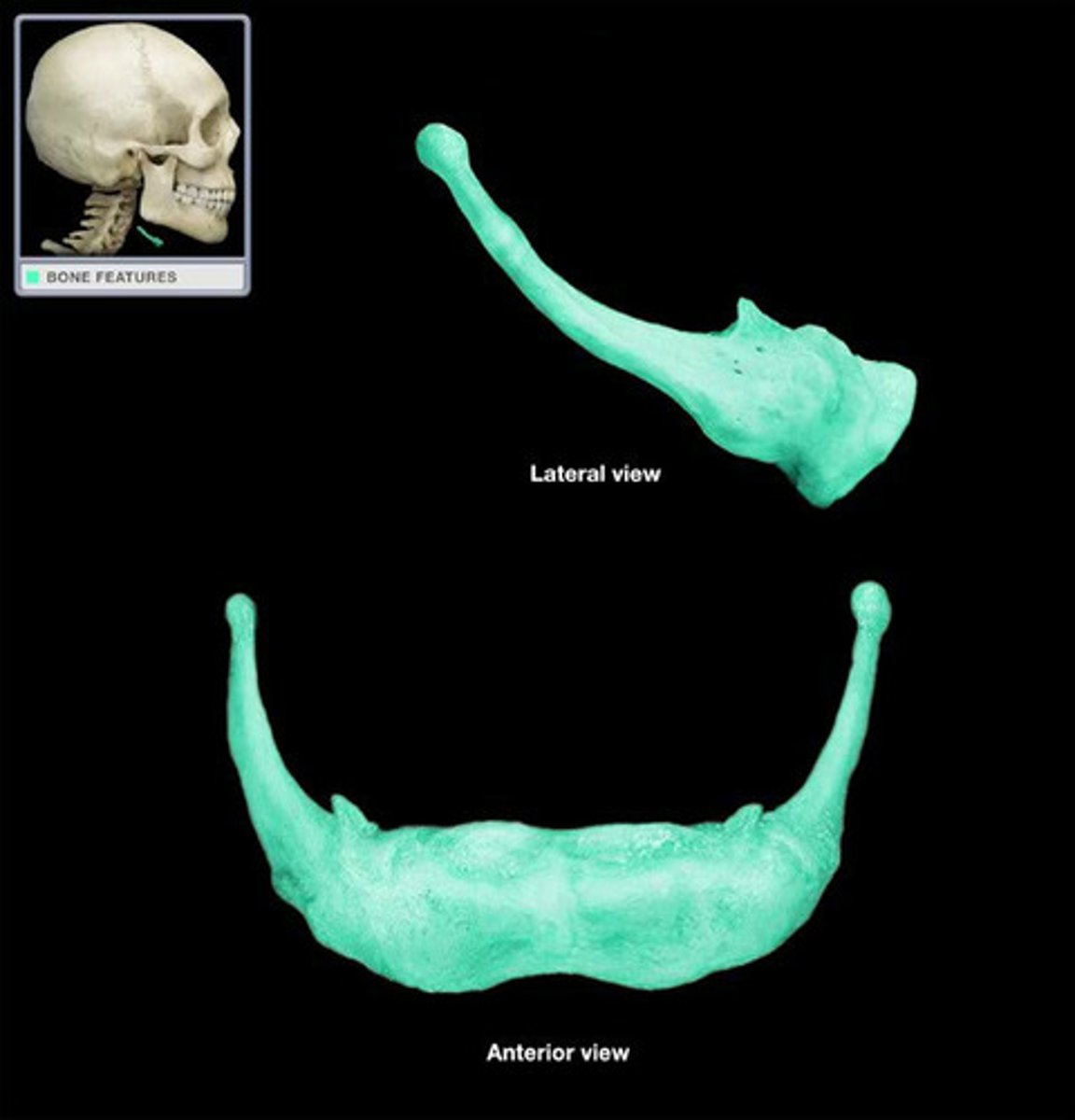
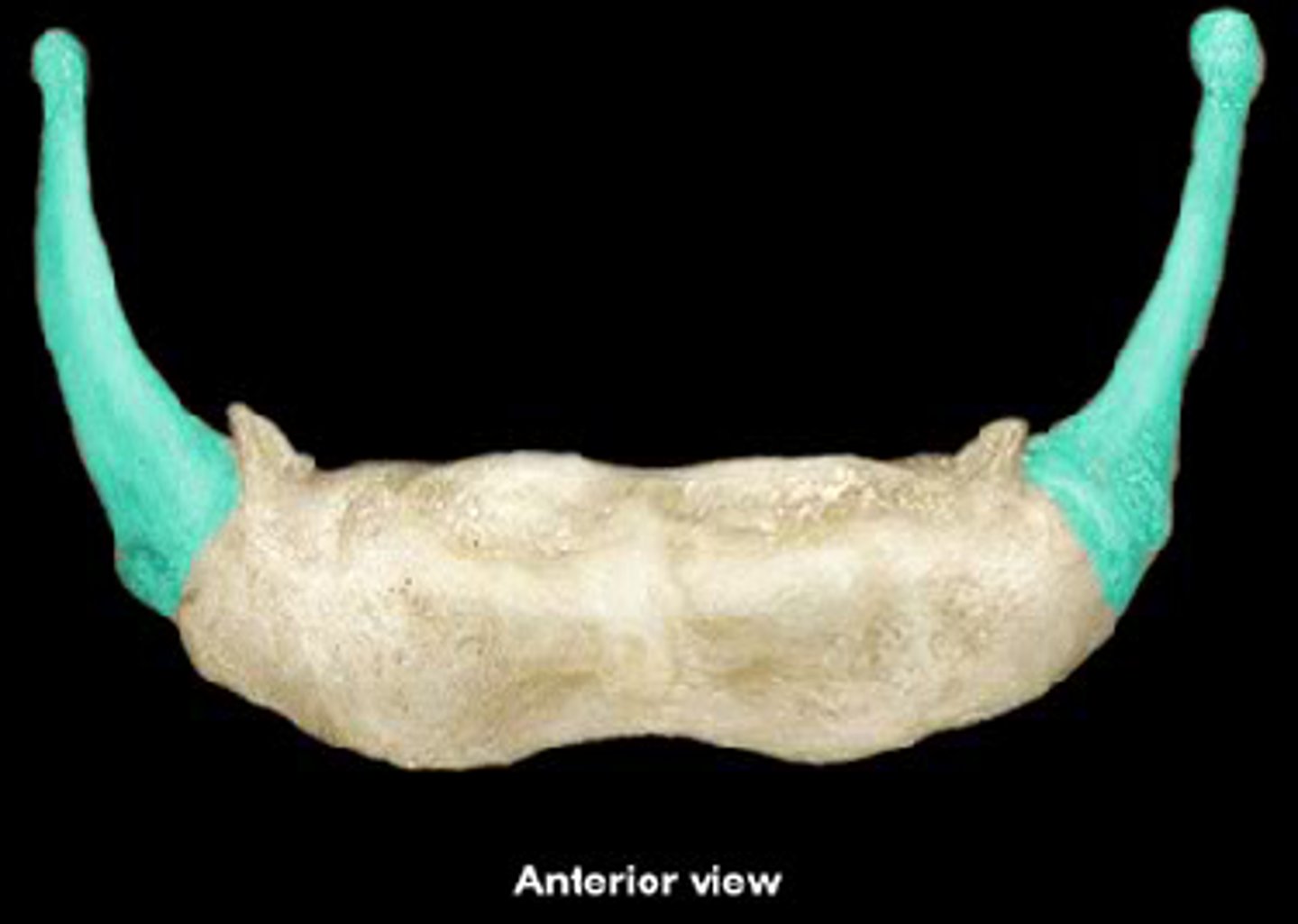
Hyoid bone
-lies in anterior part of neck at level of C-3, between mandible and thyroid cartilage
-does not articulate with any other bone
Function: attachment for anterior neck muscles and keeps airway open
Hyoid bone greater horn