Organelles and Cell Life Cycles
1/64
There's no tags or description
Looks like no tags are added yet.
Name | Mastery | Learn | Test | Matching | Spaced | Call with Kai |
|---|
No study sessions yet.
65 Terms
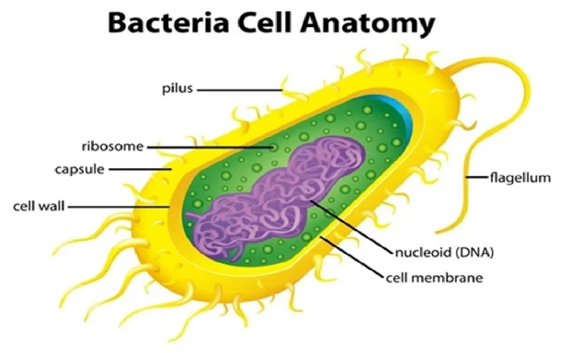
Cells
Definition: Basic unit of life
Functions: carry out life processes, provide structure, reproduce, respond to environment
Comes in all shapes and sizes
Key Structures:
Nucleus: DNA/control center
Mitochondria: energy (ATP)
Ribosomes: protein production
Cell membrane: protection & transport
Cytoplasm: holds organelles
Examples: animal cells, plant cells, bacterial cells
💡 = tiny factories keeping the body alive
True or false? Structure determines function
True! Structure → Function
Concept: the shape or structure of a cell or organelle helps it do its job.
Examples:
Mitochondria: folded inner membrane → more surface area → makes more ATP
Ribosomes: small & round → efficiently make proteins
Cell membrane: flexible & selectively permeable → controls what enters/exits
💡 Quick tip: How it’s built determines what it can do!
Organelle
Definition: tiny “mini-organs” inside a cell that do specific jobs
Functions / Examples:
Nucleus: control center, holds DNA
Mitochondria: makes energy (ATP)
Ribosomes: make proteins
Endoplasmic Reticulum (ER): transports & folds proteins/lipids
Golgi Apparatus: packages & ships proteins
Lysosomes: breaks down waste
Cell membrane: protects cell & controls entry/exit
Importance: each organelle’s structure is related to its function → keeps cell alive
💡 tiny factories inside the cell, each with a job
Plasma Membrane
Definition: outer layer of the cell that separates inside from outside
Structure: phospholipid bilayer with hydrophilic heads & hydrophobic tails, plus proteins
Functions:
Controls what enters/exits the cell (selectively permeable)
Protects the cell
Allows communication with other cells
Importance: maintains homeostasis and cell survival
Examples: cell membranes of all animal and plant cells
💡 = gatekeeper of the cell
Semipermeable (Selectively Permeable)
Definition: allows some substances to pass through but blocks others
Example / Situation:
A plasma membrane lets oxygen and water enter the cell to keep it alive but blocks bacteria or large toxins from coming in
Importance / Function:Controls what enters/exits the cell
Maintains homeostasis
Other info: usually allows small or nonpolar molecules (like oxygen, water) but blocks large or charged molecules
💡 = selective gate for the cell
Phospholipids
Definition / Structure: made of glycerol + 2 fatty acid tails (hydrophobic) + phosphate head (hydrophilic)
Function / Importance:
Forms cell membranes (phospholipid bilayer)
Controls what enters/exits the cell
Protects the cell & keeps homeostasis
Example / Situation: in a plasma membrane, heads face water, tails face inward → barrier forms to protect cell
💡 = building blocks of the cell membrane
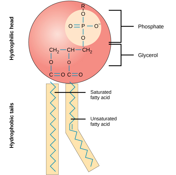
Phospholipid - Hydrophilic and Hydrophobic
Hydrophilic (“water-loving”): phosphate head → faces water (outside & inside the cell)
Hydrophobic (“water-fearing”): fatty acid tails → face inward, away from water
Function / Importance: this arrangement forms the phospholipid bilayer, controls what enters/exits the cell, protects the cell, maintains homeostasis
Example / Situation: in a cell membrane, heads touch water inside/outside the cell, tails hide in the middle → forms a selective barrier
💡 Quick tip: Heads love water, tails hate water → bilayer barrier!
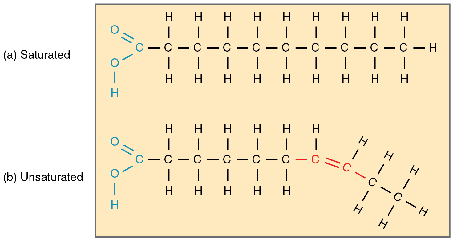
Saturated vs Unsatured Fatty Acids
Saturated Fatty Acid:
Structure: no double bonds, straight chains
State at room temp: solid (butter, lard)
Health note: can raise cholesterol if eaten in excess
Unsaturated Fatty Acid:
Structure: has ≥1 double bond, bent chains
State at room temp: liquid (olive oil, avocado, nuts)
Health note: generally healthier, can lower cholesterol
💡 Quick tip: Saturated = straight & solid, Unsaturated = bent & liquid
Membrane Proteins
Definition: proteins embedded in the cell membrane
Types / Functions:
Transport proteins: move substances in/out of cell
Receptor proteins: receive chemical signals
Enzymes: speed up chemical reactions at the membrane
Structural proteins: give support and shape to the cell
Importance: help the cell communicate, transport, and maintain homeostasis
Example / Situation: glucose enters the cell through a transport protein, hormones bind to receptor proteins
💡 = workers in the cell gate, delivery, and communication system
Passive Transport
Definition: movement of substances across the cell membrane without using energy
How it works / Types:
Simple Diffusion: molecules move from high → low concentration
Osmosis: diffusion of water
Facilitated diffusion: uses transport proteins to help molecules cross
Importance / Function: balances concentrations inside/outside the cell → maintains homeostasis
Example / Situation: oxygen enters a cell from blood, water moves in/out to balance cell volume
💡 = no energy needed, molecules go with the flow
Facilitated Diffusion
Definition: passive transport of molecules with the help of transport proteins
How it works: molecules move from high → low concentration using channel or carrier proteins
Importance / Function: allows molecules that can’t pass through the lipid bilayer (like glucose or ions) to enter/exit the cell
Example / Situation: glucose enters cells via a glucose transporter protein
💡 = “helped” passive transport
Osmosis
Definition: diffusion of water across a semipermeable membrane
How it works: water moves from high → low water concentration (or low → high solute concentration)
Importance / Function: balances water inside/outside the cell → maintains homeostasis
Example / Situation: plant roots absorb water from soil; water moves into red blood cells in the body
💡 = water moving to where it’s needed
Glycoprotein
Definition: protein with a carbohydrate chain attached
Location: cell membrane
Function / Importance:
Acts as an ID tag so cells can recognize each other
Helps with cell communication
Plays a role in the immune system (recognizing friendly vs. foreign cells)
Example / Situation: blood types (A, B, AB, O) are determined by specific glycoproteins on red blood cells
💡 = cell’s ID badge for recognition & communication
Glycolipid
Definition: lipid with a carbohydrate chain attached
Location: cell membrane
Function / Importance:
Acts as a recognition marker (like glycoproteins)
Helps cells stick together
Maintains stability of the membrane
Example / Situation: glycolipids on red blood cells help determine blood type and allow immune recognition
💡 = lipid + sugar → recognition & stability
Membrane Proteins – Process
Transport proteins: move molecules in/out (ex: glucose channel)
Receptor proteins: receive signals (ex: insulin receptor)
Enzymes: speed up reactions
Structural proteins: give support & connect cells
💡 Quick tip: Membrane proteins = workers of the cell gate
Cytoplasm
Definition: jelly-like fluid inside the cell that surrounds organelles
Composition: mostly water + salts + proteins
Function / Importance:
Holds organelles in place
Site of many chemical reactions (like glycolysis in cellular respiration)
Helps transport materials within the cell
Site of Anaerobic Respiration
Liquid portion is called Cytosol
Example / Situation: like the “soup” where all the ingredients (organelles) float and reactions happen
💡 = the cell’s soup where reactions happen
Endoplasmic Reticulum
Definition: network of folded membranes near the nucleus
Types:
Rough ER: has ribosomes → makes & transports proteins
Smooth ER: no ribosomes → makes lipids, detoxifies, stores calcium
Function / Importance: pathway for making, folding, and moving molecules in the cell
Helps build essential molecules (proteins & lipids), moves them around, keeps the cell healthy.
Where is it? Found in the cytoplasm, right next to the nucleus.
Example / Situation: like a factory assembly line → Rough ER builds proteins, Smooth ER makes lipids & cleans toxins
💡 ER = cell’s factory → Rough = proteins, Smooth = lipids & detox
Rough ER
What it is: Folded membranes in the cytoplasm, covered with ribosomes (looks rough)
What it does: Makes, modifies & transports proteins; sends them out or to the membrane
Importance: Essential for growth, repair, enzymes, hormones, transport
Where: Cytoplasm of eukaryotic cells, abundant in protein-making cells
Example: Pancreas cells → make insulin
💡 = protein factory (ribosomes are the workers)
Smooth ER
Definition: type of ER without ribosomes
Functions / Importance:
Makes lipids → used for cell membranes
Detoxifies chemicals
Stores calcium for cell signaling
Example / Situation: like a cell’s oil & cleaning department → produces fats for membranes and removes toxins
💡 = lipid factory → builds membranes + detox + calcium storage
Golgi Apparatus
Definition: stack of flattened membranes in the cell
Function / Importance:
Modifies, sorts, and packages proteins and lipids from the ER
Prepares molecules for transport inside/outside the cell
Example / Situation: like a post office or shipping center → labels packages (proteins/lipids) and sends them to their destination
💡 = cell’s shipping & packaging center
Ribosomes
Definition: small organelles that make proteins
Location:
Free in cytoplasm: make proteins for the cell itself
Attached to Rough ER: make proteins for export or membranes
Function / Importance:
Build proteins by linking amino acids together (protein synthesis)
Site of protein synthesis
Example / Situation: like a cell’s kitchen, cooking up proteins needed for the cell or to send elsewhere
💡 = protein factories of the cell
Endocytosis
Definition: process where the cell engulfs substances to bring them inside the cell
How it works:
Cell membrane wraps around material → forms a vesicle → brings it into the cytoplasm
Function / Importance: lets the cell take in nutrients, liquids, or large molecules
Example / Situation: white blood cells engulf bacteria to fight infection; cells take in large nutrients
💡 = cell “eating” or “drinking” things
Exocytosis
Definition: process where the cell releases substances to the outside
How it works:
Vesicle containing molecules fuses with the cell membrane → contents are expelled
Function / Importance: removes waste, sends out hormones or proteins
Example / Situation: pancreas cells release insulin into the blood; neurons release neurotransmitters
💡 = cell “spitting out” or exporting things
Vesicles
Definition: small membrane-bound sacs in cells
Function / Importance:
Transport proteins, lipids, or waste within or out of the cell
Can store materials temporarily
Example / Situation:
Carry proteins from Rough ER → Golgi Apparatus → cell membrane
Transport neurotransmitters in neurons
Relation to Endo/Exocytosis: vesicles are the “packages” brought in or sent out
💡 = cell’s delivery trucks or packages
Phagocytosis
Definition: type of endocytosis where the cell engulfs large particles or cells
How it works: cell membrane wraps around a particle → forms a phagosome → particle is digested
Function / Importance: allows the cell to eat bacteria, debris, or dead cells → important for immune defense
Example / Situation: white blood cells engulf bacteria to fight infection
💡 = cell “eating” big stuff like bacteria
Pinocytosis
Definition: type of endocytosis where the cell engulfs liquids or dissolved substances
How it works: cell membrane folds inward → forms a vesicle → brings fluid into the cell
Function / Importance: lets the cell take in nutrients or extracellular fluids
Example / Situation: cells in the intestine absorb nutrients dissolved in water
💡= cell “drinking” liquids
Mitochondria
Definition: double-membrane organelle known as the cell’s powerhouse
Function / Importance:
Produces ATP (energy) through cellular respiration
Breaks down glucose and other nutrients to release energy
Structure:
Outer membrane: protects organelle
Inner membrane (cristae): folded → more surface area for energy production
Matrix: fluid inside where reactions occur
Example / Situation: muscle cells have many mitochondria → need lots of energy for movement
💡 = energy factory of the cell
Lysosomes
Definition: small organelles containing digestive enzymes
Function / Importance:
Surrounds and break down waste, damaged organelles, and foreign particles
Helps recycle materials for the cell
Structure: membrane-bound sac
Example / Situation: white blood cells use lysosomes to digest engulfed bacteria
💡 = cell’s recycling & cleanup crew
Nucleus
Location: Inside the cytoplasm of the cell
What it does:
Gives the cell shape & support
Moves organelles and vesicles
Helps the cell move/divide
(like a skeleton + highway for transport)
Overall framework of cells
Structures: Microtubules, microfilaments, intermediate filaments
Example: Moves vesicles around the cell like a conveyor belt
💡 = the cell’s skeleton + transport system inside the cytoplasm
Microfilaments
Definition: thin protein fibers in the cytoskeleton made of actin
Function / Importance:
Maintain cell shape
Part of Cytoskeleton + Smallest/thinnest fibers
Help with cell movement (like crawling or changing shape)
Assist in cytoplasm streaming and cell division
Example / Situation: white blood cells use microfilaments to move toward infection sites
💡 = thin threads that shape the cell and help it move
Intermediate fibers
Definition: medium-thickness protein fibers in the cytoskeleton
Function / Importance:
Provide mechanical strength
Help maintain cell shape
Medium thickness fibers
Anchor organelles in place
Example / Situation: skin cells have strong intermediate filaments to withstand stretching
💡 = sturdy ropes that support and stabilize the cell
Microtubules
Definition: thick, hollow protein tubes in the cytoskeleton made of tubulin
Function / Importance:
Maintain cell shape
Largest/thickest fibers
Act as tracks for organelle and vesicle movement
Form cilia, flagella, and spindle fibers during cell division
Example / Situation: vesicles use microtubules like highways to reach different parts of the cell
💡 = cell’s highways + structural support
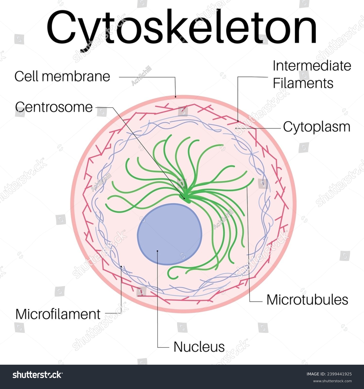
Cytoskelton structure
Microfilaments: thin, flexible fibers → shape + movement (actin)
Intermediate Filaments: medium thickness → strength + anchor organelles
Microtubules: thick hollow tubes → shape, transport, cilia/flagella, cell division
💡 = framework of microfilaments + intermediate filaments + microtubules
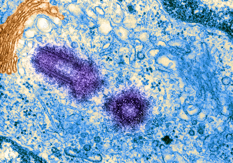
Centrosome - (micrograph structure of this pictured below)
Definition: region near the nucleus that organizes microtubules
Function / Importance:
Controls microtubule growth
Forms spindle fibers during cell division
Helps maintain cell shape and structure
Plays important role in cell divison + can only be seen during CD
General location of this is idenitifed by the centrioles
Example / Situation: during mitosis, centrosomes pull chromosomes apart using spindle fibers
💡 Quick tip: Centrosome = cell’s microtubule organizer & division helper
Centrioles
Definition: cylindrical structures made of microtubules
Location: found in the centrosome
Function / Importance:
Help organize spindle fibers during cell division
Assist in forming cilia and flagella
Example / Situation: during mitosis, centrioles help pull chromosomes apart
💡 = cell’s spindle organizers + cilia/flagella helpers
Cell extension
Definition: structures that stick out from the cell surface to help with movement or sensing
Types:
Cilia → short, hair-like, move substances across cell surface (ex: in respiratory tract)
Flagella → long, tail-like, move the whole cell (ex: sperm)
Microvilli → tiny finger-like folds, increase surface area for absorption (ex: intestines)
Importance: help cells move, sense, or absorb nutrients
💡 Quick tip: Cilia = sweep, Flagella = swim, Microvilli = absorb
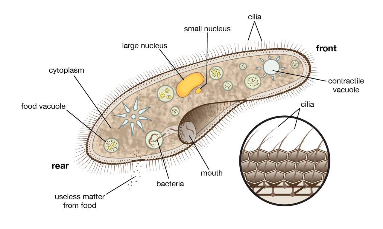
Cilia
Definition: short, hair-like extensions on cell surface
Function: move substances across the cell surface
Shorter + more numerous than flagella
All of __ have sensory functions
Location / Example: lining the respiratory tract → sweep mucus & dust out of airways
Importance: keep airways clear; help movement in some cells
💡 = tiny hairs that sweep
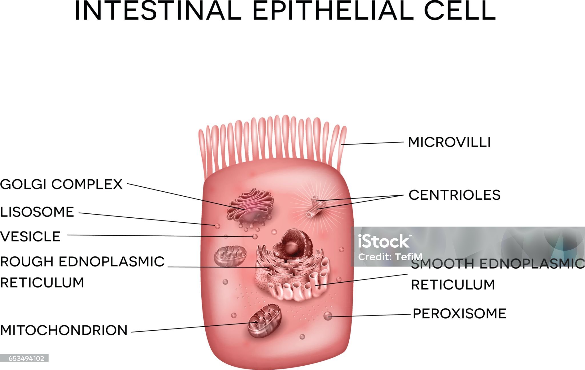
Microvilli
Definition: tiny finger-like projections on cell surface
Function: increase surface area for absorption
Location / Example: found in small intestine → absorb nutrients from food
Importance: make absorption faster and more efficient
💡 = “absorption fingers”
Flagella
Definition: long, tail-like extension of a cell
Function: moves the entire cell
Only found on human sperm cells
Location / Example: sperm cell → flagellum helps it swim to the egg
Importance: allows mobility in certain cells
💡 = “tail for swimming”
Cell Process Movement
Signal → Cell gets a signal to move (chemicals, environment, etc.).
ATP provides energy → Needed for movement.
Cytoskeleton = engine
Microtubules → act like tracks that guide cilia & flagella.
Microfilaments → help the cell crawl or change shape.
Movement types:
Cilia → microtubules inside beat back & forth → move mucus/dust (like oars).
Flagella → microtubules whip tail-like → move the whole cell (like a fish tail).
Microvilli → supported by microfilaments → don’t move, but increase absorption (like a sponge).
Importance: lets cells move substances, travel (sperm), or absorb nutrients (intestines).
💡 Cytoskeleton = engine → powers cilia, flagella, microvilli.
Cilia = oars paddling water, Flagella = fish tail swimming, Microvilli = sponge soaking up nutrients
Nucleus
Definition: control center of the cell
Function: stores DNA (genetic info) and controls cell activities (growth, metabolism, protein synthesis)
Site of transcription
Structures inside:
Nuclear envelope → protects nucleus, controls what enters/leaves
Nucleolus → makes ribosomes
Chromatin → DNA + proteins
Importance: without it, the cell can’t function or reproduce
💡 = cell’s brain (controls & stores DNA)
Chromatin vs Chromosomes
Definition: loose, uncoiled form of DNA + proteins
Function: allows easy access to DNA for transcription & replication
When: found when the cell is not dividing
Chromosomes
Definition: tightly coiled, condensed form of DNA
Function: keeps DNA organized & safe during cell division
When: visible only when the cell is dividing
Both are located inside the nucleus
💡 Quick tip: Chromatin = relaxed (normal life), Chromosomes = condensed (cell division)
Nucleus enveleope
Definition: double membrane that surrounds the nucleus
Structure:
Inner membrane: supports nucleus structure
Outer membrane: continuous with rough ER, may have ribosomes
Function:
Protects DNA
Controls what enters and leaves the nucleus via nuclear pores
Location: around the nucleus
💡 = nucleus’ protective barrier + gatekeeper
Nucleous
Definition: dense structure inside the nucleus
Function / Importance:
Makes ribosomal RNA (rRNA)
Assembles ribosome subunits to send to the cytoplasm
Location: inside the nucleus, floating in nucleoplasm
Example / Situation: think of it as a ribosome factory inside the nucleus
💡= ribosome-making factory in the nucleus
Cell connections
Definition: structures that link cells together in tissues
Types & Functions:
Tight junctions: seal cells → prevent leaks (ex: lining of intestines)
Desmosomes: anchor cells → resist stretching (ex: skin)
Gap junctions: channels → allow communication & molecule exchange (ex: heart cells)
Importance: maintain tissue integrity, communication, and protection
💡 = bridges & gates between cells
Densosomes
Definition: strong cell-to-cell anchoring junctions
Structure: protein fibers on outer surface interlock with fibers of neighboring cells
Function / Importance:
Hold cells together
Resist stretching and mechanical stress
Permeability: not fully sealed, some substances can pass
Location / Example: skin, heart tissue
Analogy: like spot welds or Velcro keeping cells tightly connected
💡 = “interlocking anchors” for strength + some permeability
Gap Junctions
Definition: channels that electrically and physically connect neighboring cells
Structure: gaps/tunnels that join the cytoplasm and fuse plasma membranes
Function / Importance:
Allow ions, nutrients, small molecules, and signals to pass directly between cells
Enable rapid communication and coordination (ex: heartbeat)
Location / Example: cardiac muscle and some smooth muscle
Analogy / Quick tip: tiny tunnels or bridges that connect cells for communication
= cell-to-cell tunnels for communication
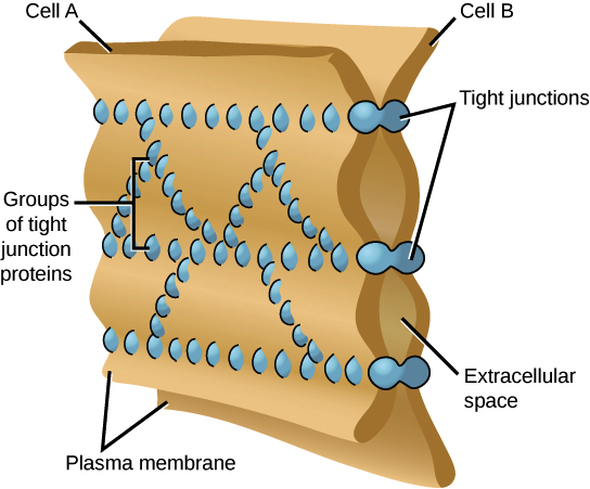
Tight Junction
Definition: seal neighboring cells together to prevent leaks
Structure: proteins fuse plasma membranes at contact points
Function / Importance:
Create a barrier that blocks movement of substances between cells
Maintain tissue integrity and control what enters/exits
Location / Example: lining of intestines → prevent digestive fluids from leaking
Analogy / Quick tip: Tight junctions = waterproof seal between cells
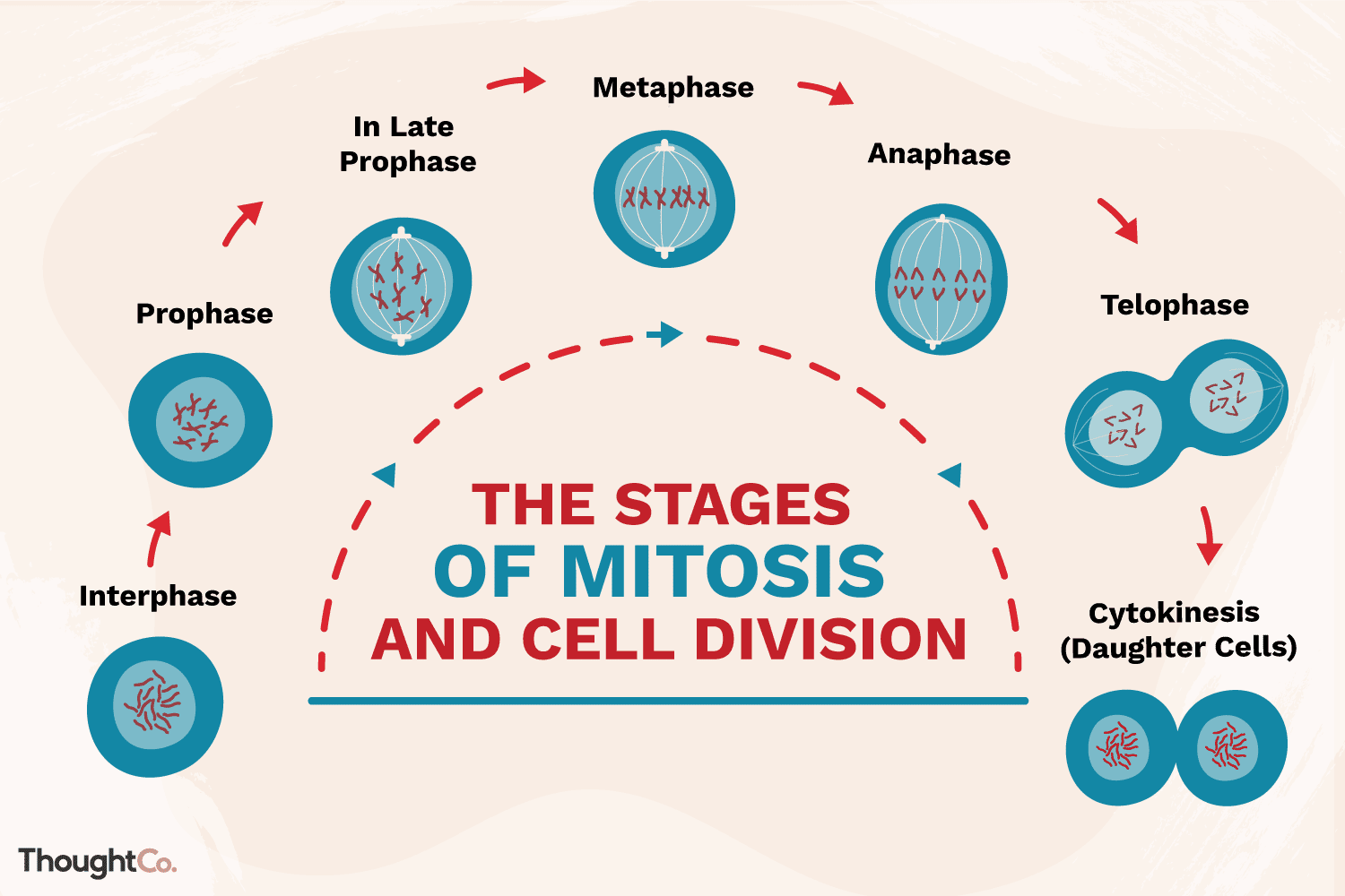
Cell Life Cycle
Definition: series of stages a cell goes through from formation to division
Main Phases:
Interphase – cell grows, performs normal functions, and prepares for division
G1: growth
S: DNA replication
G2: preparation for mitosis
Mitotic Phase (M phase) – cell divides
Mitosis: division of nucleus (prophase, metaphase, anaphase, telophase)
Cytokinesis: division of cytoplasm → 2 daughter cells
Importance: ensures growth, repair, and replacement of cells
Example / Situation: skin cells constantly divide to replace dead cells
💡 = grow, copy DNA, divide
Interphase
Definition: the longest phase of the cell cycle where the cell grows and prepares for division
Subphases:
G1: cell grows, performs normal functions
S: DNA replication (copies genetic material)
G2: prepares for mitosis, makes organelles & proteins needed for division
Importance: ensures the cell is ready to divide accurately and efficiently
Example / Situation: skin cells growing and copying DNA before dividing
💡 = cell’s prep & growth stage
M Phase
Definition: phase of the cell cycle where the cell actually divides
Subphases:
Mitosis: division of the nucleus
Prophase, Metaphase, Anaphase, Telophase
Cytokinesis: division of the cytoplasm, forming 2 daughter cells
Cell divides through creating new offsprings/daughters cells
Importance: ensures each new cell gets a complete set of DNA and organelles
Example / Situation: skin cells divide to replace dead cells
💡 Quick tip: M Phase = the “splitting” stage of the cell cycle
DNA Replication
Definition: process of copying DNA before cell division
Location: occurs in the nucleus during S phase of interphase
Function / Importance:
Ensures each daughter cell gets an exact copy of DNA
Maintains genetic continuity
Process (simplified):
Helicase unwinds the DNA double helix
DNA polymerase builds new complementary strands (A + T / C + G)
Two new complete identical DNA molecules are formed
Example / Situation: skin cells replicating DNA before mitosis
💡 = copying the cell’s instruction manual
Central Dogma
Definition: the flow of genetic information in a cell
Process:
DNA → RNA (Transcription)
RNA → Protein (Translation)
Function / Importance:
DNA stores information
RNA carries the instructions
Proteins perform cell functions and structure
Example / Situation: DNA in your cells codes for insulin protein through transcription and translation
💡 = DNA → RNA → Protein
Why is DNA Replication so important?
If there’s a problem in DNA, then there will also be a problem in RNA —— leading to mutations in proteins
Because proteins are used structurally and functionally in the body, which can cause body wide problems
When interphase completes cell divison can begin..
Concept: Once interphase (growth + DNA replication) is complete, the cell has enough resources and copied DNA to begin cell division (M Phase).
Importance: ensures accurate division so each daughter cell gets a complete set of DNA and organelles.
Cellular Respiration
Definition: process by which cells break down glucose to produce energy (ATP)
Location: occurs in cytoplasm (glycolysis) and mitochondria (Krebs cycle + electron transport chain)
Purpose / Importance: provides energy for all cell activities
Process (simplified):
Glycolysis – glucose → pyruvate + 2 ATP (cytoplasm)
Krebs Cycle – pyruvate → CO₂ + 2 ATP + electron carriers (mitochondria)
Electron Transport Chain – electrons → 34 ATP + water (mitochondria)
Examples / Situation: powers muscle contraction, nerve signals, and active transport
💡 = turning sugar into usable energy (ATP)
Mitosis
Definition: division of the nucleus to produce two identical daughter cells (diploid)
Location: occurs in the nucleus during M phase of the cell cycle
Purpose / Importance: ensures each new cell gets a complete set of DNA
Vast majority of cells undergo this type of replication
Phases (simplified):
Prophase – chromosomes condense, spindle forms
Metaphase – chromosomes line up in the center
Anaphase – sister chromatids are pulled apart
Telophase – nuclear envelope reforms around separated DNA
Example / Situation: skin cells dividing to replace dead cells
💡 = copying and splitting DNA for growth & repair
Meiosis
Definition: type of cell division that produces gametes (sperm or egg) with half the DNA of the parent cell
Results in four genetically unique haploid daughter cells
Location: occurs in reproductive organs (ovaries and testes)
Purpose / Importance: ensures genetic diversity and correct chromosome number in offspring
Phases (simplified):
Meiosis I – homologous chromosomes separate → 2 cells
Meiosis II – sister chromatids separate → 4 haploid gametes
Example / Situation: sperm and egg cells formation for sexual reproduction
💡 = making gametes with half the DNA
Prophase
Definition: first stage of mitosis
Key Events / Structures:
Chromosomes condense and become visible
Spindle fibers start forming from centrosomes
Nuclear envelope begins to break down
Function / Importance: prepares chromosomes for alignment and separation
Example / Situation: skin cell preparing to divide
💡 = chromosomes condense, spindle forms, nuclear envelope disappears
Metaphase
Definition: second stage of mitosis
Key Events / Structures:
Chromosomes line up at the cell’s equator (metaphase plate)
Spindle fibers attach to chromosome centromeres
Function / Importance: ensures each daughter cell will get an identical set of chromosomes
Example / Situation: skin cell lining up chromosomes before being pulled apart
💡 = chromosomes line up in the middle
Anaphase
Definition: third stage of mitosis
Key Events / Structures:
Centrosome of each chromosome splits to form two chromatids
Sister chromatids are pulled apart toward opposite poles of the cell
Spindle fibers shorten to move the chromatids
Function / Importance: ensures each daughter cell receives an identical set of chromosomes
Example / Situation: skin cell separating its DNA before division
💡 Quick tip: Anaphase = sister chromatids are pulled apart
Telophase
Definition: last stage of mitosis before the cell splits
Key Events:
Chromosomes loosen back into chromatin
Nuclear envelope forms around DNA
Spindle fibers disappear
Function: gets the cell ready to divide into two daughter cells
Example: skin cell finishing division
💡 = DNA relaxes, nuclei form, ready to split
Cytokinesis
Definition: division of the cytoplasm after mitosis
Key Events:
Cell membrane pinches in (animal cells) or cell plate forms (plant cells)
Two daughter cells are formed, each with its own nucleus and organelles
Function / Importance: completes cell division so each cell can function independently
Example / Situation: skin cell splitting into two new cells
💡 = cytoplasm divides, forming two new cells
True or false? During Mitosis one diploid parent cell divides to form two identical diploid daughter cells
True — During mitosis, one diploid parent cell divides to form two identical diploid daughter cells with the same number of chromosomes as the parent.
Similarites vs Differences between Mitosis and Meiosis
Similarities:
Both are types of cell division
Both replicate DNA before dividing
Both go through stages: prophase, metaphase, anaphase, telophase
Differences:
Purpose:
Mitosis → growth, repair, asexual reproduction
Meiosis → produce gametes for sexual reproduction
Number of divisions: Mitosis = 1, Meiosis = 2
Number of daughter cells: Mitosis = 2, Meiosis = 4
Chromosome number: Mitosis = diploid, Meiosis = haploid
Genetic variation: Mitosis = identical, Meiosis = unique
Location: Mitosis = body cells, Meiosis = reproductive organs
💡 Mitosis = 2 identical cells, Meiosis = 4 unique gametes