Chapter 19: The Extracellular Matrix
1/73
Earn XP
Description and Tags
Cell Junctions
Name | Mastery | Learn | Test | Matching | Spaced |
|---|
No study sessions yet.
74 Terms
5 major cell to cell junction types
desmosomes
plasmodesmata
tight junctions
gap junctions
adherens
connective and epithelial tissue junctions
see that epithelial tissue has very little matrix
the epithelial cells themselves resist stress via cell to cell and cell to matrix adhesions
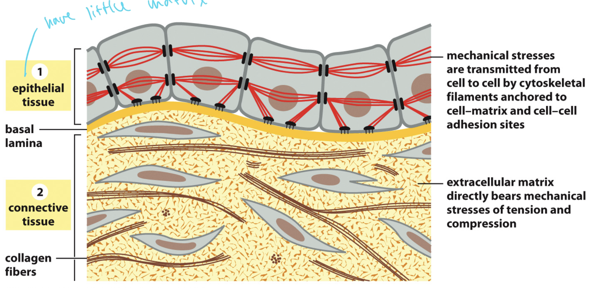
image of diff cell junctions
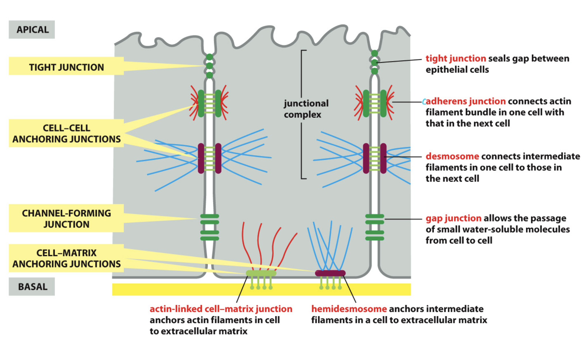
tight junctions
seal the gap between epithelial cells
adherens
connect actin filament bundles in one cell to actin filament bundles in the next cell
gap junctions
allow the passage of small water-soluble molecules between cells
hemidesmosomes
anchor IFs in a cell to extracellular matrix and binds to basal lamina and provide stability to epithelial cells.
actin-linked cell-matrix junction
anchors actin filaments in the cell to the ECM
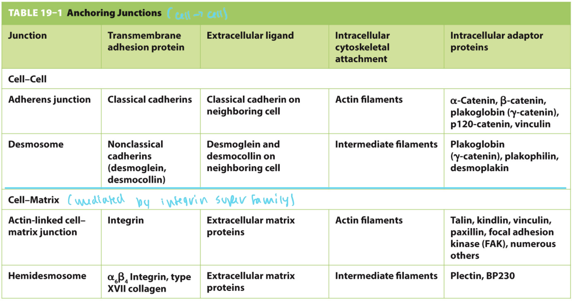
anchoring junctions
hold two cells together
each of the two cells has a transmembrane protein that connects to each other’s

things mediated by anchoring junctions summary table
notice cell to cell vs cell to matrix
cadherin and integrin superfamilies
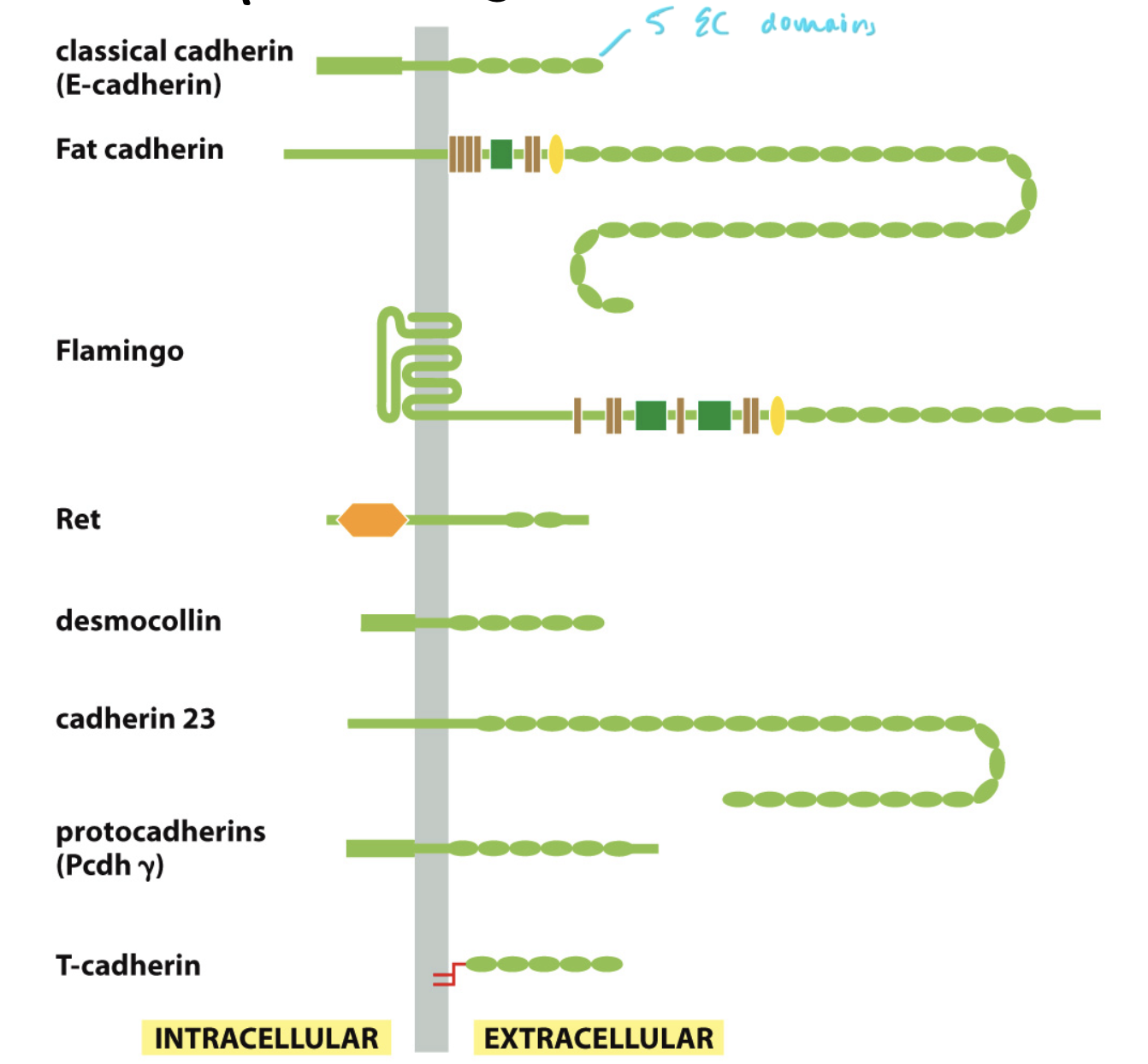
cadherins
have repeating extracellular domains
these domains are homophilic binders which bind to other ECs
ex: flamingo binding to another flamingo
in all animals, but not all eukaryotes
in some fungi, but not in plants
has an extracellular and intracellular domain
structure of cadherins
see how two cadherin EC domains bind to each other at junctions
calcium maintains the structure of cadherins to they can extend far enough to interact
calcium binds at the hinge regions
the cadherins can only bind if calcium is bound to the cadherins
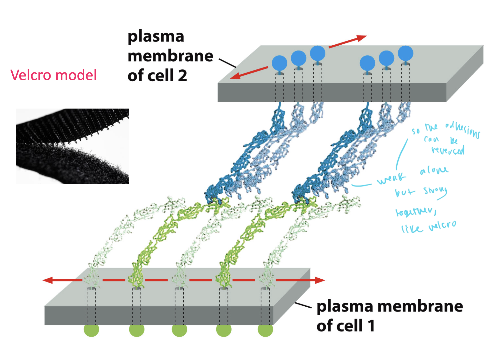
cadherins are homophilic binders
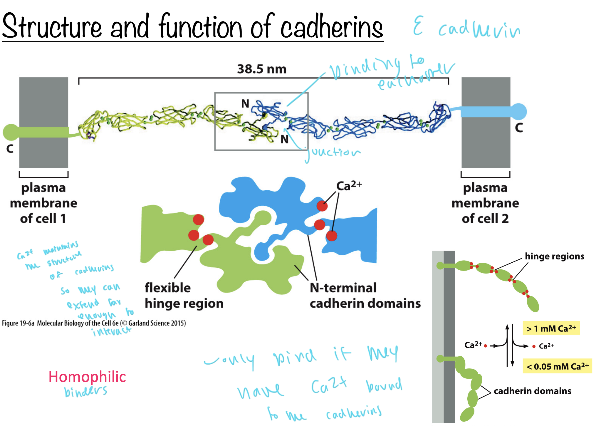
cadherins bind to each other like
velcro
the adhesions are weak alone but strong together
cadherin homophilic binding assists in development
layers of homophiic binding cadherins form the layers of the 3 embryonic germ layers
the layers reconstitute after being mixed up due to the cadherins, this shows which cell types go where
cadherinds sort according to homophilicness

catenins
adherin junctions
link cadherins to the actin cytoskeleton
are adapter proteins
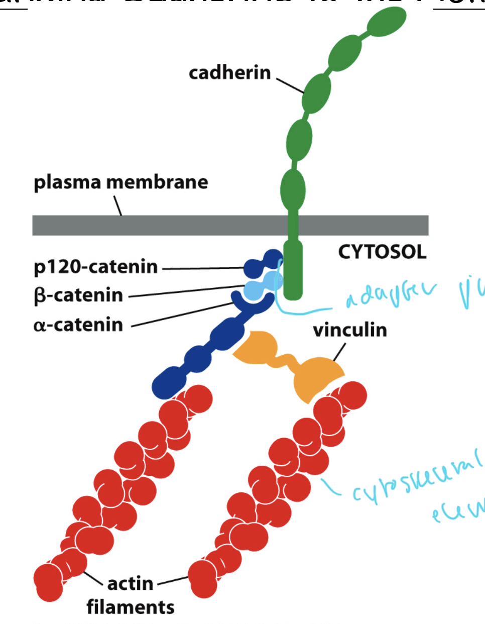
types of catenins
catenins: adapter proteins between cadherins and actin filaments
p120
beta-catenin
alpha catenin
alpha connects to beta, which connects t p120

adherens junction
connection between two cells
cadherin and catenin cluster between two cells
areas closest to each other bind so if the cadherins are close enough to bind each other, they will

Rac at adherens junctions
causes branched actin to form
promotes additional protrusions to expand the zone of contact between two cells
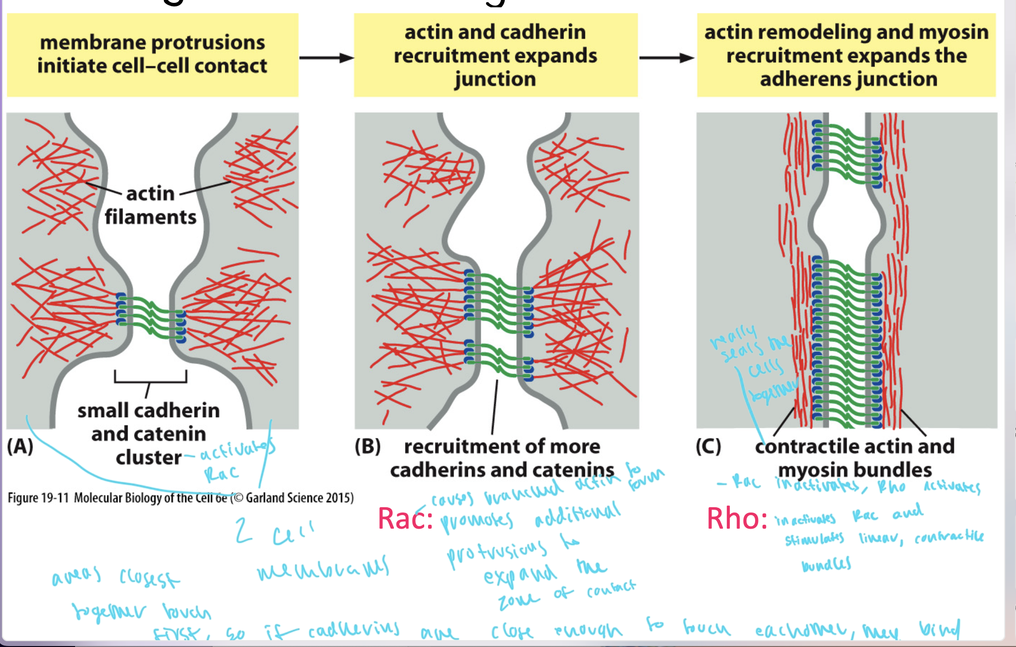
Rho at adherens junctions
inactivates Rac and stimulates the formation of linear contractile bundles
this contractile actin and myosin bundles are what seals the cells together

steps of adherens junction formation
1) a cluster of cadherins and catenins forms, starting to bring the two adjacent cells together
2) more and more cadherins and catenins get recruited, bringing the two cells closer together and Rac promotes branched actin to form
3) Rho inactivates Rac and causes linear contractile bundles to form of actin and myosin
4) the two cells seal together

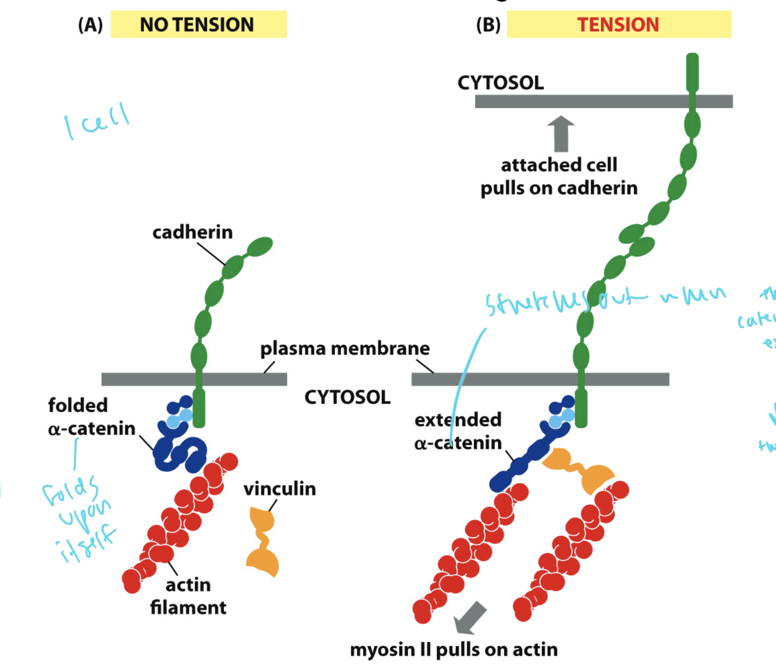
mechanotransduction in adherens junctions
when the cells feel more tension, they form tighter adhesions in response to stay stable
the cadherins get stretched out, pulling on the alpha catenin
when the cell feels tension, the alpha catenin goes from folded to stretched out
where the alpha catenin stretches out, vinculin builds more actin off of it and myosin pulls on the actin in the oppo direction to stabilize
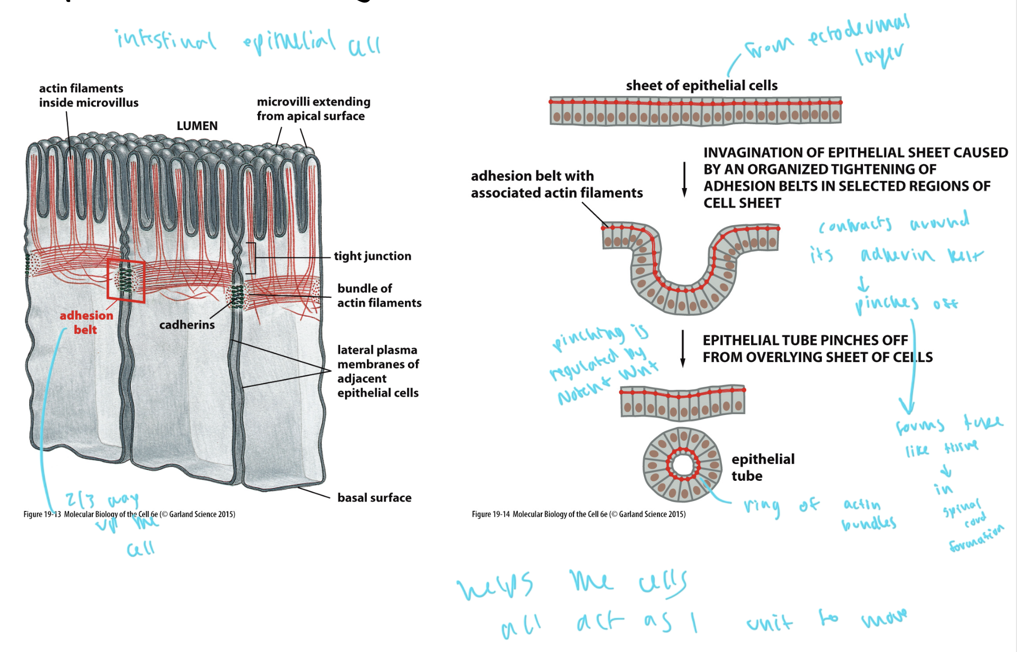
adhesion belts
when adherens junctions in epithelial cells form a loop so that all of the cells can function as one unit to move
from the ectodermal layer of cells
forms a ring of actin bundles when the adherin belt contracts and pinches off
these rings/belts form 2/3 the way up on epithelial cells
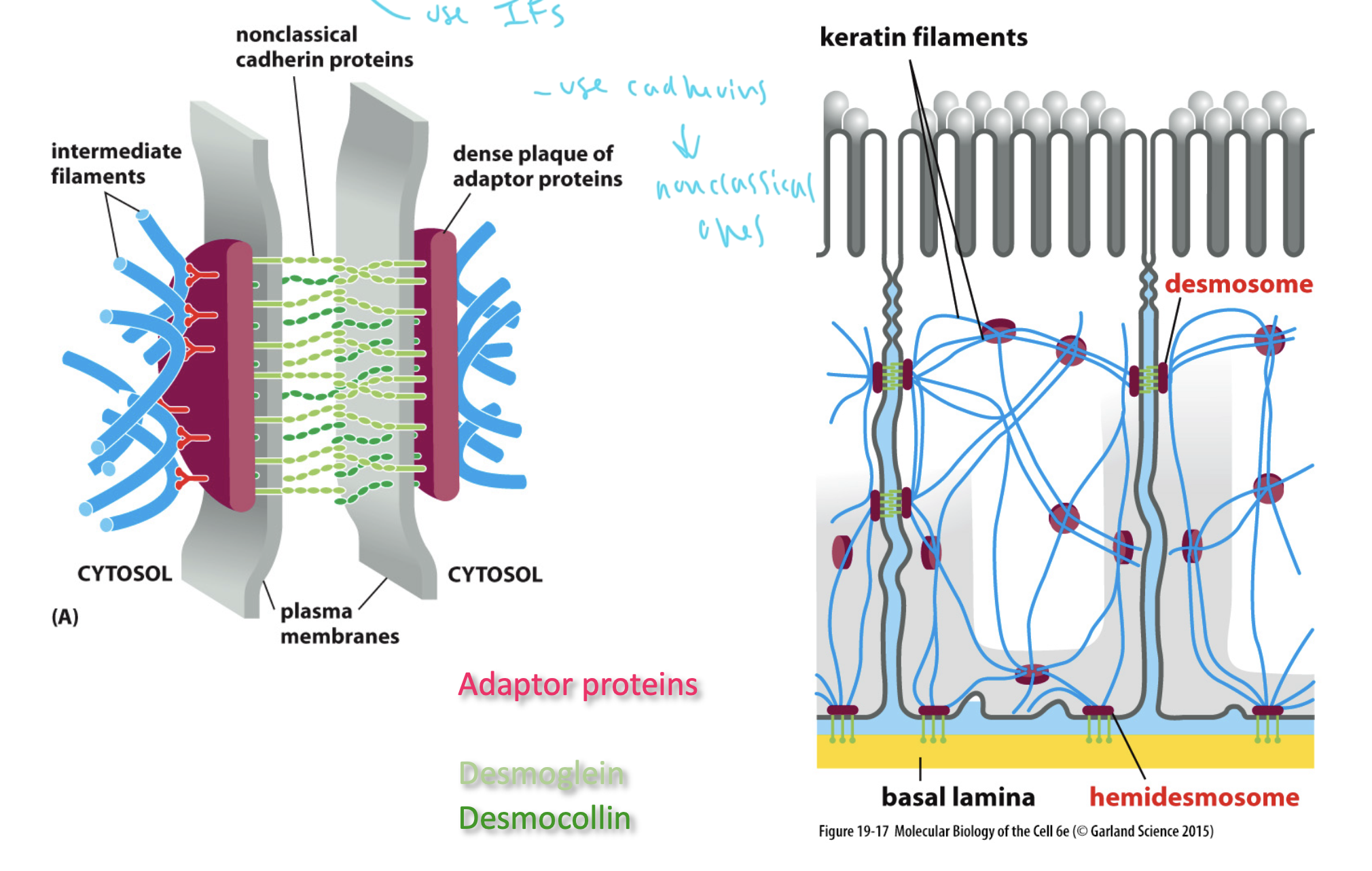
desmosomes
provide mechanical strength for the cell
form “buttons”
use nonclassical cadherins to seal between two cells
cadherins connect intermediate filaments to desmosomes
desmocollins and desmogleins are cadherins that help form desmosomes
IFs are on the outside and connect to the button adpater proteins, which connect to desmogleins and desmocollins, the cadherins that hold the two sides of the cadherins together
tight junctions
partitions the body and lines organs by forming sheets of epithelial cells
act as permeability barriers in cells
seal cells together so that molecules are moved selectively between cells (allows for regulation)
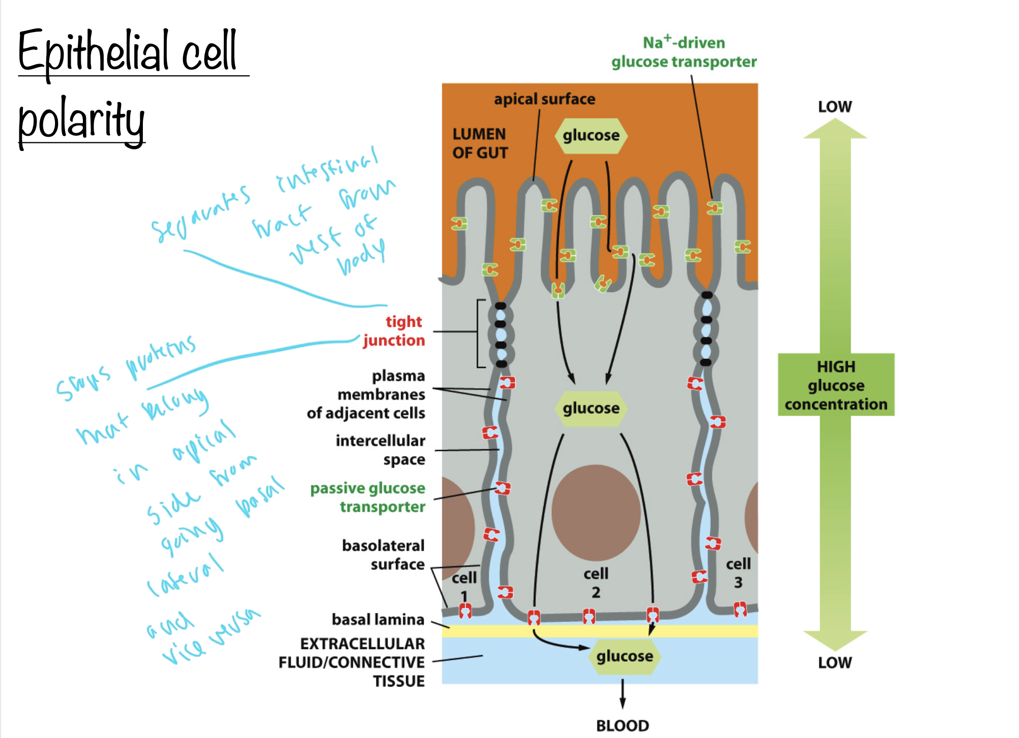
polarity of epithelial cells
see how tight junctions separate the outside of the body from the area between two cells
tight junctions stop proteins that belong in the apical side from going to the basal side and vice verse
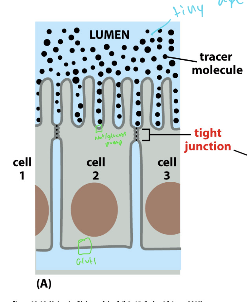
experiment that shows the function of tight junctions
tiny dye molecules placed on either side of the tight junctions (inside or outside of the cell) could not get in to the other side
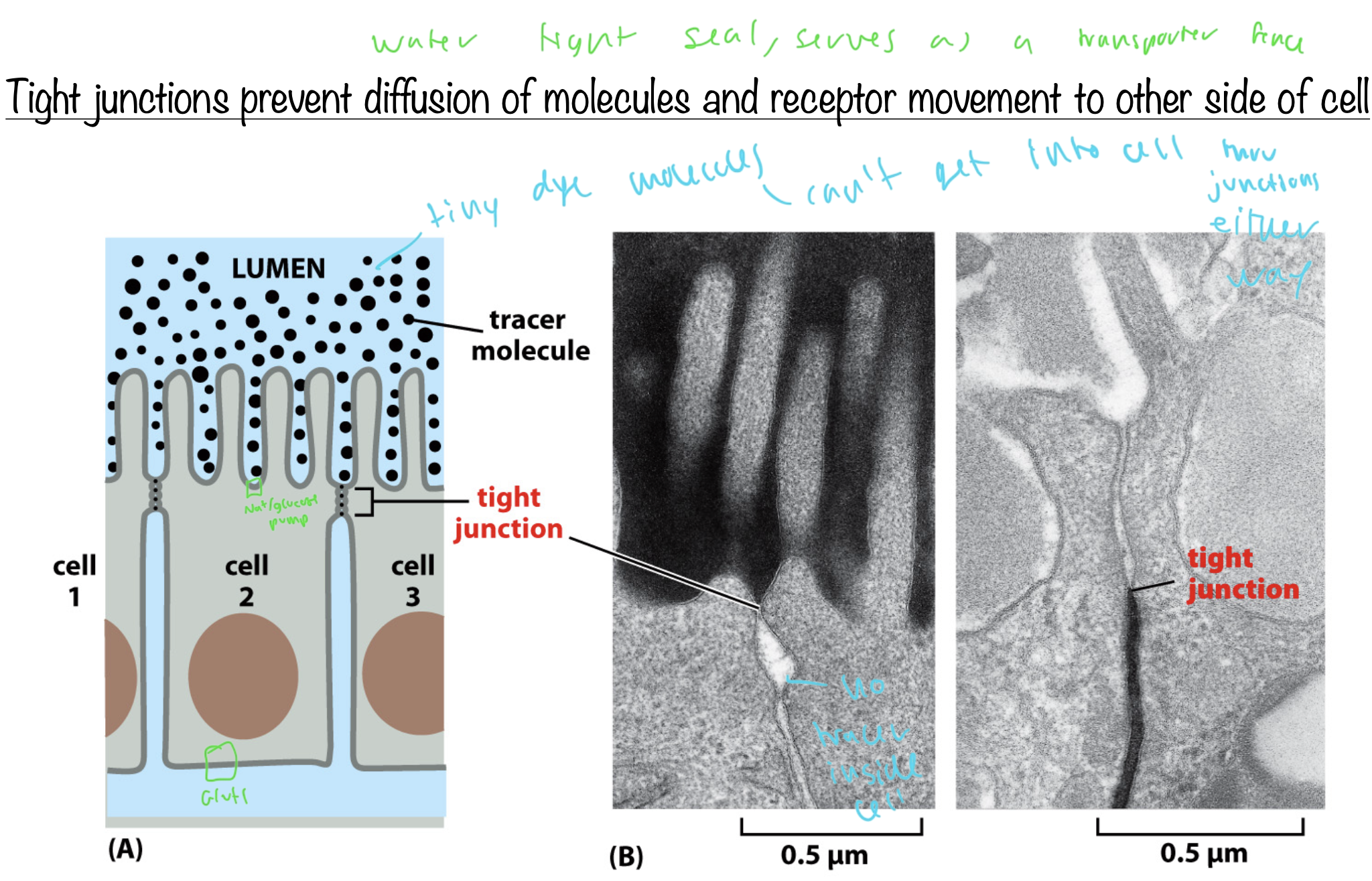
where GLUT1 and the sodium glucose pump are on epithelial cells
sealing strands of tight junctions
water tight seals between two cell membranes
like a woven material
forms when claudins and occludins bind to each other (of themselves) on opposite sides of the tight junctions
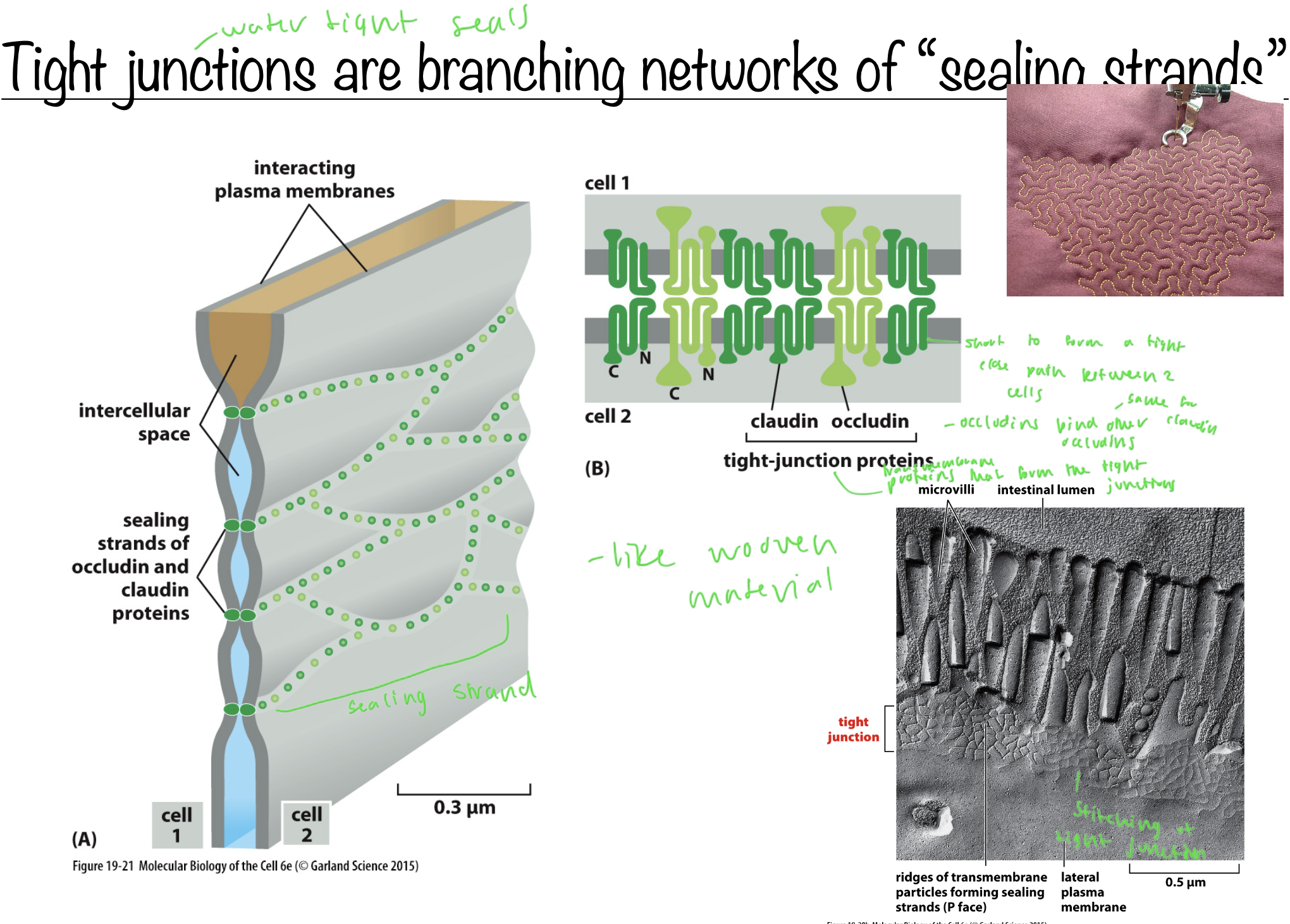
claudins and occludins
claudins bind other claudins and vice versa
transmembrane proteins that help form tight junctions
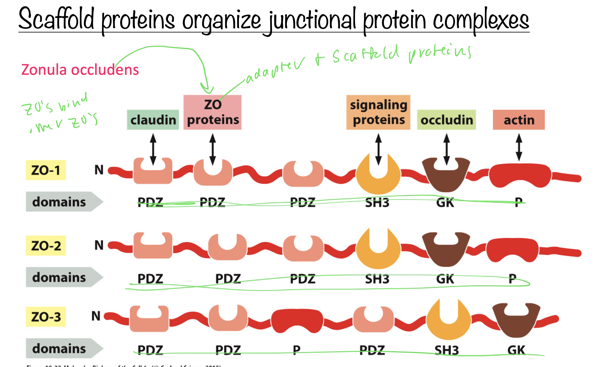
how scaffold proteins help form junction proteins
Zonula occludens (ZOs) bind to other ZOs
ZOs are adapter and scaffold proteins and are next to other claudins in a line
gap junctions overview
bridge gaps between cells to create and direct channels between two cells cytoplasms
made up of connexin proteins in vertebrates
connexins are large enough to allow small molecules through, but not macromolecules
gap junctions help move ATP and ions, so they “couple” cells metabolically and electrically
gap junctions can function as channels
structure of gap junctions
6 connexins form a connexon
connexons can be homomeric or heteromeric
two connexons combine to form homo or heterotypic intercullular channels
diff combos of these channels selects for diff things, allowing the gap junctions to serve as a selectivity barrier
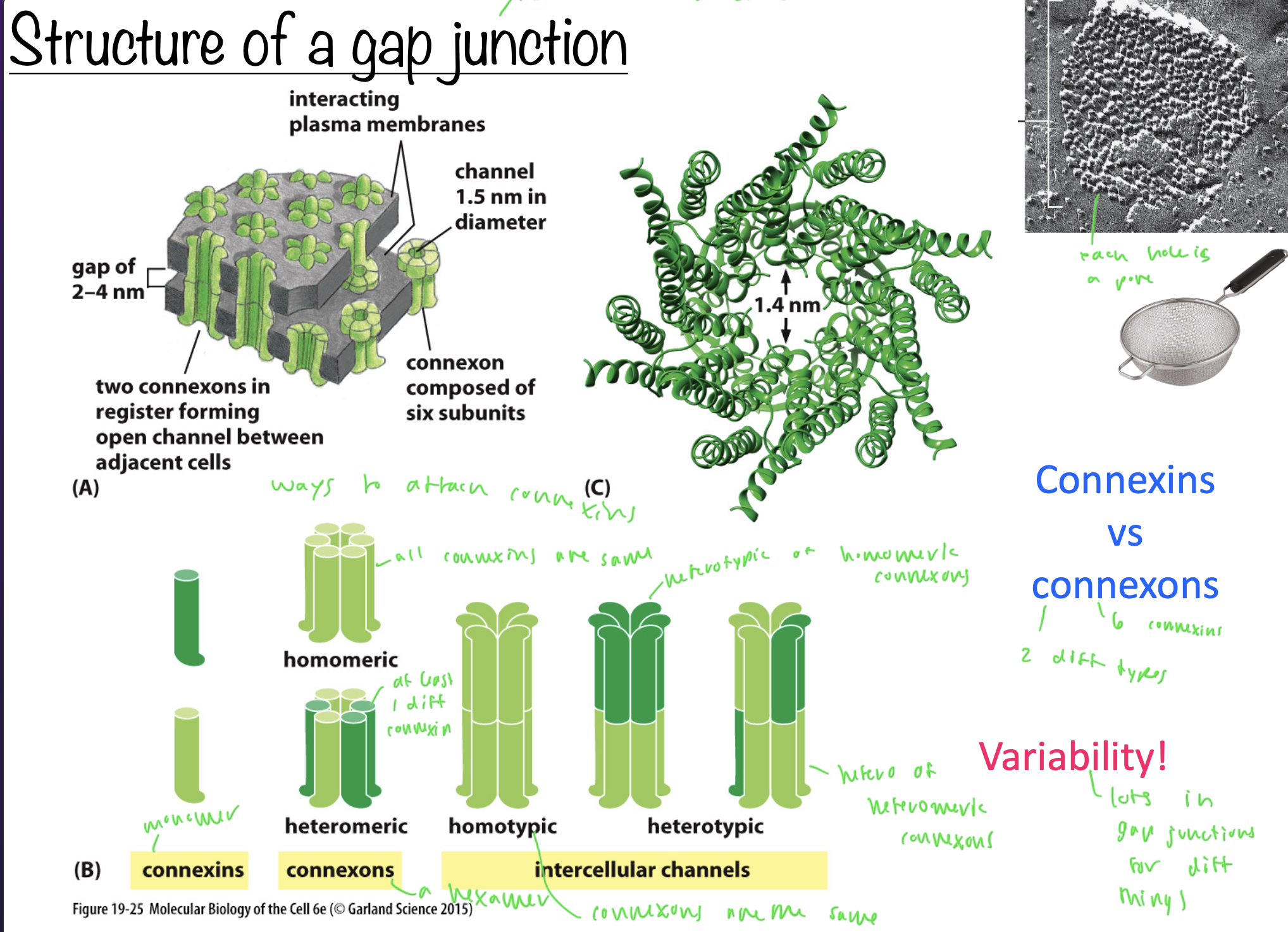
how many types of connexons are there?
2
homomeric and heteromeric
do plant cell walls have adhesion junctions?
NO, since they are already bound tightly and don’t need it
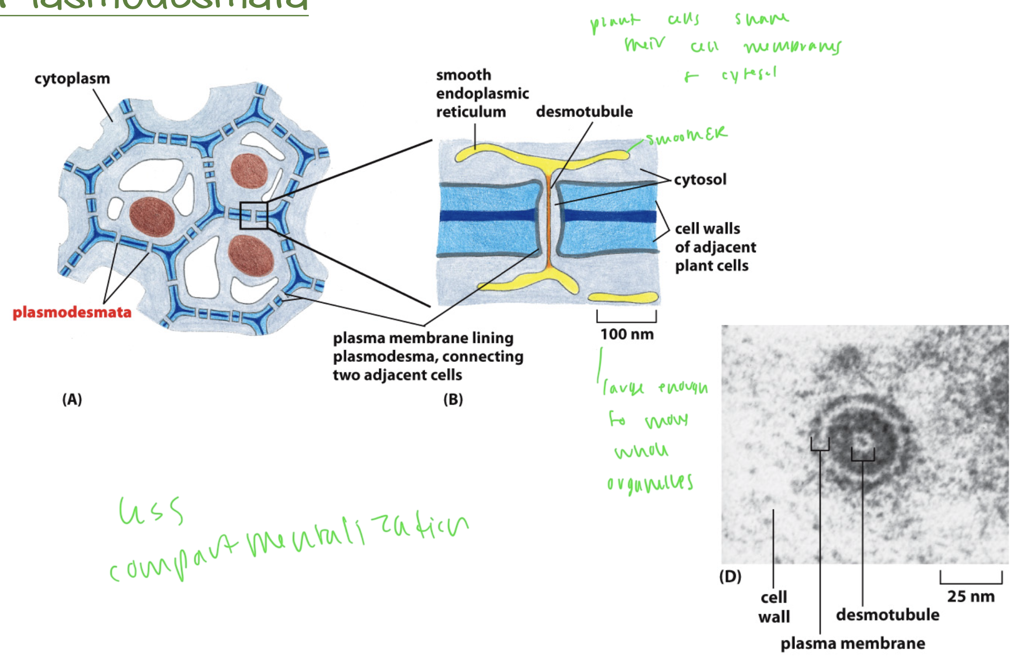
plasmodesmata
in between plant cells, similar to gap junctions in animal cells
plant cell walls are tightly
cemented together
the plasma membrane of one plant cell is continuous with that of its neighbor via plasmodesmata
the plasma membrane of one plant cell is continuous with that of its neighbor via
plasmodesmata
plasmodesmata structure
the area between two plant cells
allows plant cells next to each other to share their cell membranes and cytosol to allow solutes through, which cannot get through the cell wall
there are large enough gaps that whole organelles can get through to adjacent cells
what forms the ECM
cells and their matrix
the ECM is very dynamic and active
the ECM is made up of macromolecules
the ECM i has its own signals
things that are formed out of the ECM
teeth and bone
the transparent cornea of the eye
tendons
ALL of this is acellular
3 major classes of ECM macromolecules
proteoglycans and GAGs
fibrous proteins (collagens)
glycoproteins
proteoglycans
perlecan
decorin
aggrecan
these three also contains GAGs

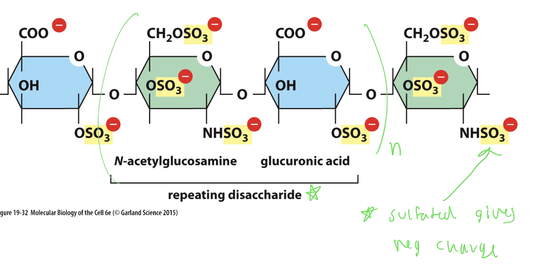
GAGs
ex: hyaluronan
this is only a GAG, not a proteoglycan
glycosaminoglycans: made up of repeating disaccharides of amino sugars
made of unbranched polysaccharide chains
mostly beta linkages between the dissacharides
all are sulfated, which gives a neg charge
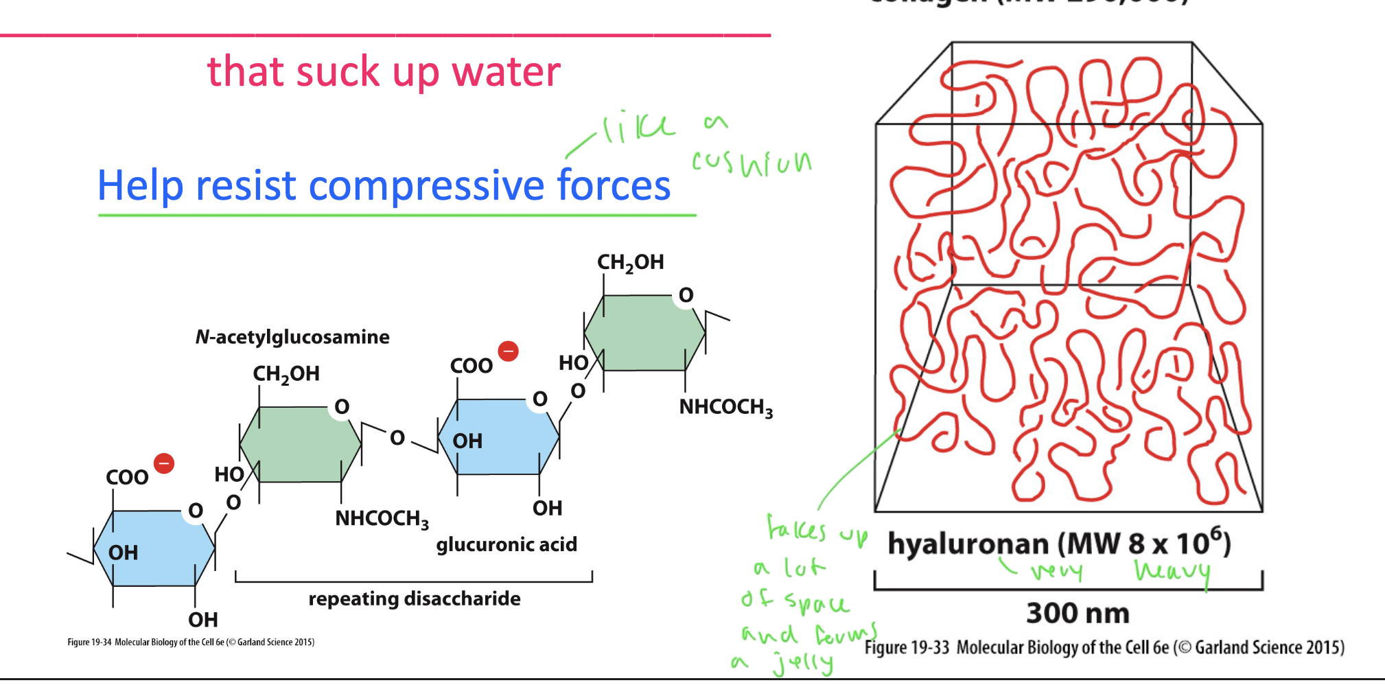
hyaluronan
a space filler GAG that forms a hydrated gel that attracts nearby ions, and therefore water, forming a cushion that takes up a lot of space
lacks sulfates, so it attracts a bit less water and is a bit more firm
forms a cushion/jelly
helps resist compressive forces
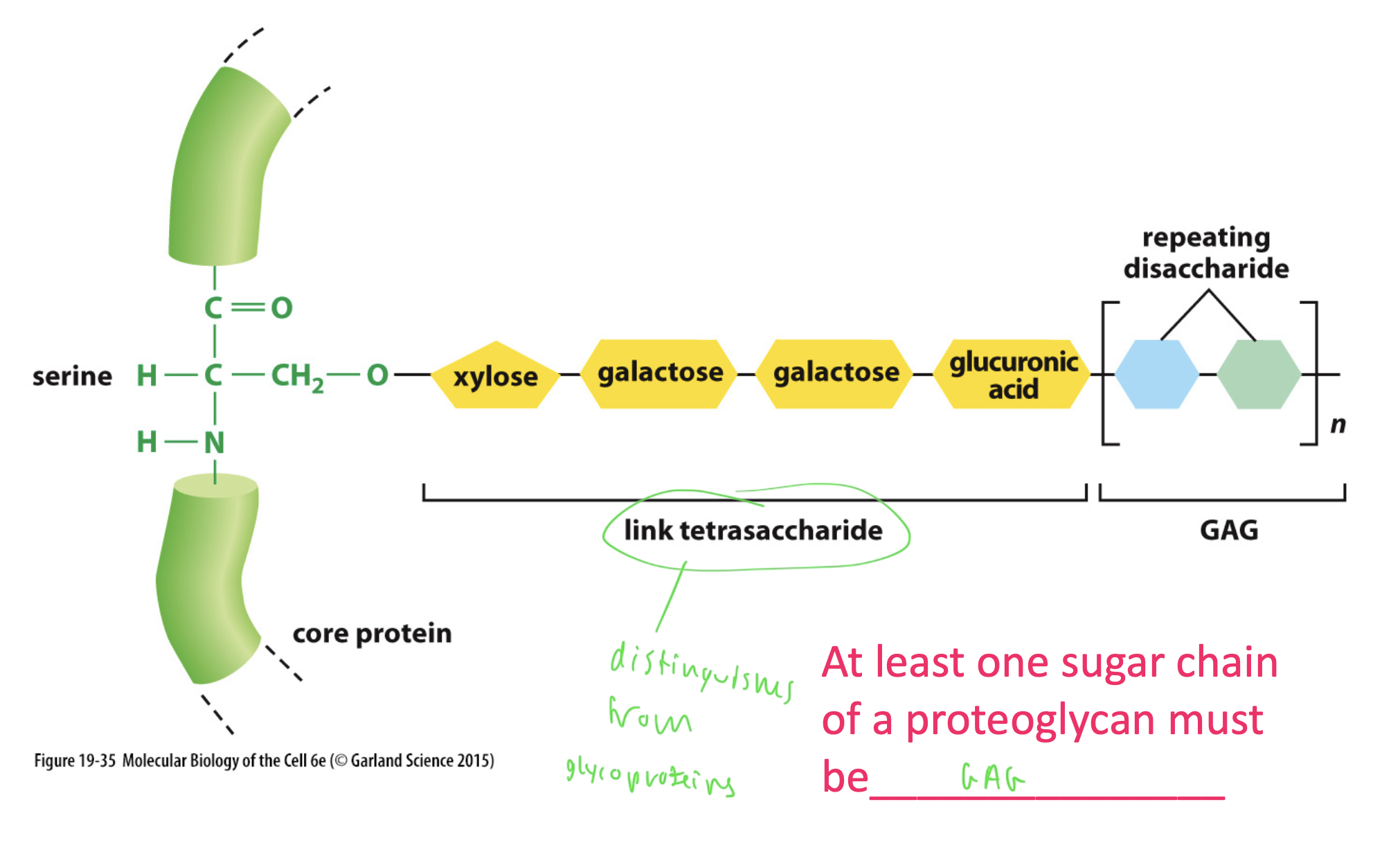
proteoglycans (that are also GAGs)
GAGs attached to a protein
have a linked tetrasaccharide, which differentiates them from glycoproteins
must have at least one sugar chain that is a GAG to be a proteoglycan
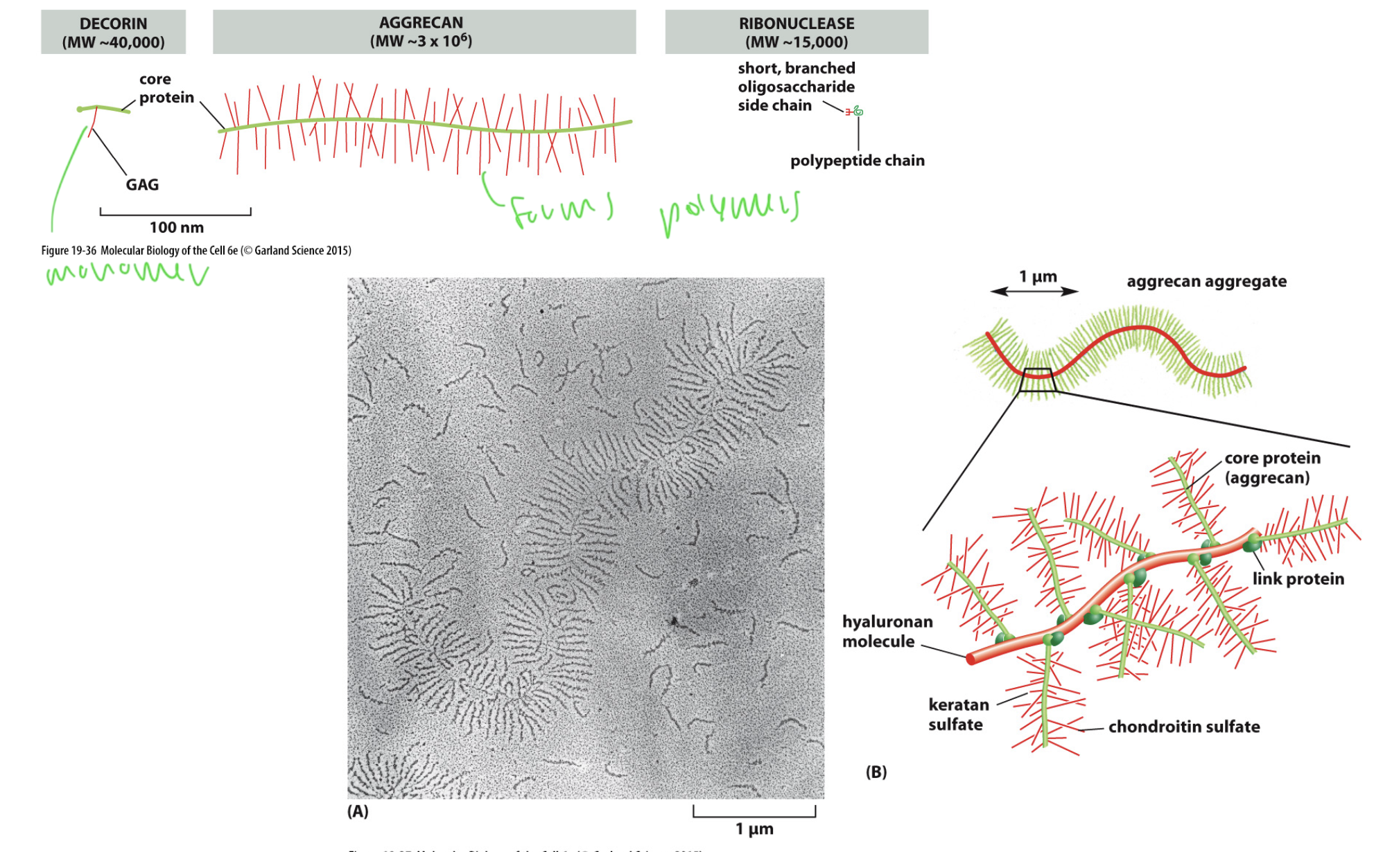
proteoglycans can be
huge
occurs when they form even larger polymeric complexes
ex: aggrecan, which polymerizes and forms long branched propteoglycans

aggrecan
forms huge proteogylcan complexes
What is the most abundant protein in mammals?
collagens (fibrous proteins)
fibrous proteins/collagens
resist tensile strength
form long, stuff helical structures to provide strength for the ECM
form triple helixes using proline and glycine which form kinks in the AA backbone to get a twisted shape
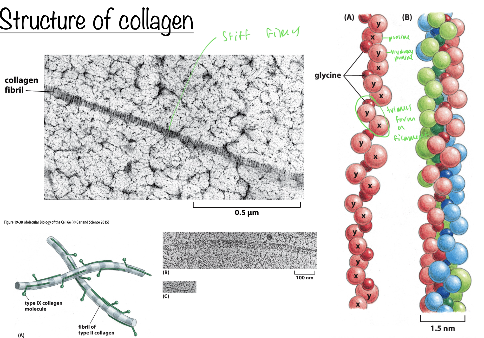
structure of collagen
long stiff fibers
collagen backbone is formed of glycine, proline, and hydroxy proline (these three form a trimer)
these trimers form a filament
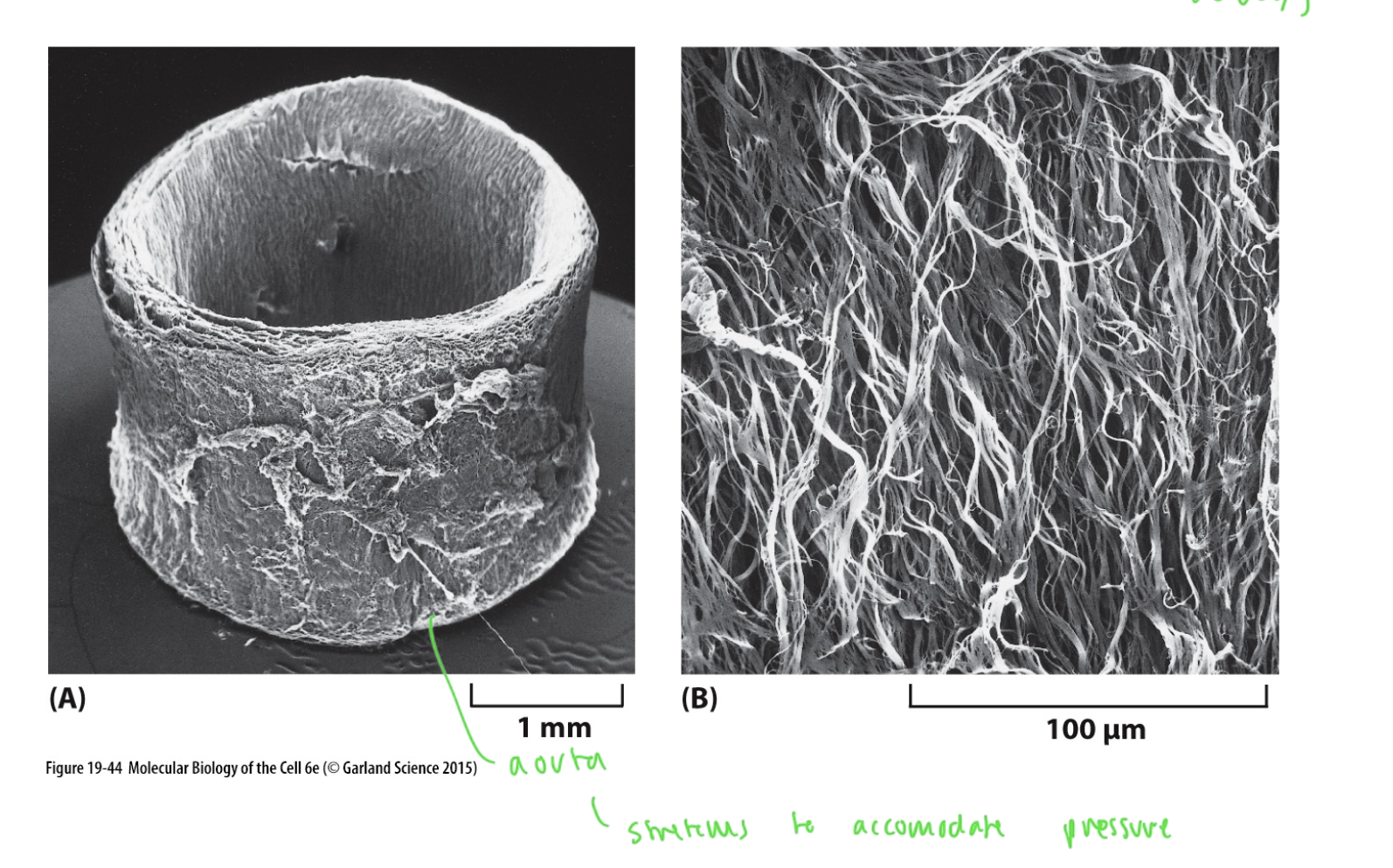
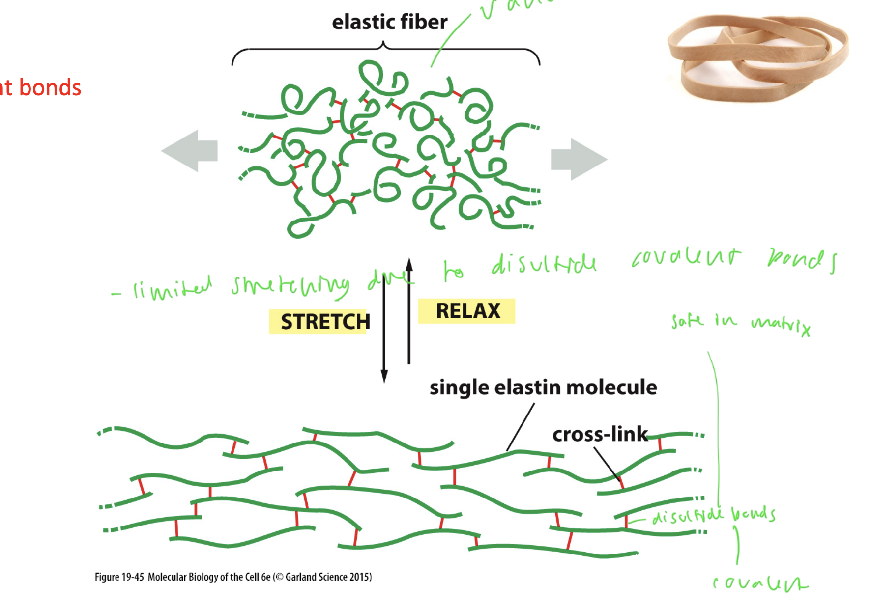
elastin
a form of fibrous protein that gives tissues their flexibility
in the skin, uterus, bladder, aorta, lungs, etc for stretching
elastin forming a network
elastin is intrinsically disordered so it forms random coils, which forms a network, stabilized by crosslinks of disulfide bonds (covalent bonds)
this is why elastin has limited stretching, as these disulfide bonds only allow the elastin to go so far
these S-S bonds are fine in the matrix, which is polar
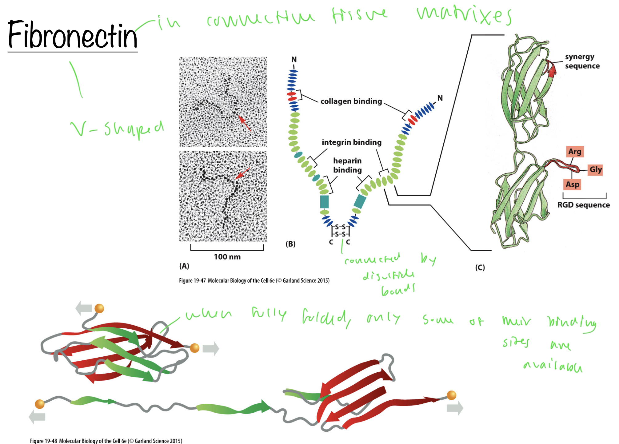
glycoproteins
ex: fibronectin
scaffold proteins with multiple binding domains
helps organize the ECM
can respond to stretch by binding to other fibronectin molecules
fibronectin
connective tissue in matrices
forms V-shapes, connected in the middle by disulfide bonds
when they are fully folded, only some of their binding sites are available
unfolds to allow more binding sites to be accessible
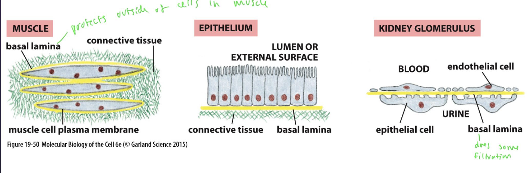
basal lamina
a specialized form of the ECM
on the underside of all epithelial cells
aka the basement membrane
protects the outside of cells and adds additional filtration into cells (kidneys)
it also gives survival signals to the cells on top of it, therefore is a part of cell survival/proliferation/etc
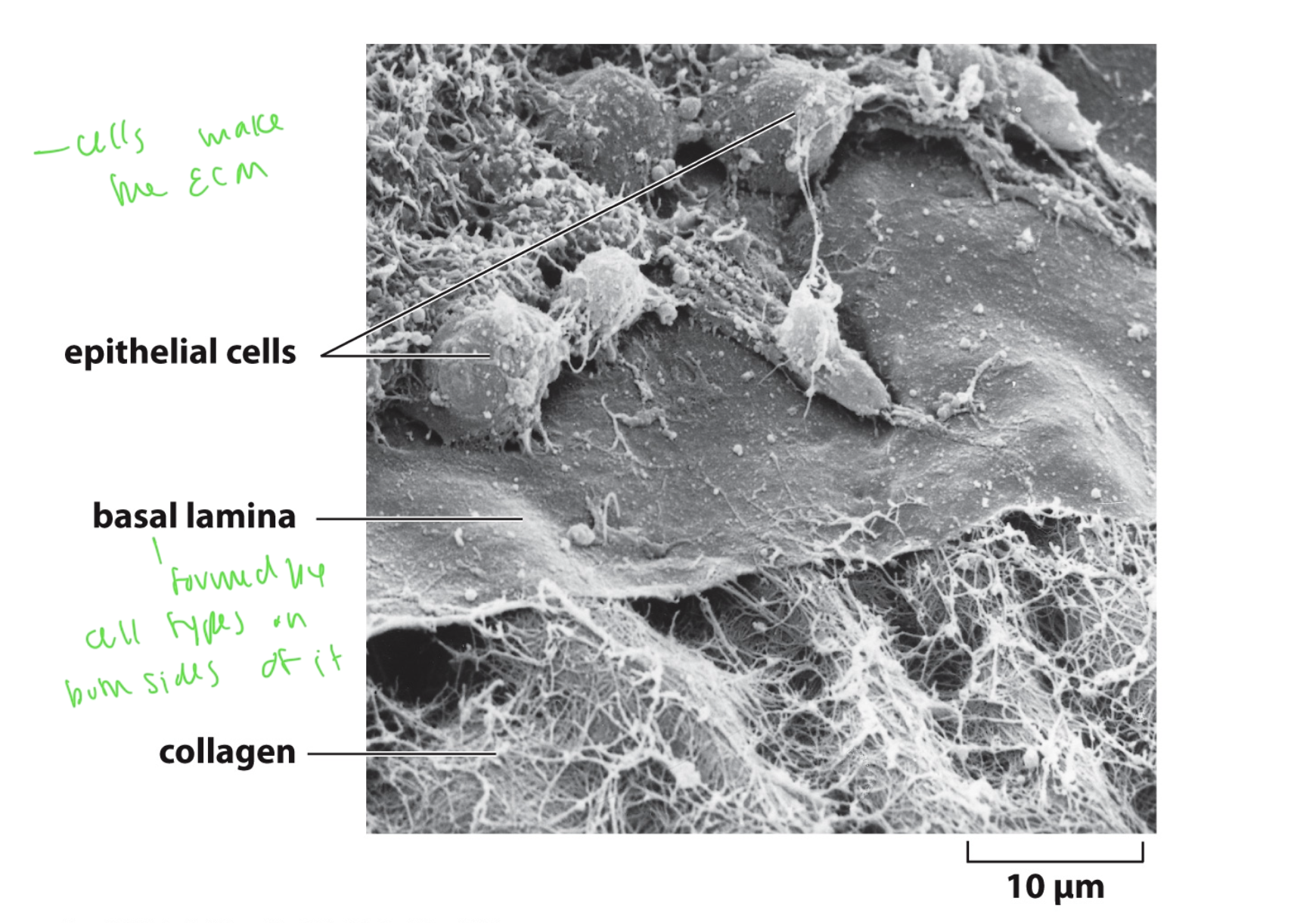
see basal lamina formation up close
the basal lamina is formed by the cells on either side of it
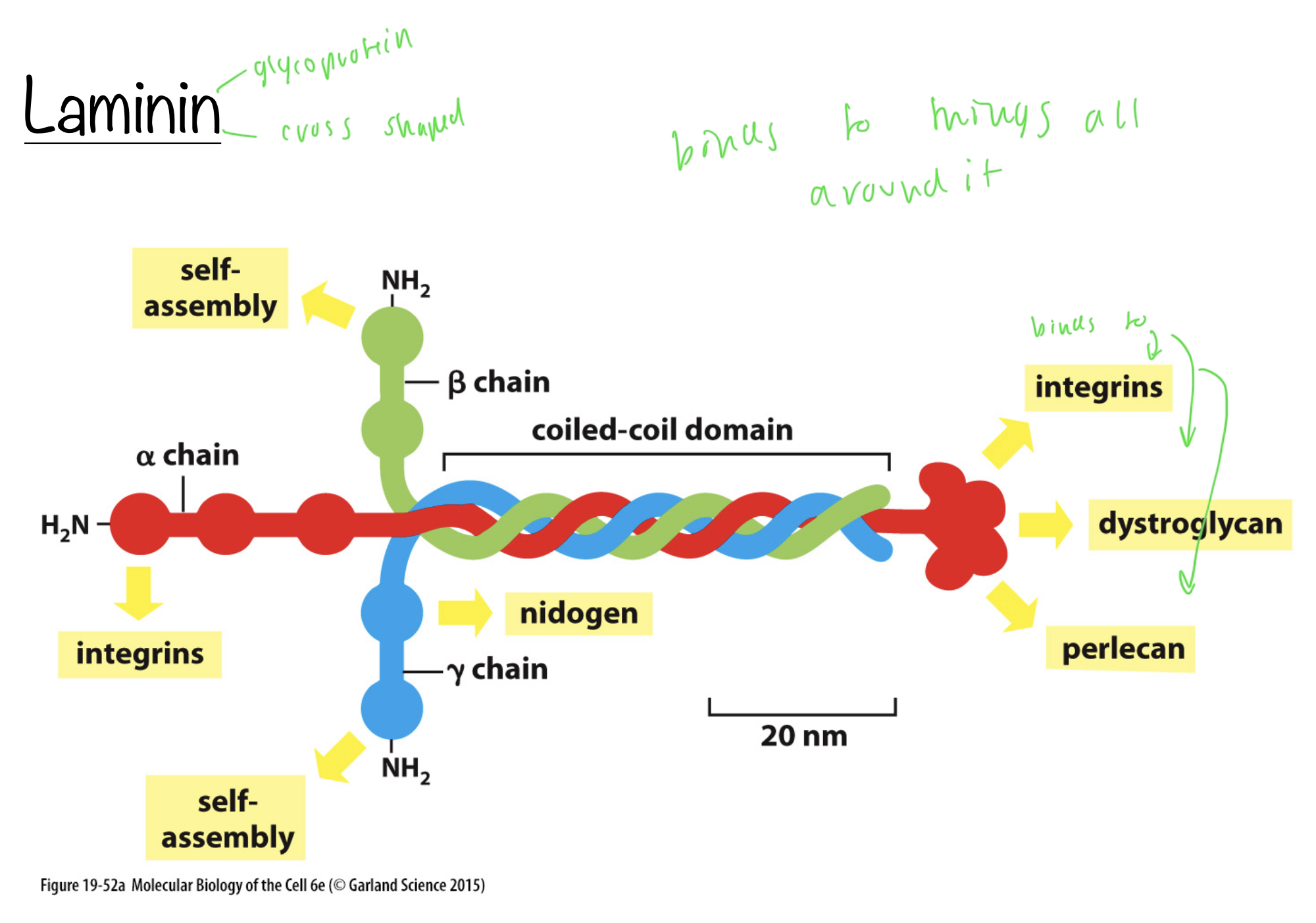
laminin
a glycoprotein
cross-shaped
binds to things all around it (integrins, dystroglycan, perlecans)
has a coiled domain and three NH2 domains that form the cross (alpha, beta, and gamma)
alpha strand forms the tip of the cross
laminin is the primary organizer of the basal lamina

What does the alpha chain of the laminin bind to?
other integrins (on both side of the alpha chain)
how are cell to matrix junctions mediated?
by matrix receptors that tie the matrix to the cytoskeleton
integrins
transmit mechanical and molecular signals and convert them to help mediate the cell-matrix junctions
structure of integrins
form heterodimers of 1 alpha and 1 beta integrin
connect ECM proteins to things inside the cell by binding to adapter proteins that bind to actin
adapter proteins that bind to actin to help integrin bind more actin: vinculin, talin, kindlin
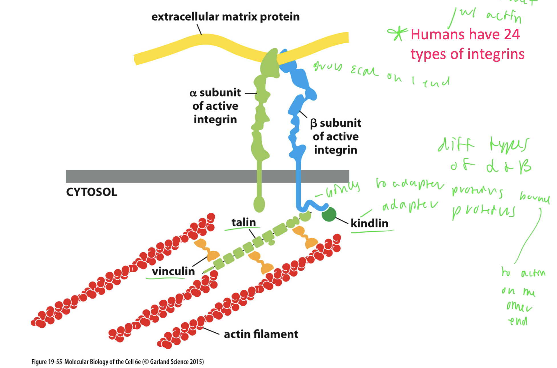
How many integrins are in humans?
24
23 of these interact with actin
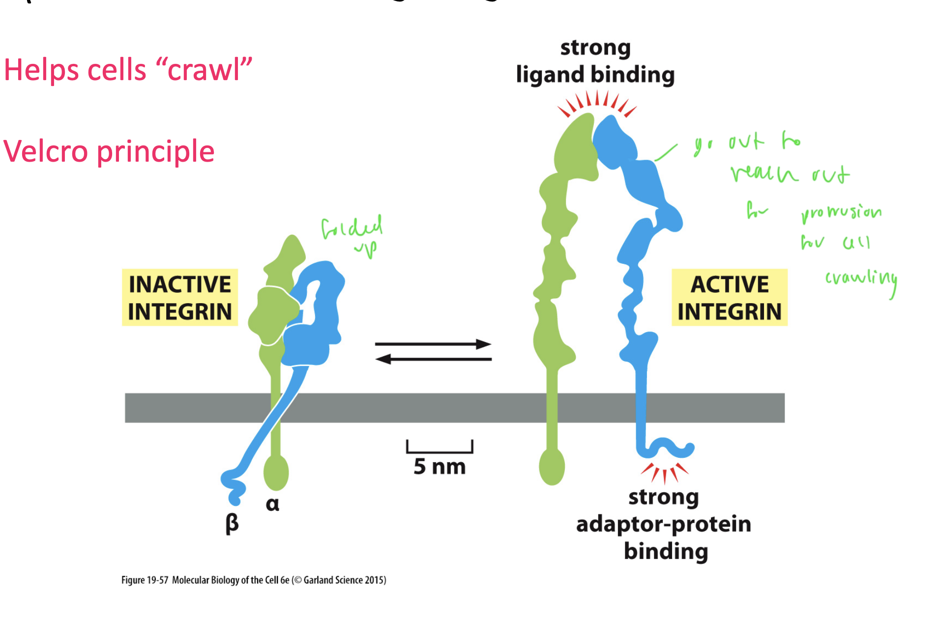
activation of integrins
inactive state when folded up on themselves
active state when they unfold and can reach out to protrusion during cell crawling
stretched out region allows for strong ligand binding
What is the most prominent cell-matrix interaction in epithelia?
hemidesmosomes
hemidesmosomes
half buttons
attach epithelial cells to the basement membrane
formed of BP230, which attaches to plectin, which attaches to integrin, which binds to collagen 12 (the adapter protein) that attaches to laminin, which attaches to collagen
the BP230 is atatched to keratin on the other side of the cell
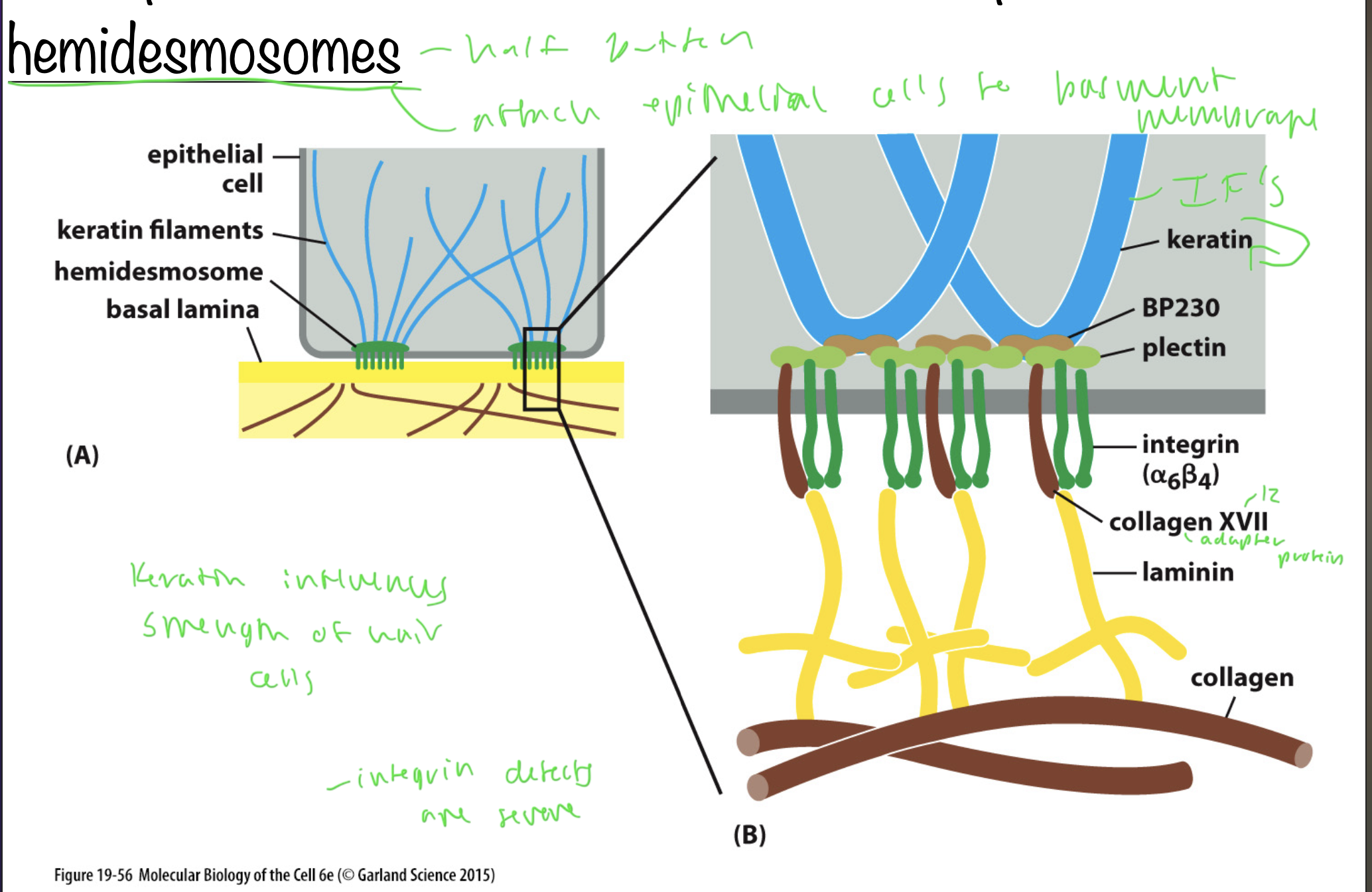
keratin
an IF that influences the strength of cells
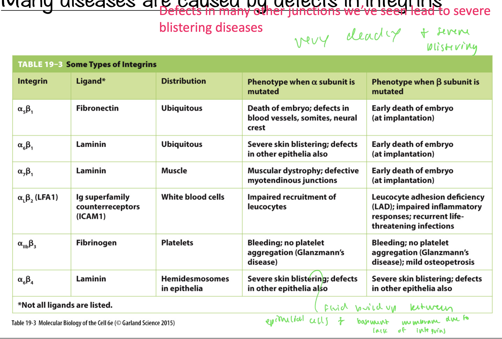
integrin defects cause
extreme defects
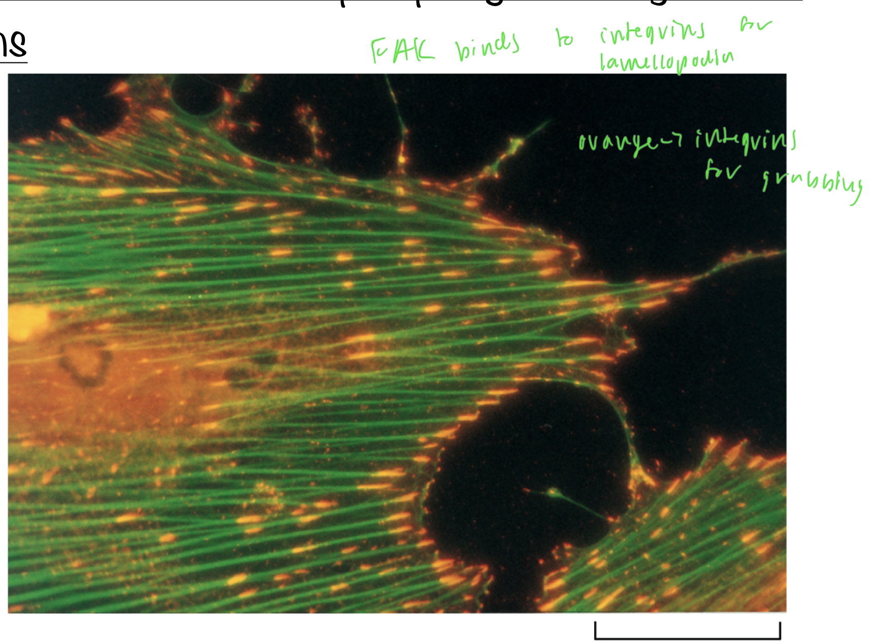
FAK
focal adhesion kinase
phosphorylates integrin focal adhesions
binds to integrins to form the lamellopodia
the cell wall resists ___ forces
tensile and compressive
the plant ECM is made up of polymers that lack ____
nitrogen
so fungi cant degrade plants for nitroigen fixation
What macromolecule do plants lack in the matrix?
proteins
use sugars instead
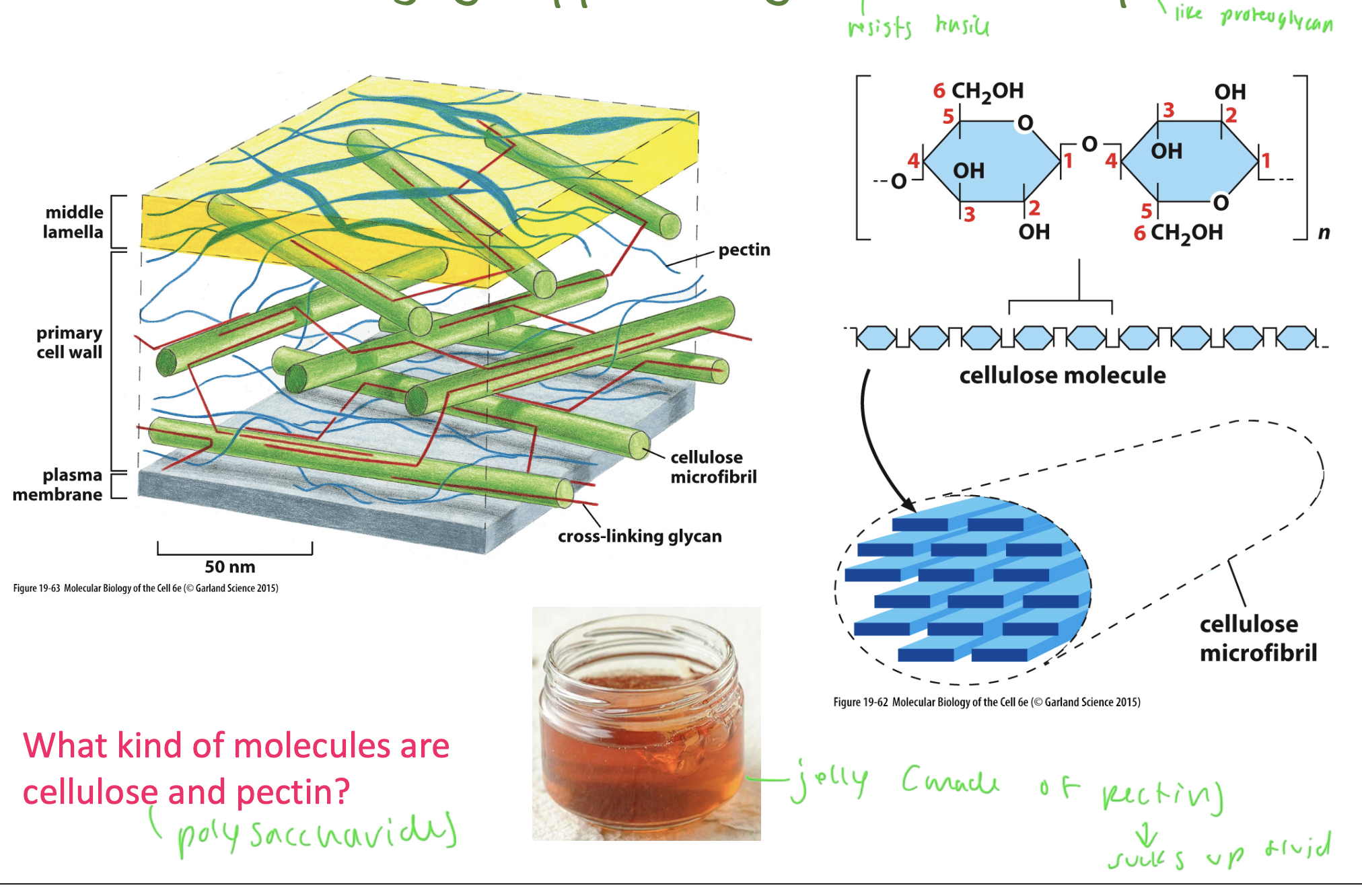
cell walls are made out of
cellulose and pectin
cellulose and pectin are made out of polysaccharides
cellulose forms perpendicular fibers (like collagen in animals)
pectin resists compression and is like the proteoglycans of animal cells
pectin is like jelly and resists compressive forces (sucks up fluid)
turgor pressure
the most solute is in the cytoplasm of plant cells, compared to the cell walls and externally, so water rushes inside the cell to accomodate, creating the pressure
large internal hydrostatic pressure that pushes outward on the cell wall, allowing plants to stand up straight
the cell wall resists the turgor pressure
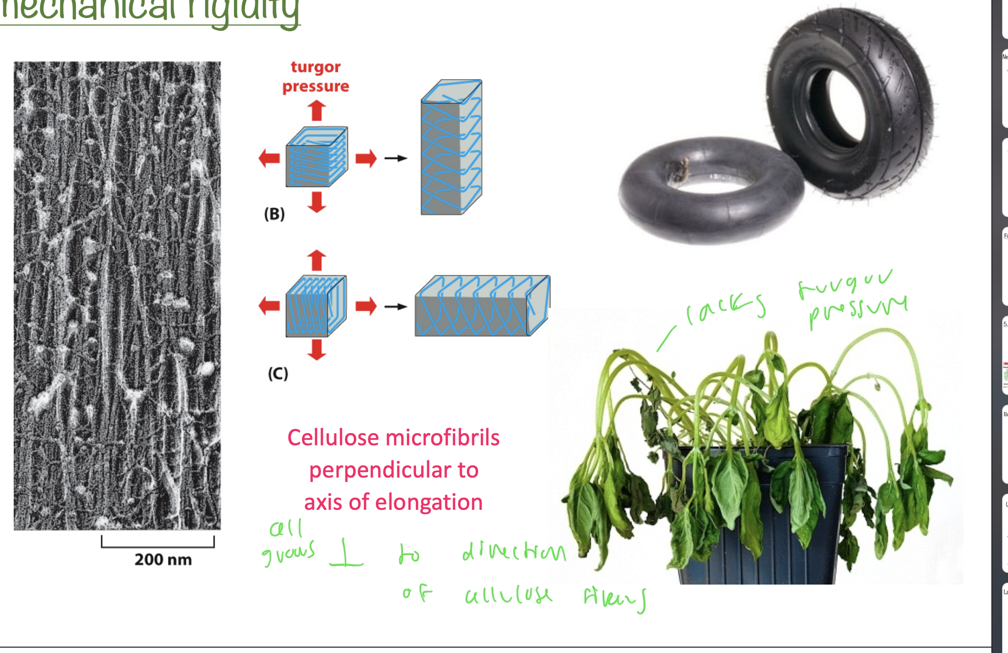
what direction do cellulose microfibrils grow?
perpendicular to the axis of elongation of the plant
creates strength for the plant