Looks like no one added any tags here yet for you.
Axial Skeleton
Bones of the skull
Accessory bones
Vertebral column
Thoracic cage (ribs, sternum, thoracic vertebrae)
Appendicular Skeleton
Upper limbs and pectoral girdle
Lower limbs and pelvic girdle
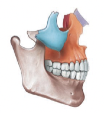
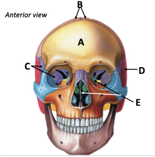
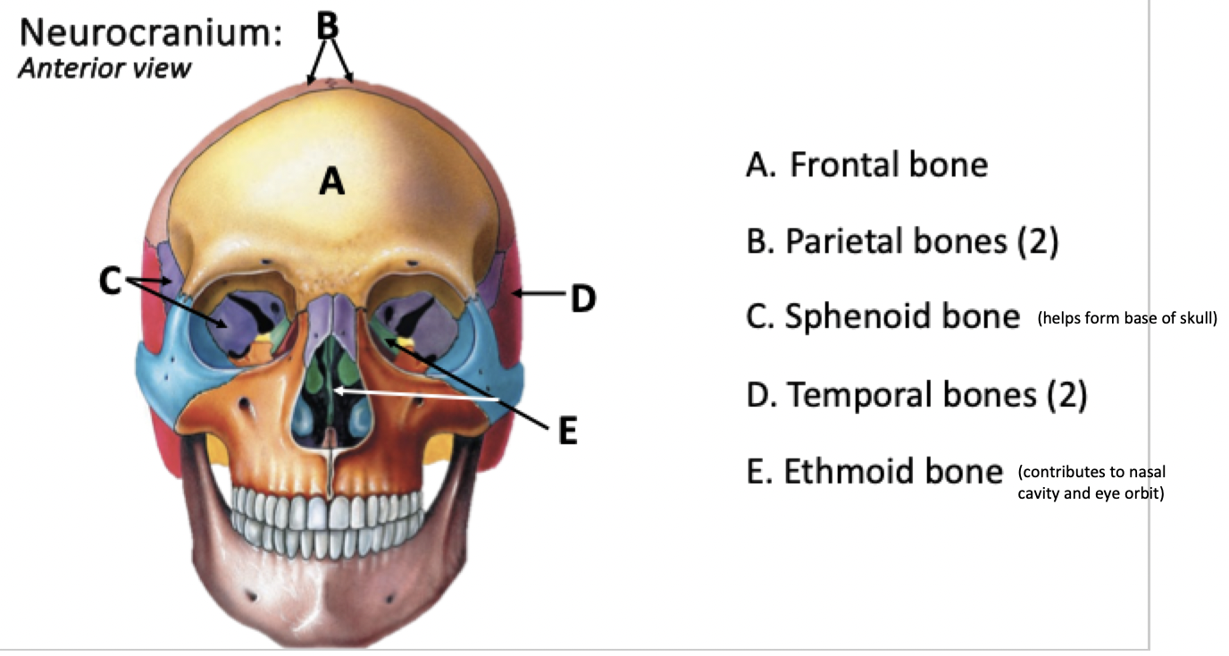
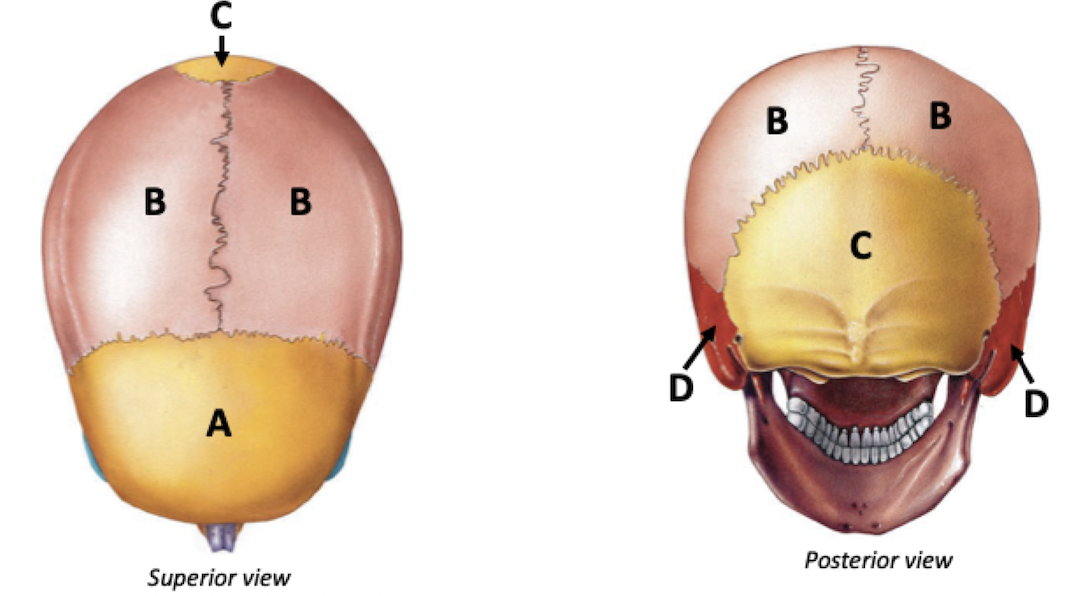
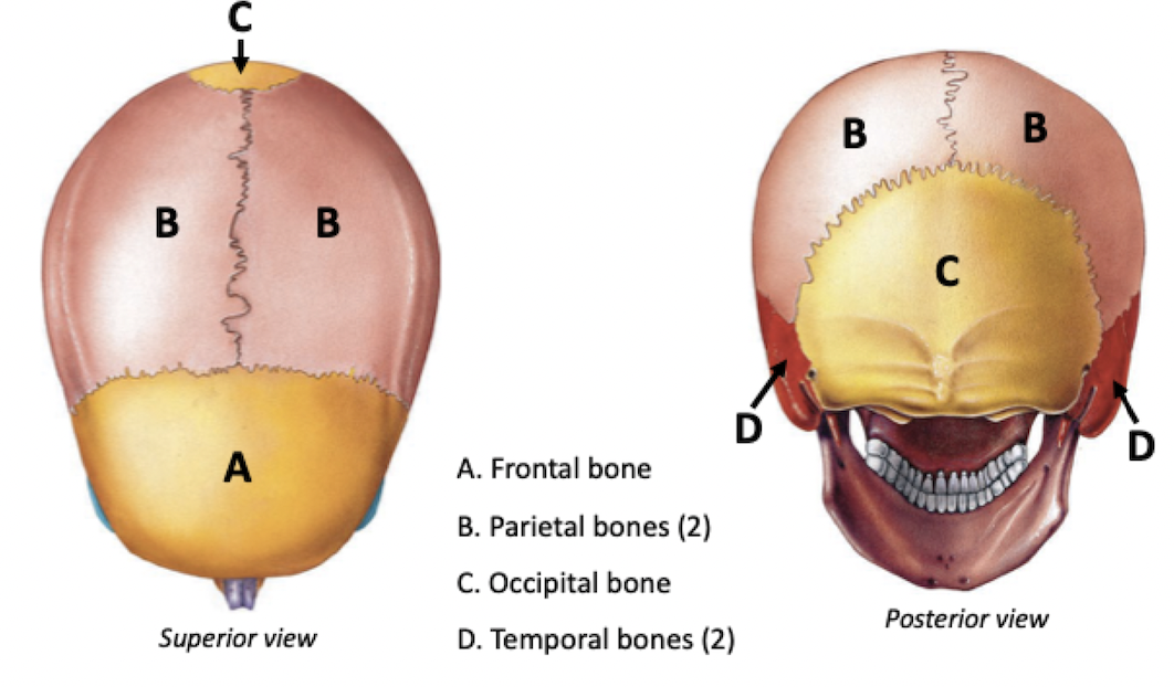
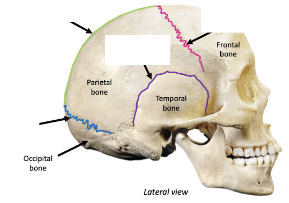
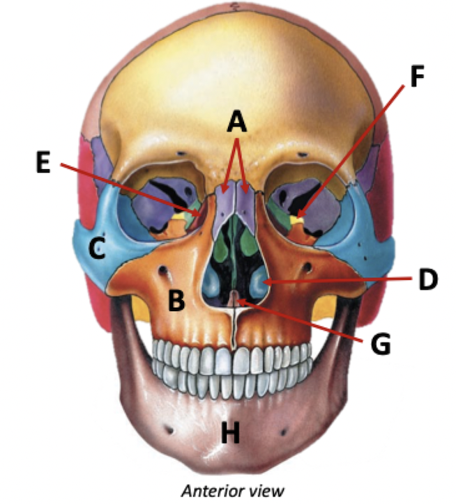
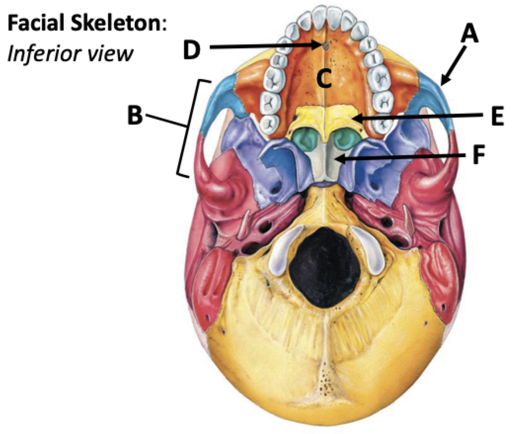
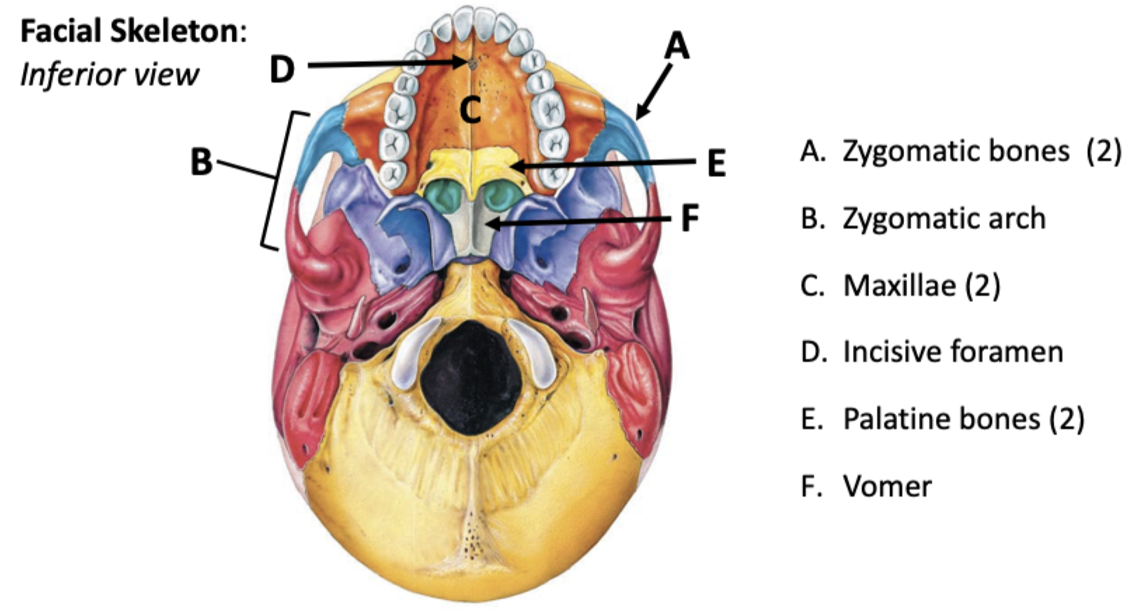
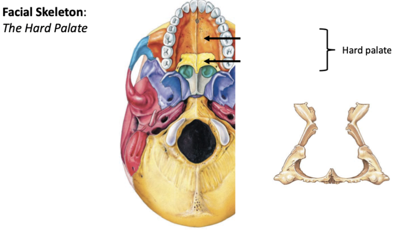
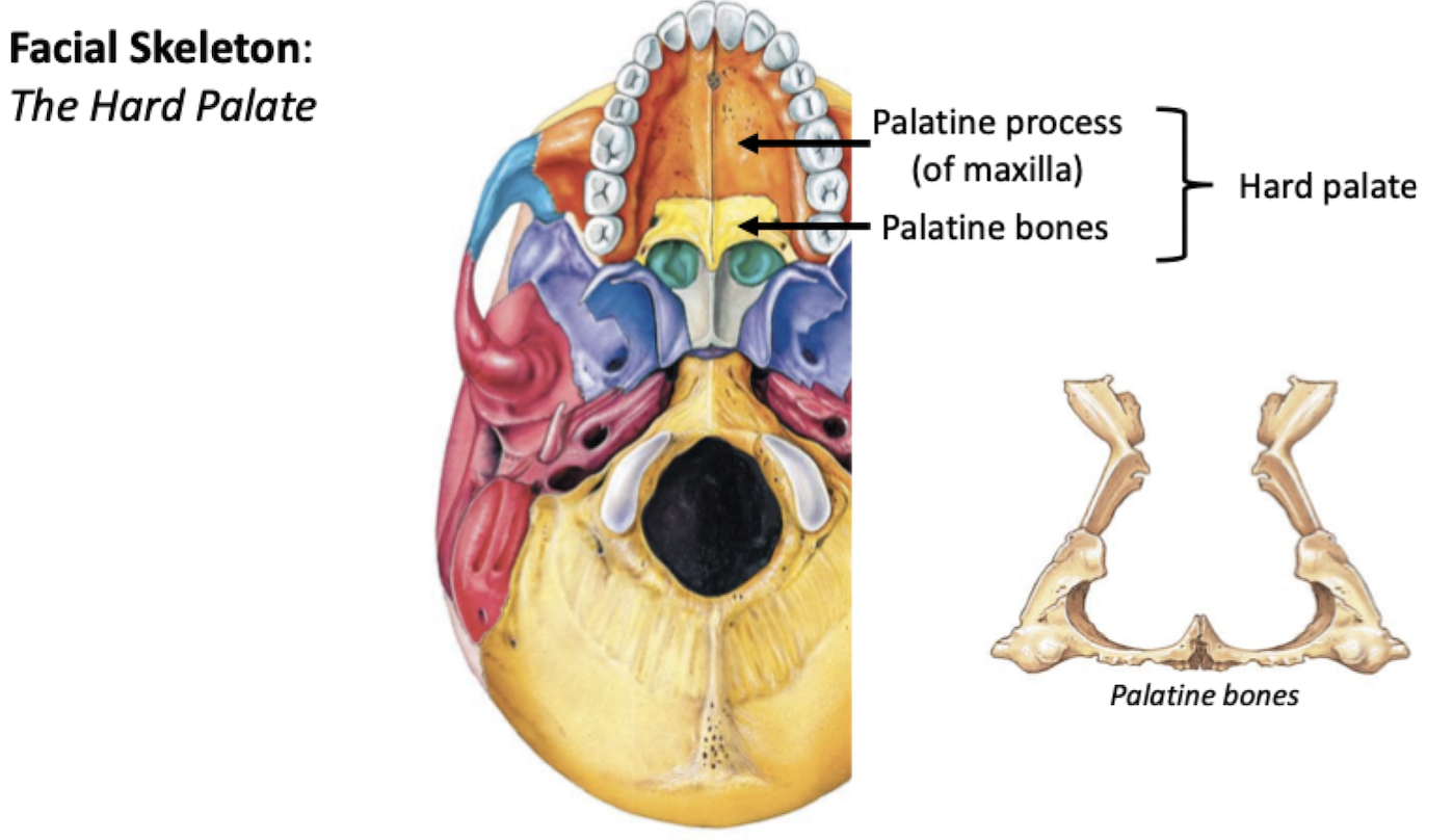
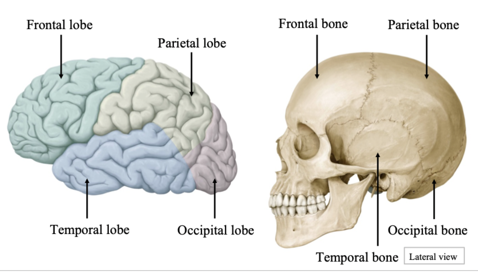
Parts of skull
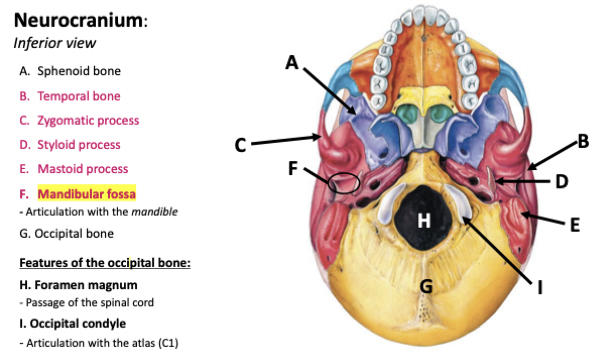
Neurocranium (braincase) (8 bones)
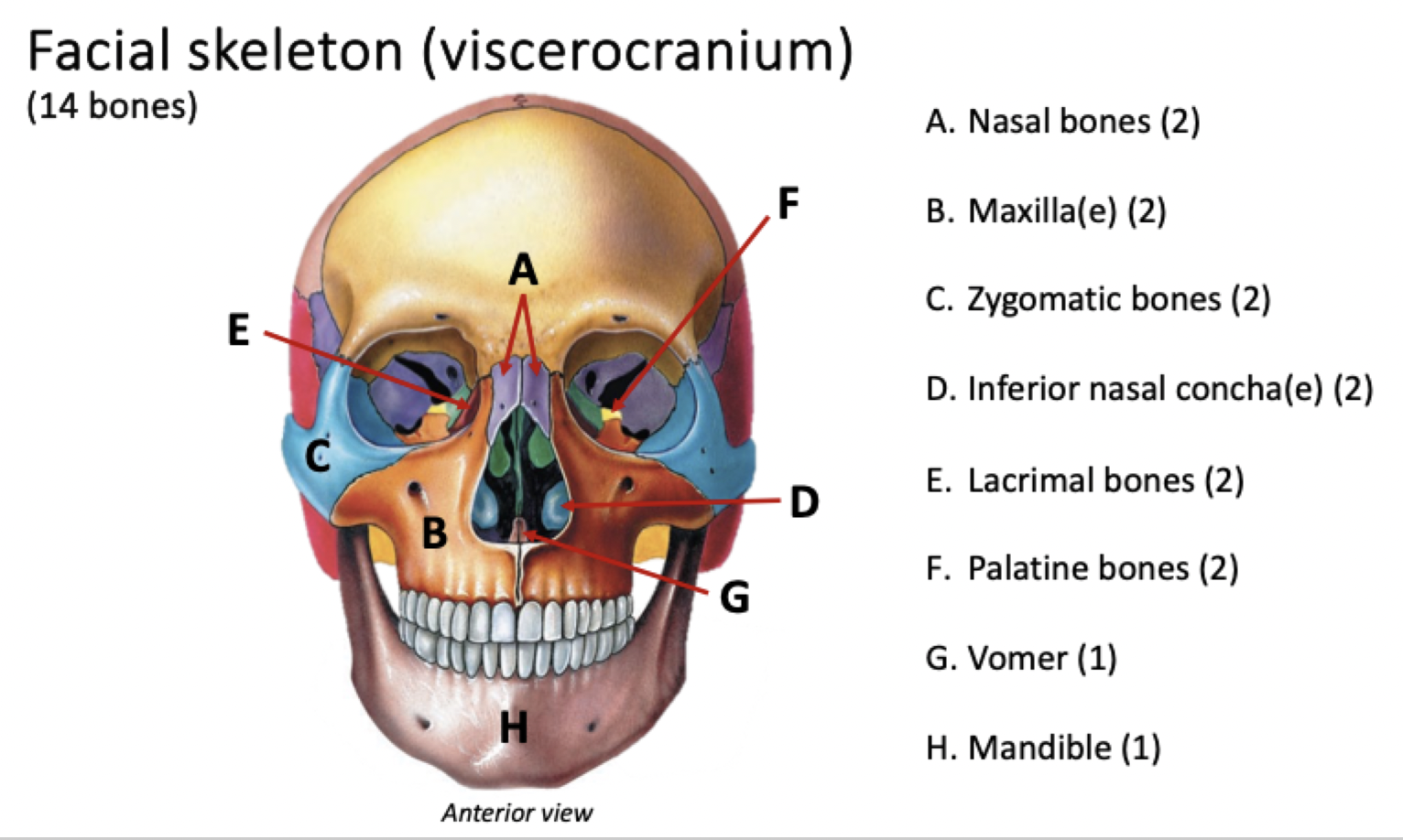
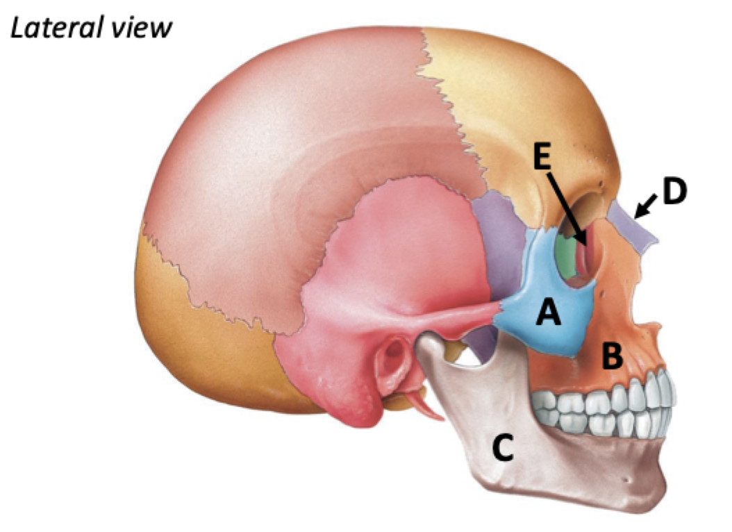
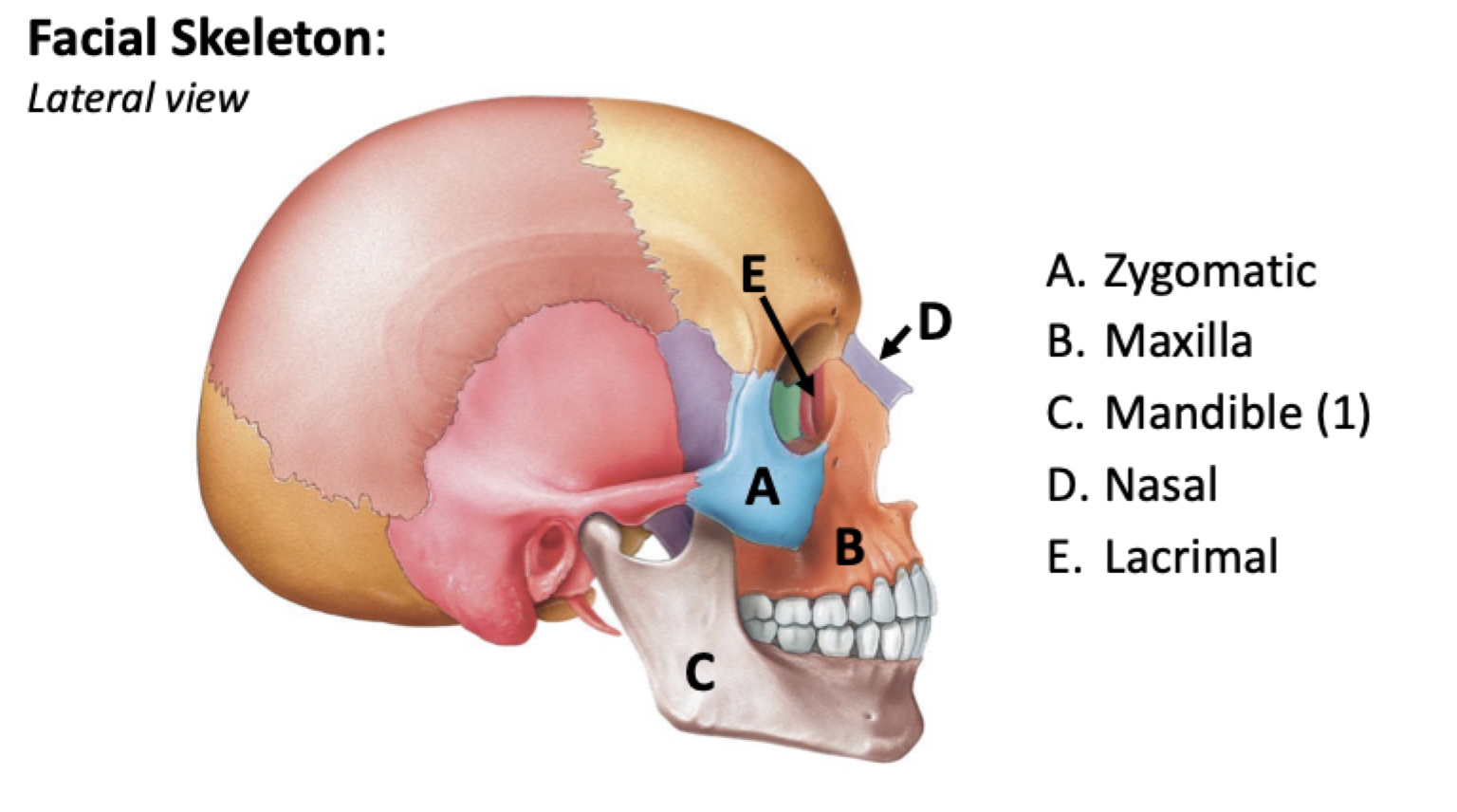
Viscerocranium (facial skeleton) (14 bones)
22 total bones
Neurocranium - functions
Surrounds and protects the brain
Articulates with the vertebral column
Viscerocranium - functions
Supports and protects entrances to the digestive and respiratory tracts
Accessory bones of the skull - functions
7 bones (hearing & muscle attachment)
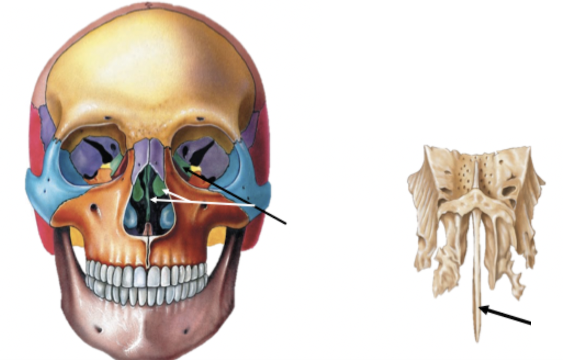
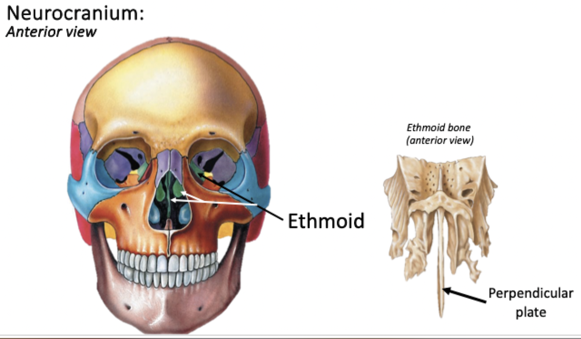
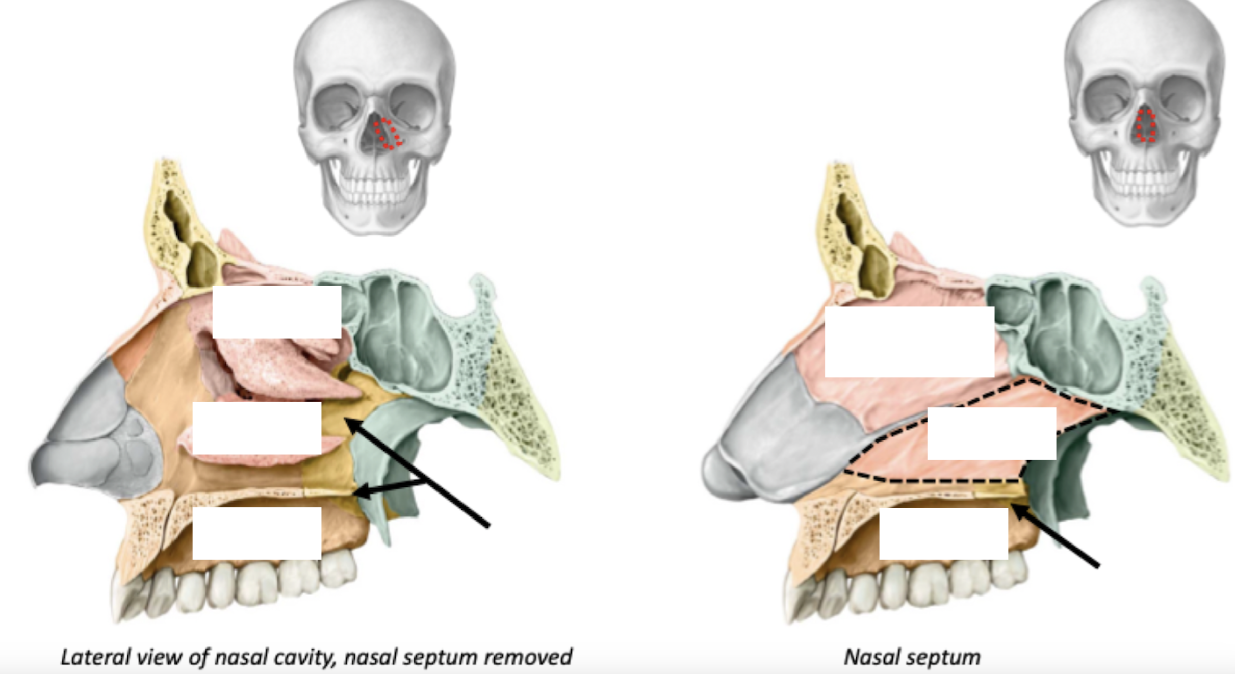
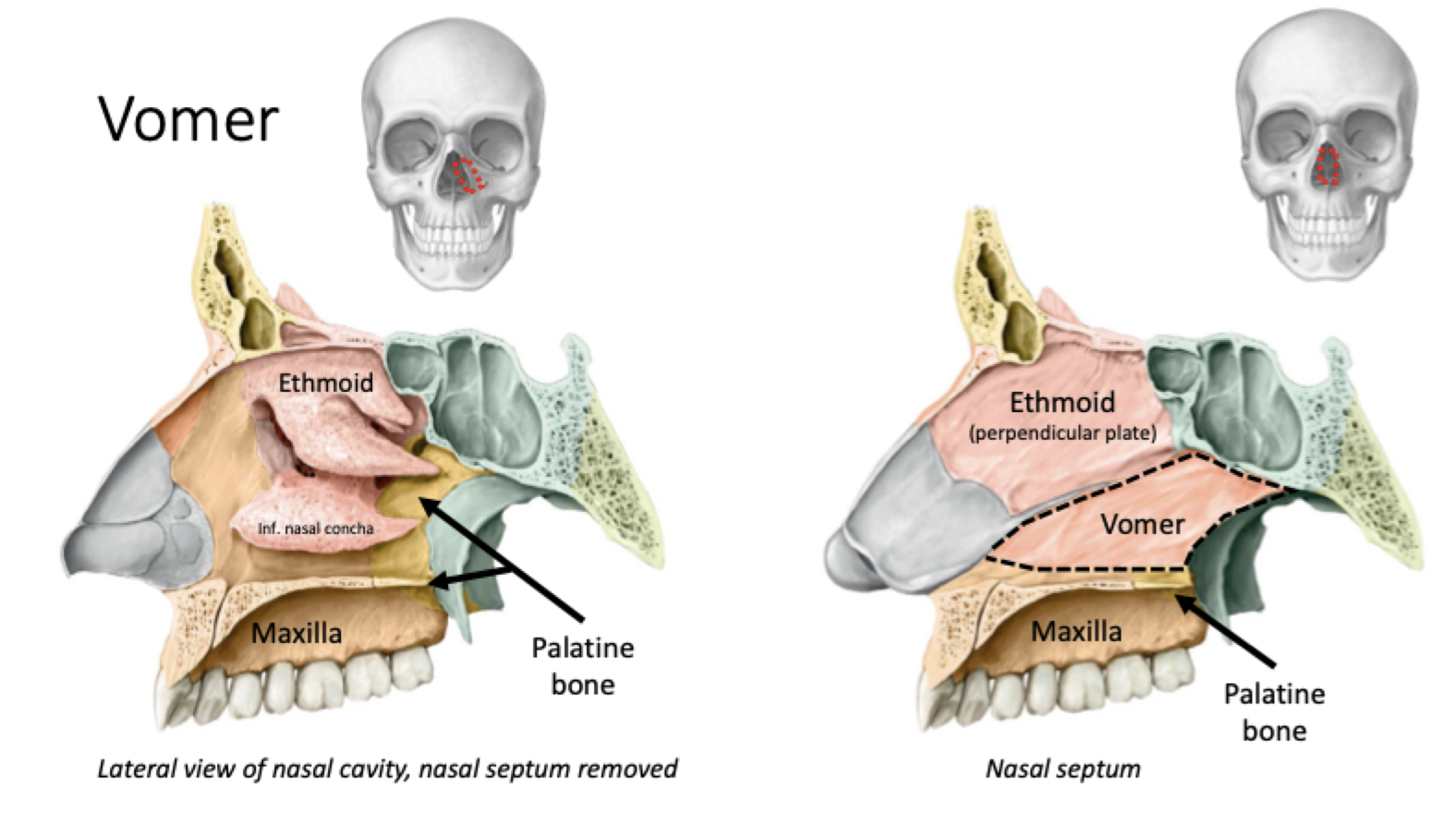
Unpaired bone, forms part of the:
Orbital wall, nasal cavity/septum
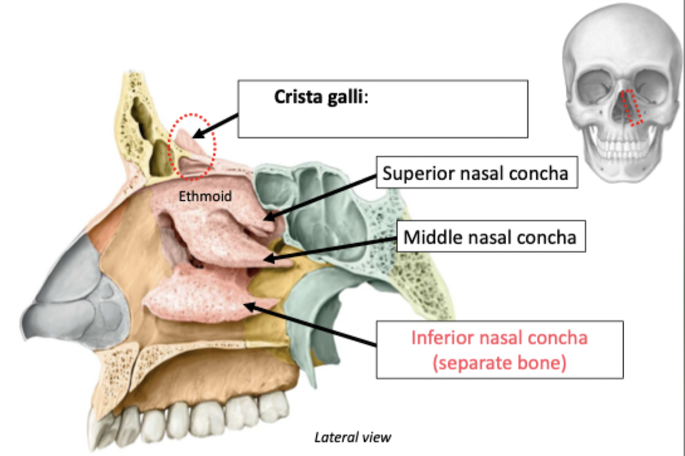
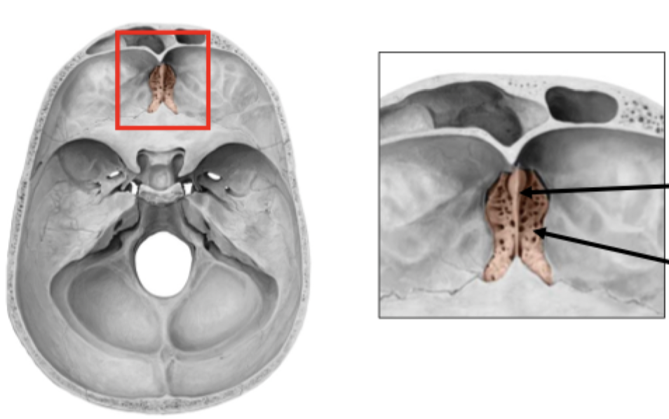
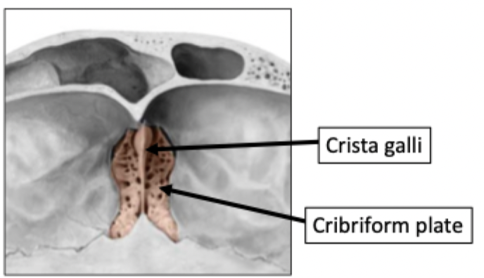
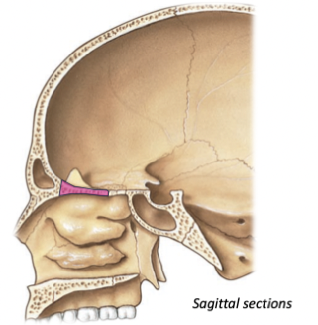
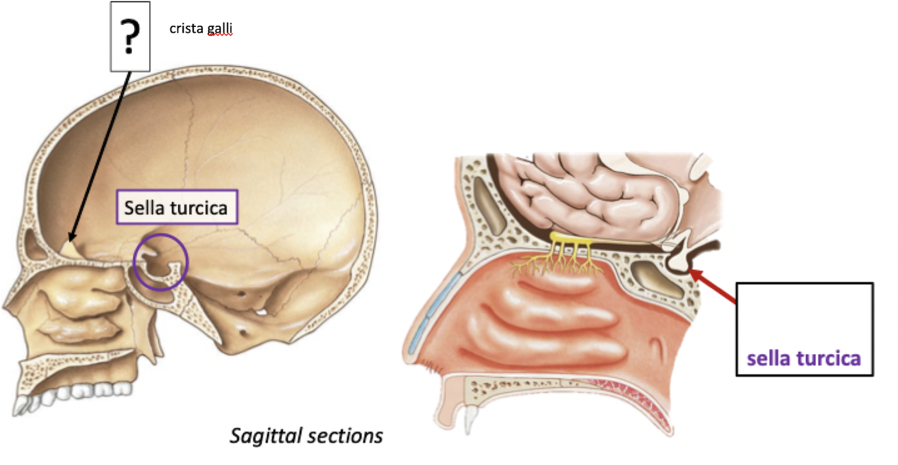
Ethmoid Features
Crista Galli
Perpendicular projection of the ethmoid bone
Acts as an anchoring point for membranes surrounding the brain (meninges)
Branches of CN I (olfactory nerve) pass through cribriform plate of ethmoid bone
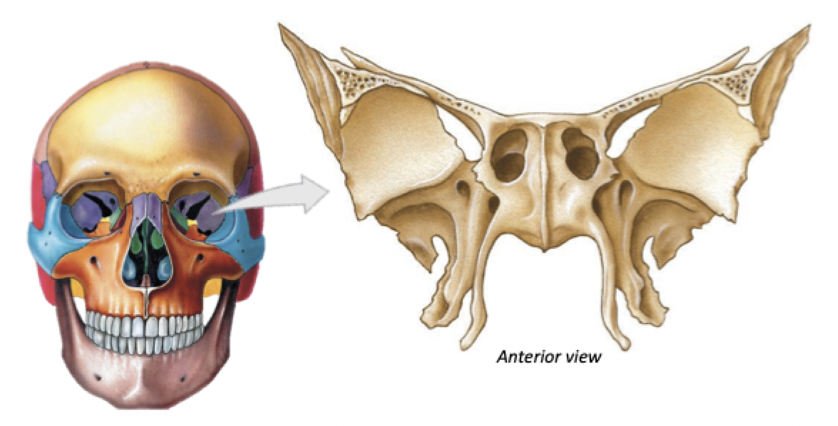
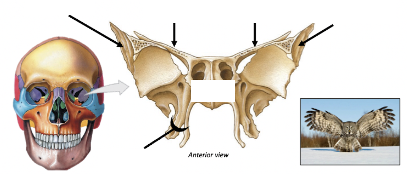
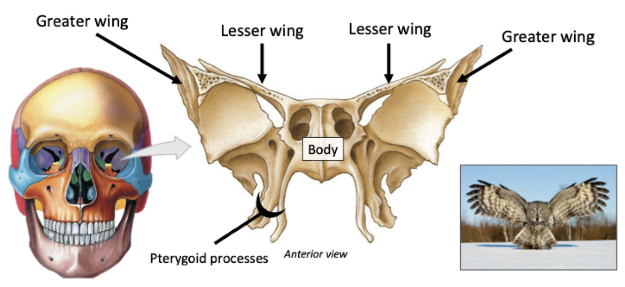
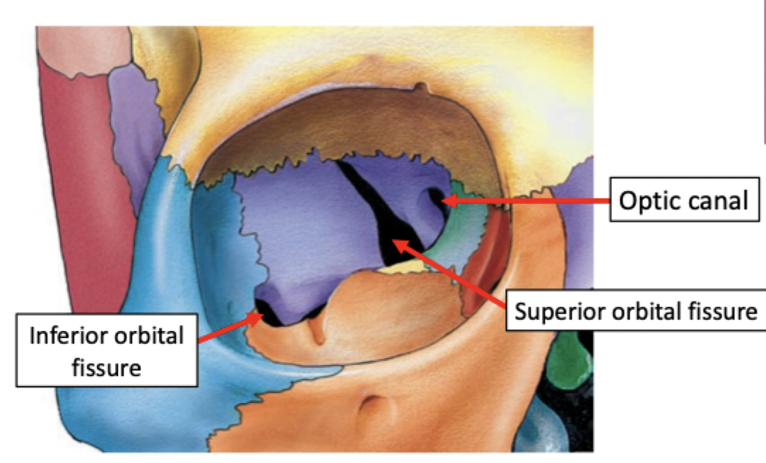
Sphenoid Bone
Unpaired bone
Forms part of the eye orbit & the base of the skull
Features of Sphenoid Bone
Pterygoid Process- attachment for muscles of mastication
Features of Sphenoid Bone
Pituitary gland in sella turcica
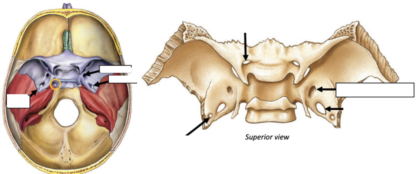
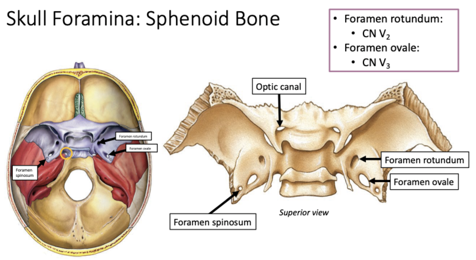
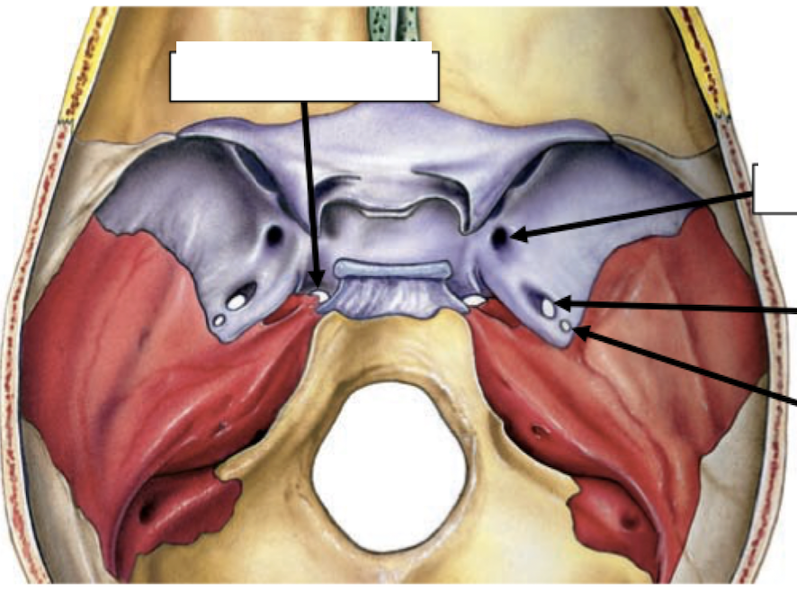
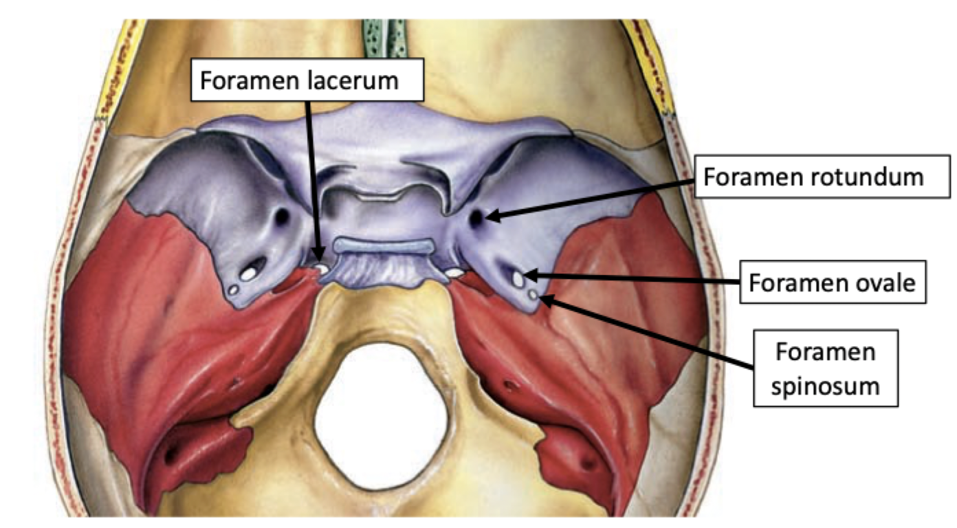
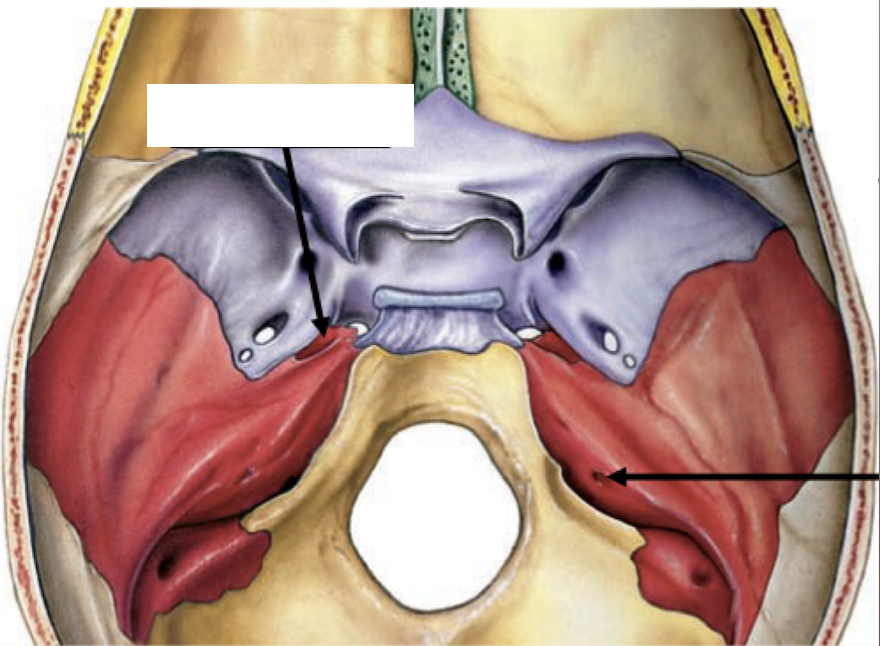
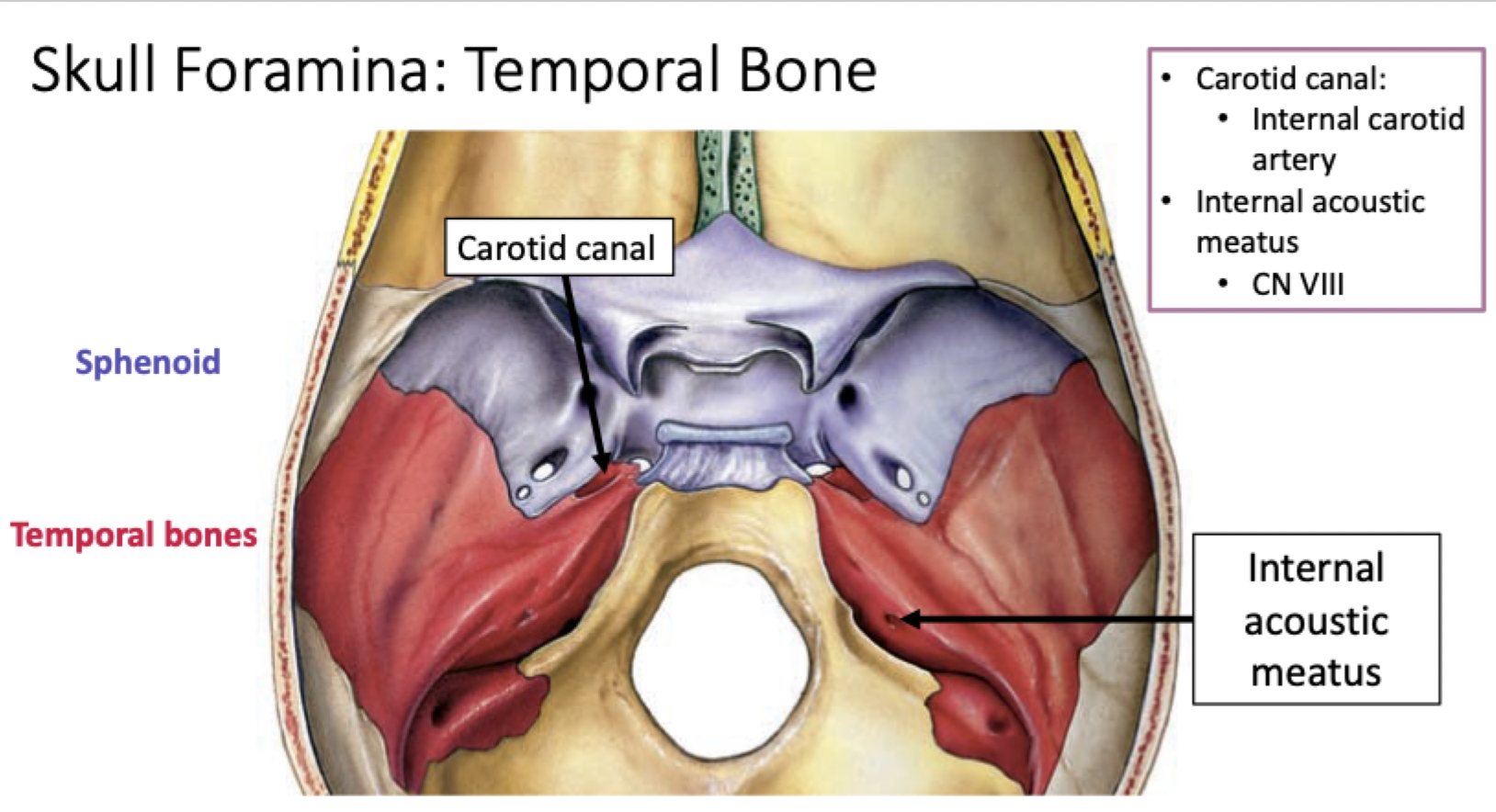
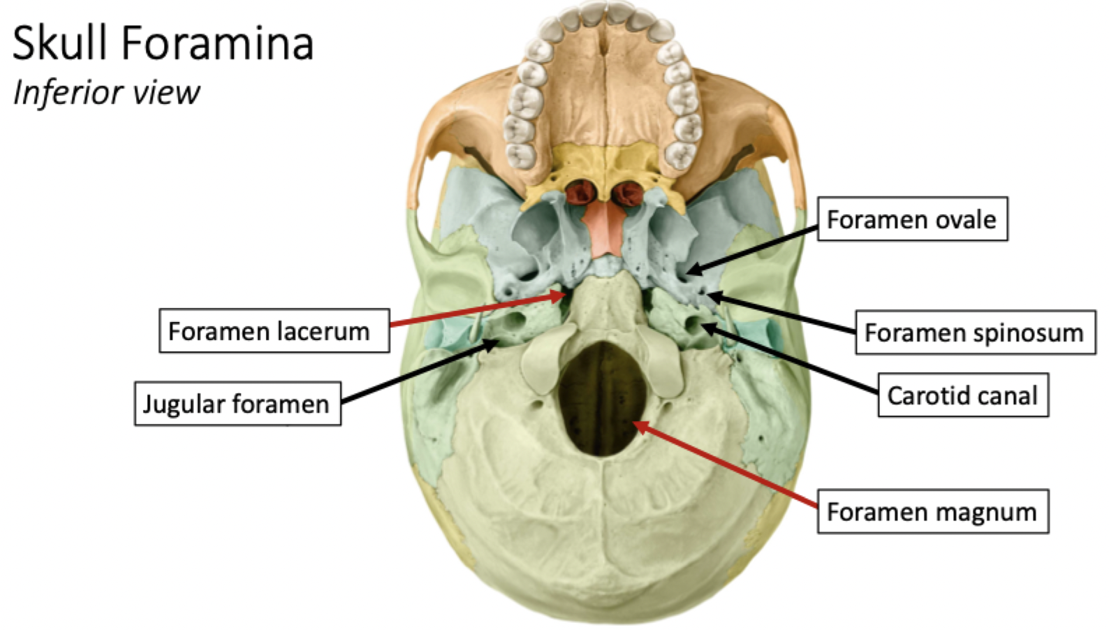
Skull Foramina: Sphenoid Bone
Skull Foramina: Sphenoid Bone
Skull Foramina: Sphenoid Bone
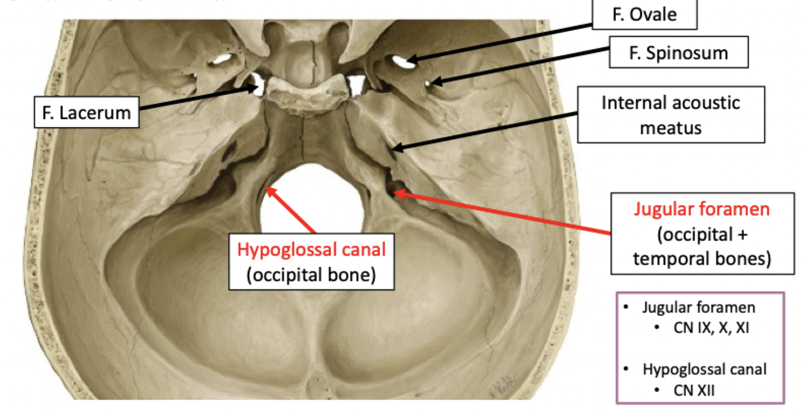
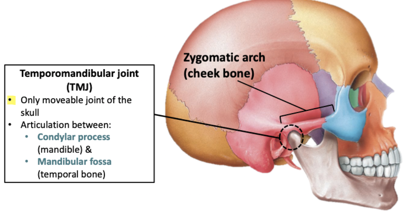
Skull Foramina: Temporal Bone
Skull Foramina
Skull Foramina
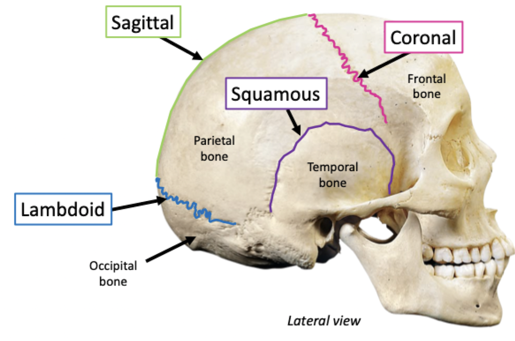
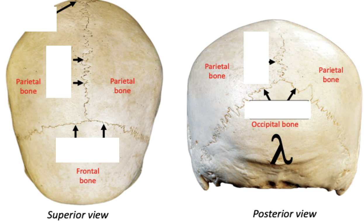
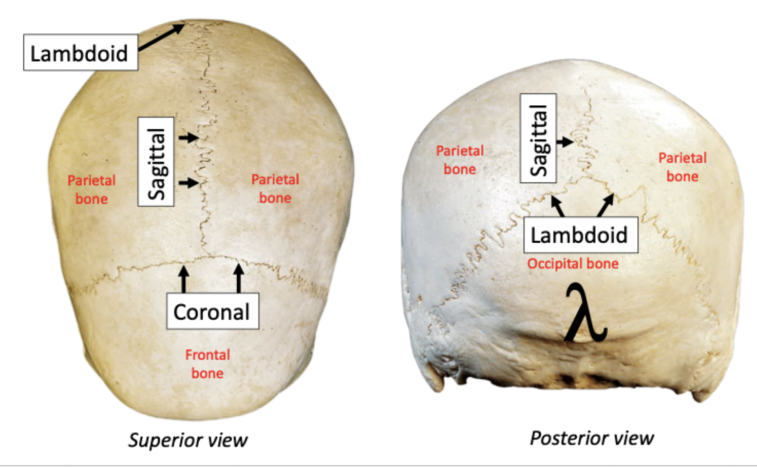
Sutures
Boundaries between skull bones
Fibrous, immovable joints
Sutures
Boundaries between skull bones
Fibrous, immovable joints
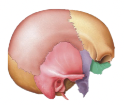
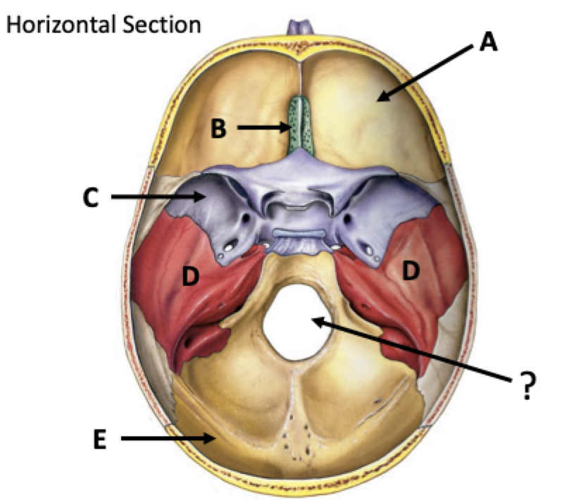
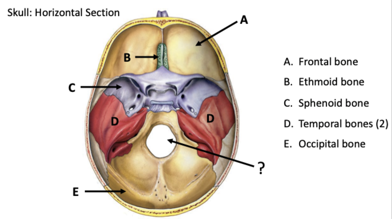
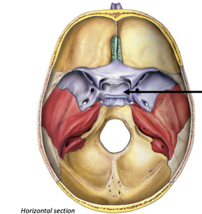
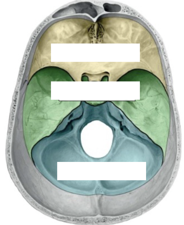
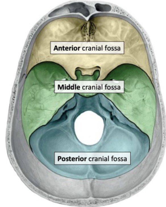
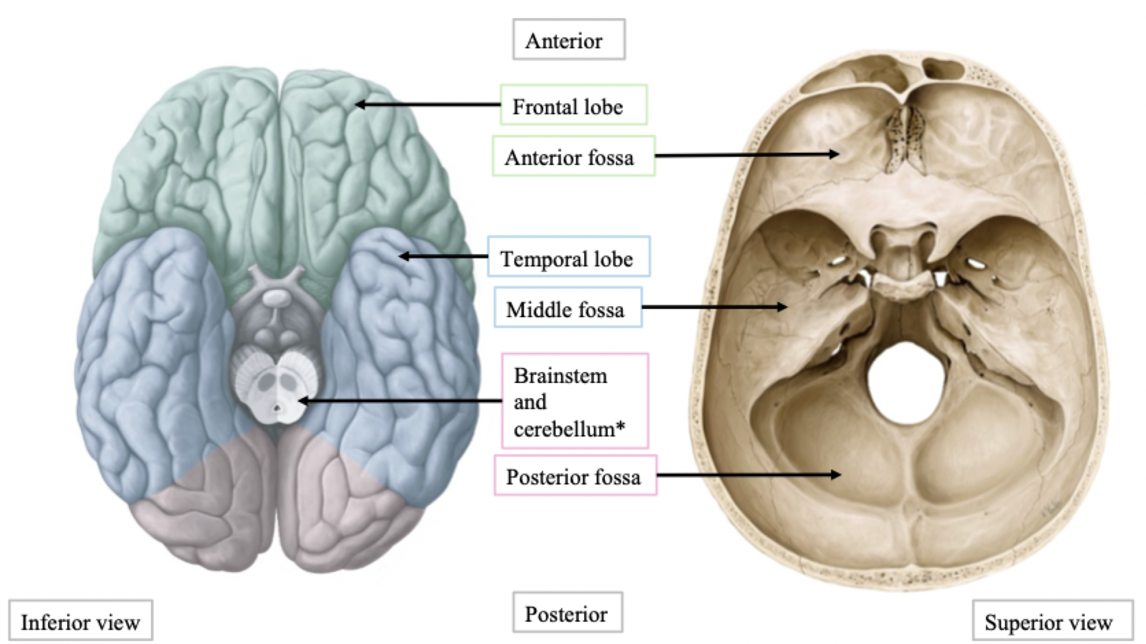
Cranial Fossae
Floor of the cranial cavity divided into 3 fossae (depressions):
Anterior cranial fossa: frontal lobe of brain
Frontal, ethmoid, sphenoid bones
Middle cranial fossa: temporal lobes of brain
Sphenoid, temporal bones
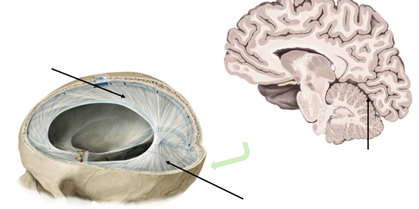
Posterior cranial fossa: cerebellum
Occipital, temporal bones
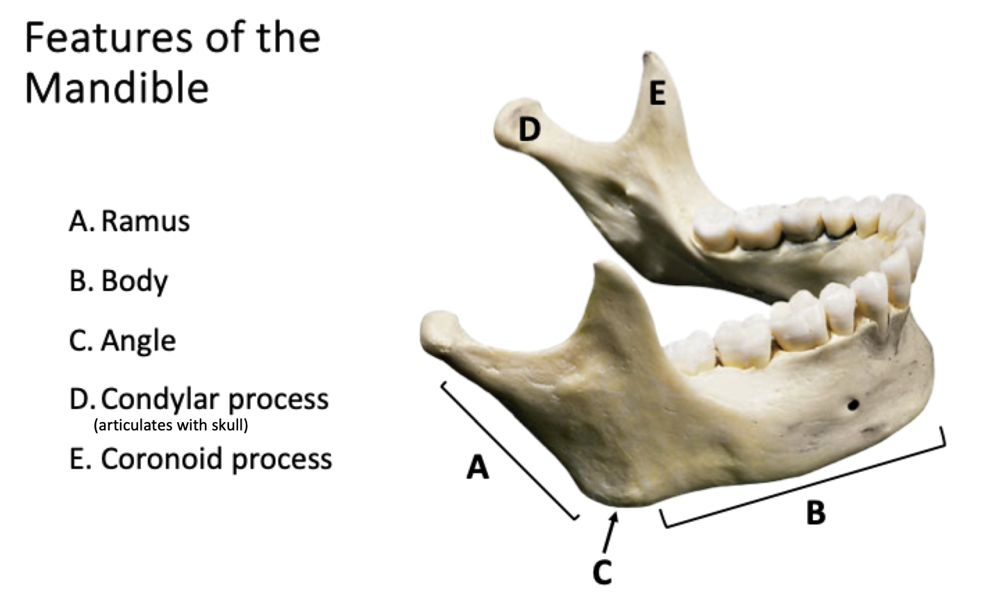
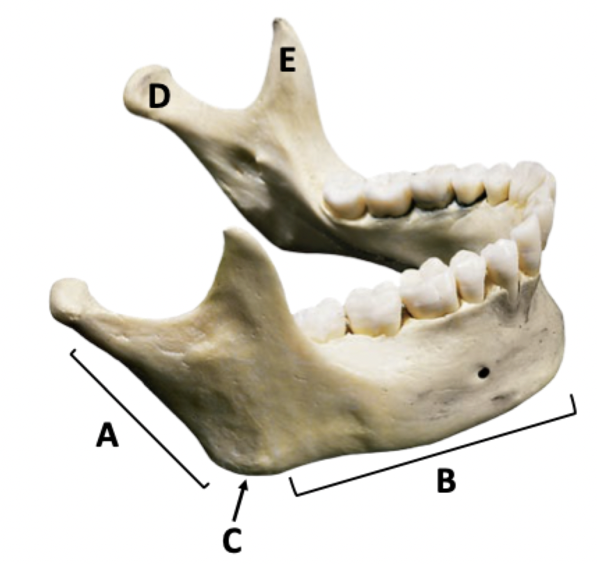
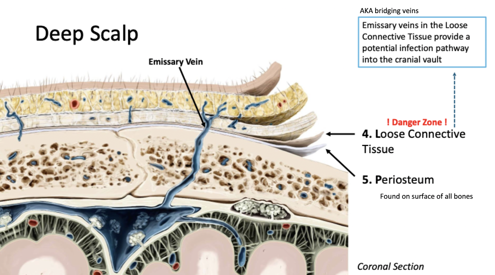
Features of the Mandible
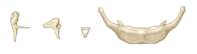
Accessory Bones of the Skull
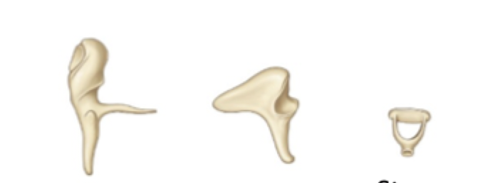
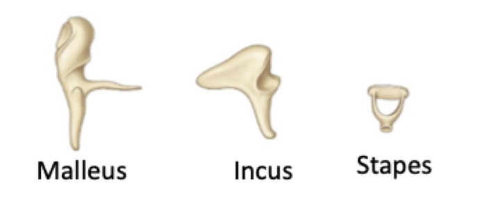
Ossicles
Small bones involved in hearing
Housed in the temporal bone
3 in each middle ear
CN VIII traveling through the internal acoustic meatus
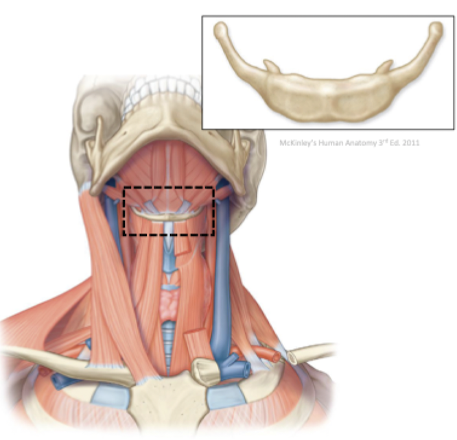
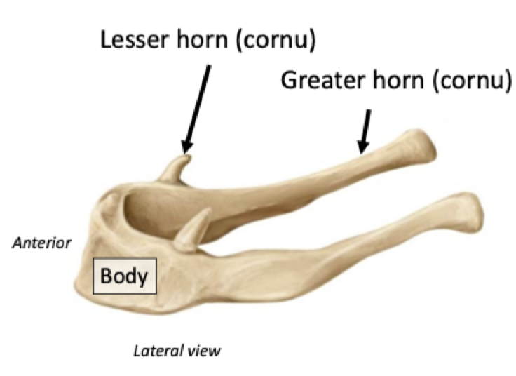
Hyoid Bone
"Floating bone" located in the neck
Important attachment site for the tongue and muscles involved in swallowing
The Nervous System
The nervous system is one of the smallest and most complex body systems:
Mass of only 2kg (~3% of total body weight)
Contains approximately 100 billion neurons
Uses more energy than any other organ (over 20%)
Highly organized network comprised of two cell types:
Neurons
Neuroglia
Major Components of the Nervous System
Brain
Spinal cord
Cranial nerves
Spinal nerves
Ganglia
Sensory receptors
Anatomical Organization of the Nervous System
Central nervous system (CNS)
Brain and spinal cord
Peripheral nervous system (PNS)
Cranial nerves and spinal nerves
Ganglia and sensory receptors
Cranial nerves
12 pairs of nerves (I-XII)
Emerge from the base of the brain
Spinal nerves
31 pairs of nerves
Emerge from the spinal cord
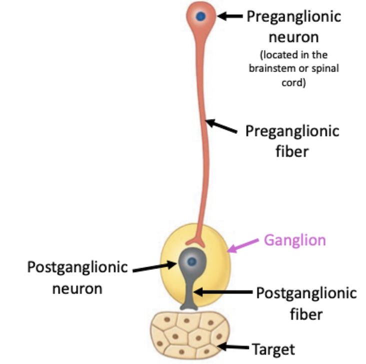
Ganglia
Relay station for neuron conversations
Clusters of neuron cell bodies
Located outside the CNS - in the PNS
Autonomic ganglia are part of the ANS
Function as relay stations between preganglionic and postganglionic neurons of the ANS
Sensory receptors
Monitor changes in environment
Detectors of external and internal stimuli
Skin, eyes, nose, muscles, etc.
Functional Organization of the Nervous System
Sensory function (input)
Integrative function (control)
Motor function (output)
NS Sensory function (input)
Sensory receptors detect internal and external stimuli
Sensory (afferent) neurons transmit information to CNS
Division of the Peripheral nervous system
NS Integrative function (control)
Interneurons analyze sensory information
Perception (Conscious awareness) of stimuli to determine whether or not a response is needed and how it will occur
Division of the Central nervous system
NS Motor function (output)
Motor (efferent) neurons respond to integration
Initiate actions in effector organs (e.g., muscles, glands)
Somatic Nervous System
Division of the Peripheral nervous system
Somatic sensory neurons (Afferent)
Convey information to the CNS from receptors for somatic senses and receptors for special senses
Input of information to the CNS for integration
Somatic motor neurons (Efferent)
Convey information from the CNS to skeletal muscles only
Output of information from the CNS for muscular contraction
Overall function: Regulates voluntary control of skeletal muscles
Autonomic Nervous System
Division of the Peripheral nervous system
Autonomic motor neurons regulate visceral activities by exciting or inhibiting activities in effector tissues
Sympathetic nervous system (SNS)
Increase in activity and metabolic rate
"Fight-or-flight" response
Examples: dilate pupils, increase heart rate, inhibit intestinal activity
Parasympathetic nervous system (PNS)
Decrease in activity and metabolic rate
"Rest-or-digest" response
Examples: constrict pupils, decrease heartrate, stimulate intestinal activity
Overall function: regulates involuntary functions such as cardiac muscle, smooth muscle, glandular tissue
Cell Types of the Nervous System
Nervous tissue is comprised of two cell types:
Neurons
Neouroglia
Neurons
Basic structural and functional units of the nervous system
Form complex processing networks
Transfer and processing of information
Neuroglia
Smaller and more abundant
Support, nourish, and protect neurons
Do not participate in transfer of information
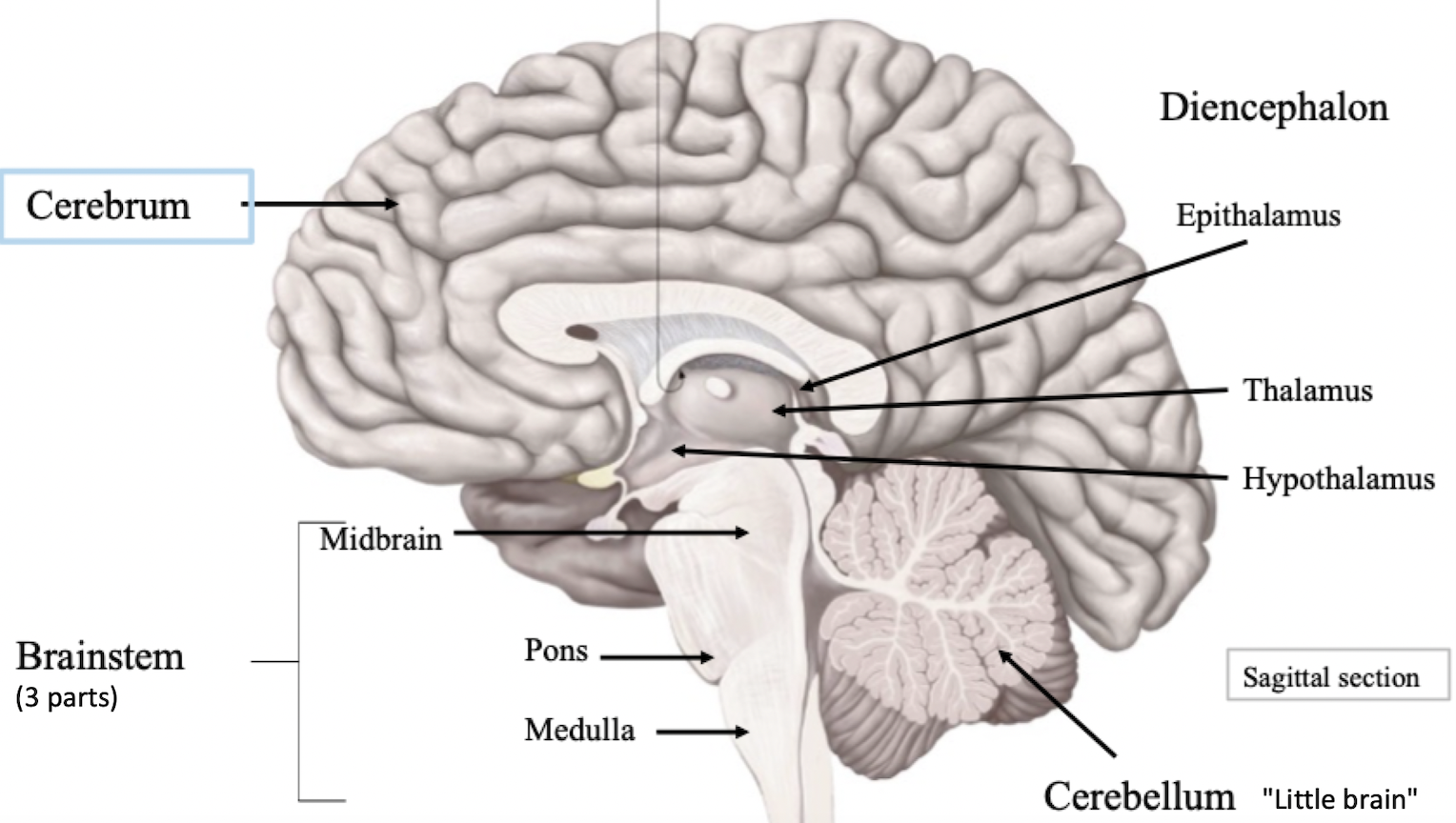
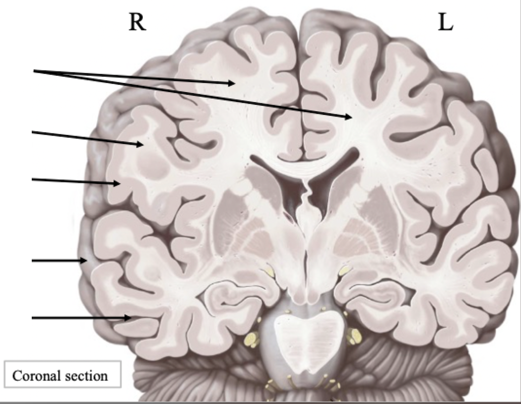
Overview of the Cerebrum (telencephalon)
The largest and most anterior part of the brain (telencephalon):
Located at the front of the skull
Consists of two hemispheres separated by a fissure
Initiates and manages conscious thoughts and actions
The cerebrum provides us with the ability to: read, write, and speak; logically make calculations and creatively make art; and remember the past, plan for the future, and imagine things that may not exist.
Label
Label + Explain
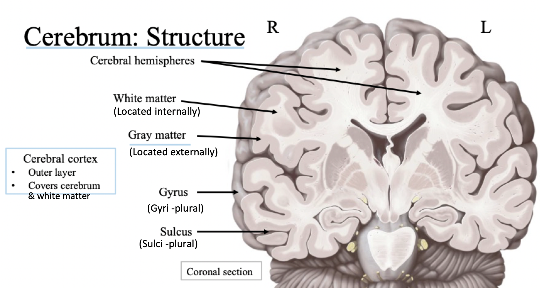
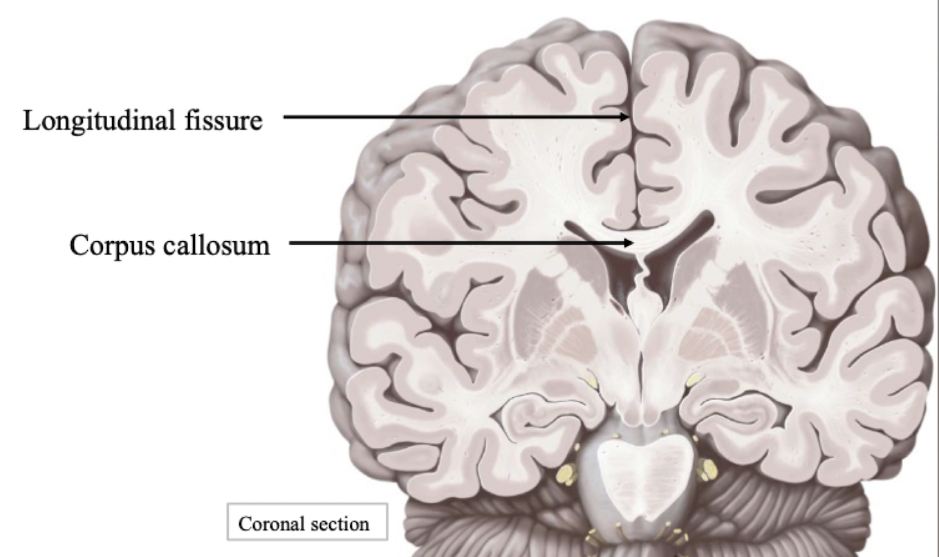
Cerebrum Features
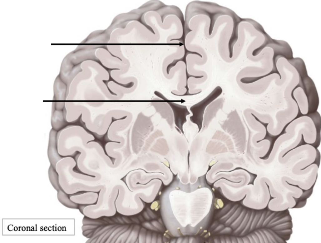
Longitudinal fissure
Separates right and left cerebral hemispheres
Travels from anterior to posterior
Corpus callosum
Connects right and left cerebral hemispheres
Comprised of white matter
Communication bridge - how left and right hemispheres communicate
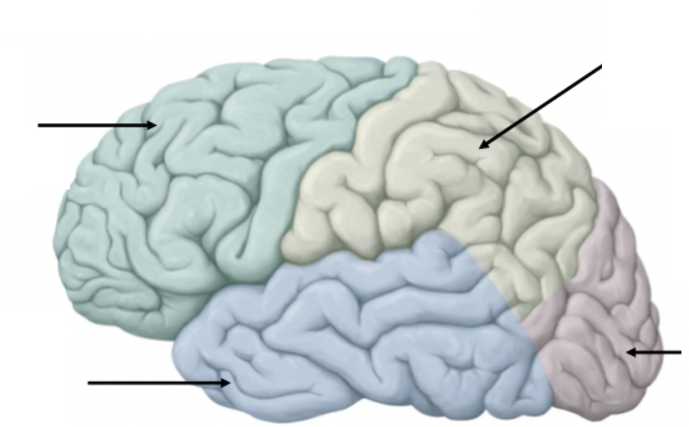
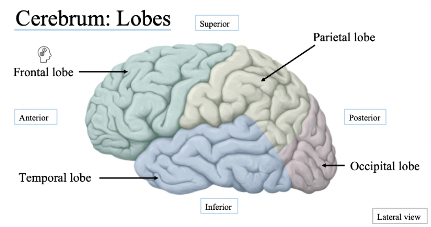
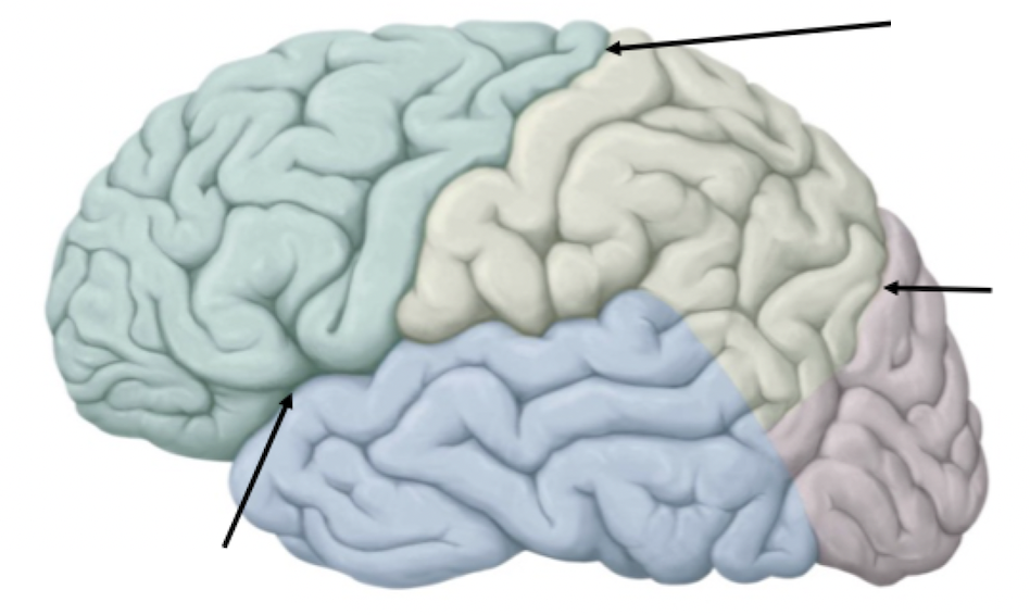
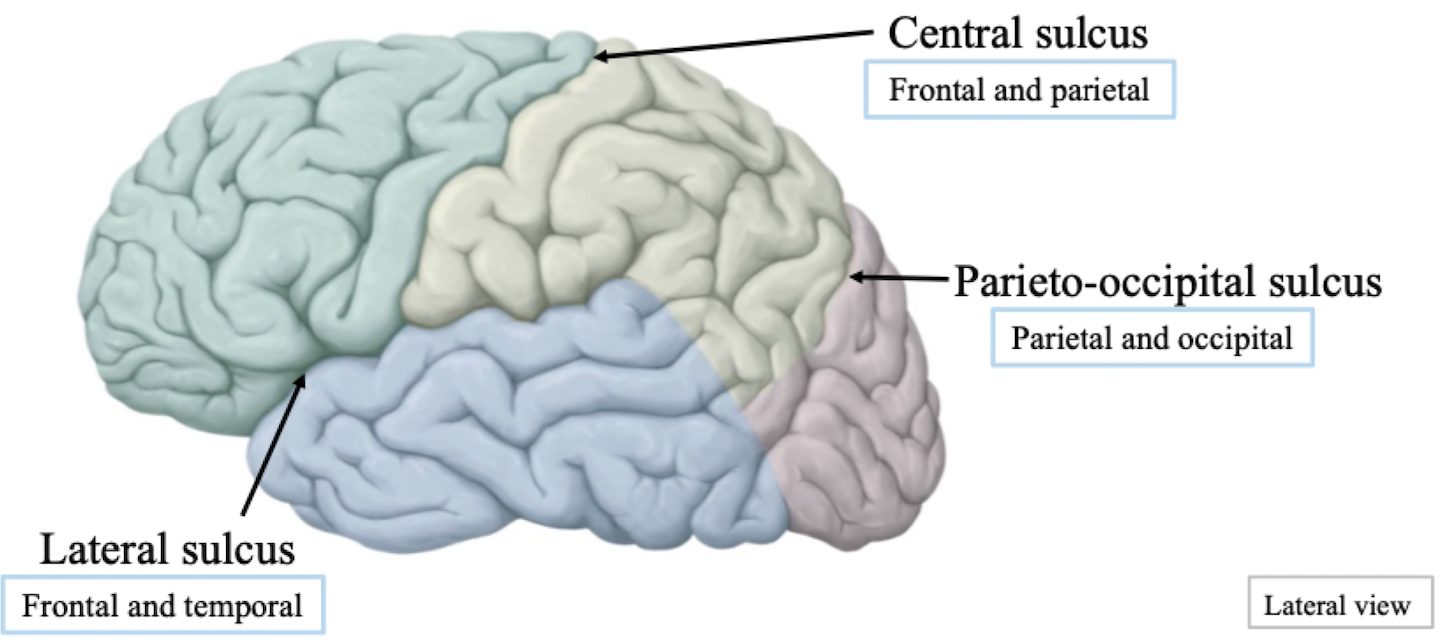
Cerebrum Lobes
Frontal lobe
Largest lobe
Executive function
Planning, mood, decisions
Integration centre
Temporal lobe
Responsible for auditory processing and speaking/verbal responses
Being able to connect thoughts and responses to speaking and hearing
Parietal lobe
Responding to stimuli from the environment
Creates mental body map of where you are in space (proprioception)
Occipital lobe
Responsible for vision
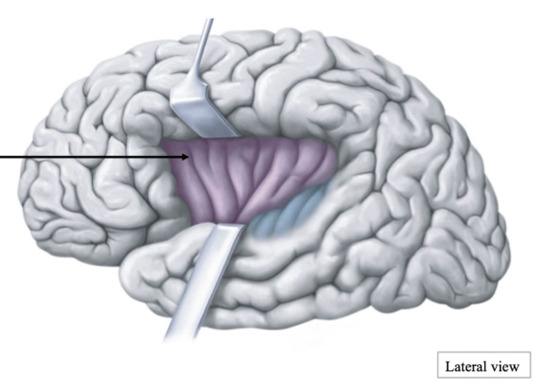
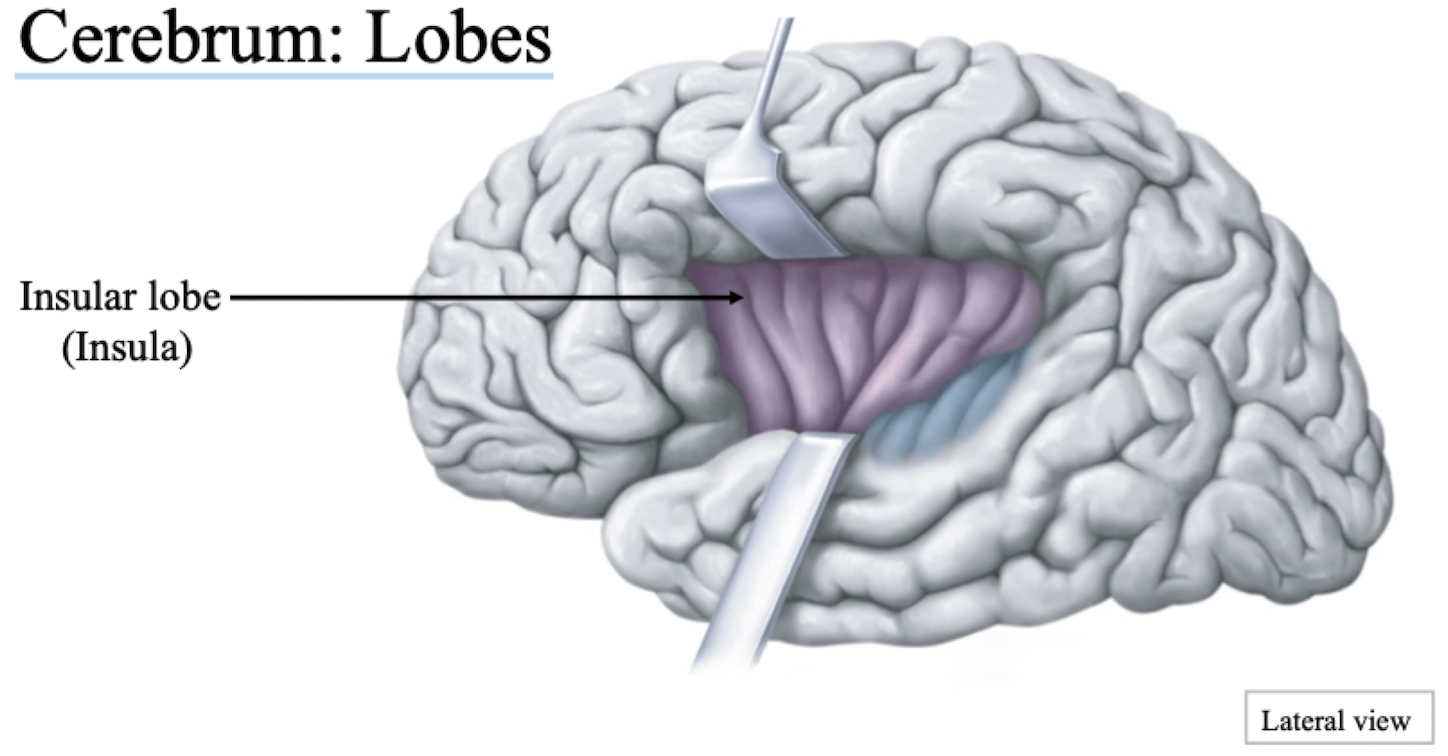
Cerebrum Lobe
Insular lobe (Insula)
If you pushed up parietal lobe and looked internally
Automatic processing responses
Which parts of the brain are housed by which parts of the skull?
Which parts of the brain articulate with which parts of the skull?
Frontal lobe articulates with anterior fossa
Temporal lobe articulates with middle fossa
Brainstem and cerebellum articulate with posterior fossa
Cerebrum: Lobe Divisions
Major sulci are usually deeper, more pronounced and travel full length/width of brain until it reaches the next lobe
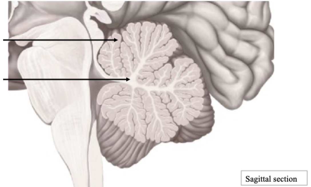
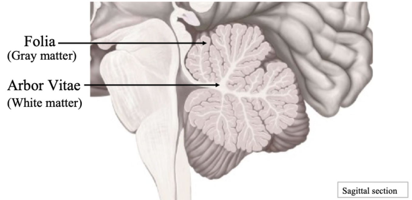
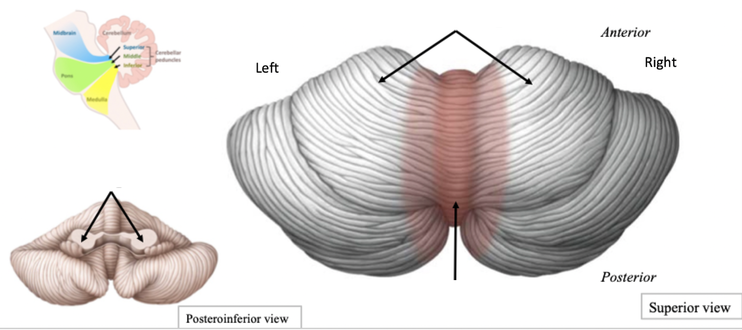
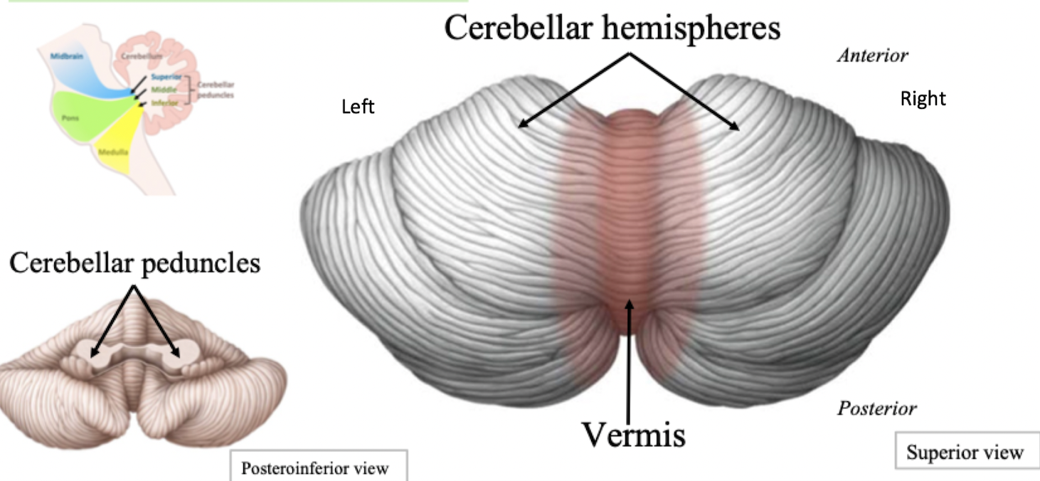
Cerebellum Features
Coordinates voluntary movements
Regulates posture and balance
Cerebellum Structure
Label + Explain
Label + Explain
Label + Explain
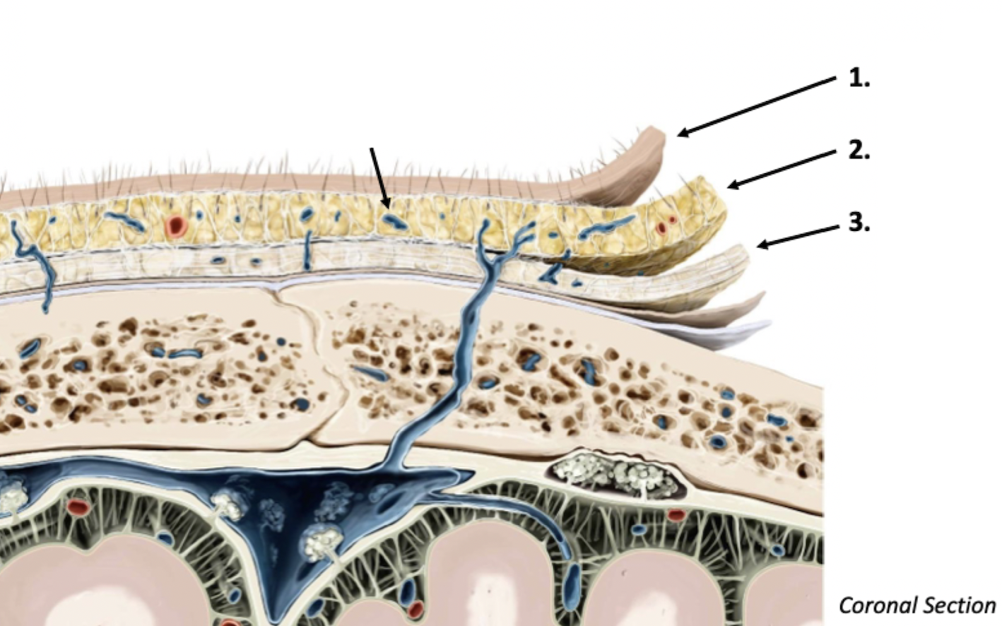
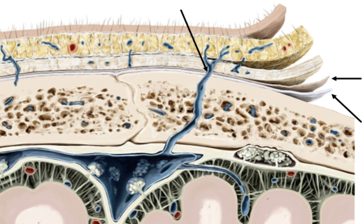
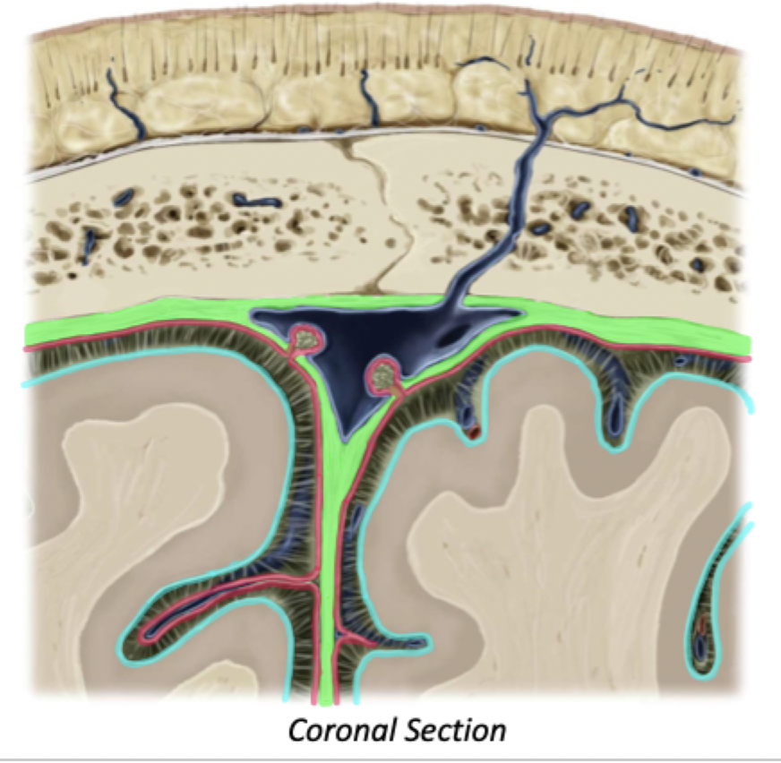
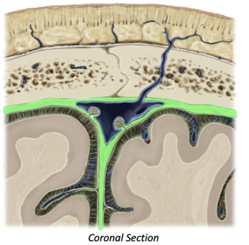
Meninges
Label + Explain
3 layers of tissue to provide protection and support to the CNS: (Brain and spinal cord)
From superficial to deep:
Dura Mater
Arachnoid Mater
Pia Mater
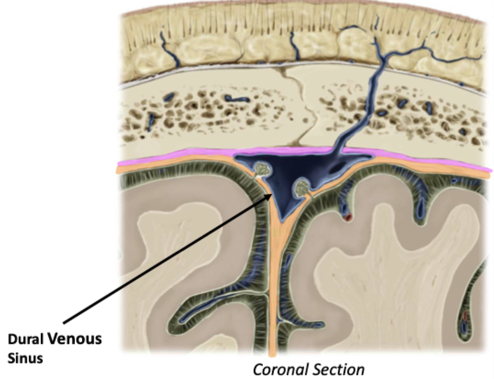
Dura Mater
Thick layer of meninges deep to the calvarium (skull cap)
Encloses dural venous sinuses, major structures that drain the cranial vault
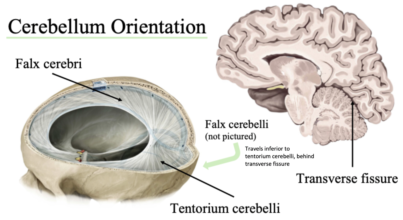
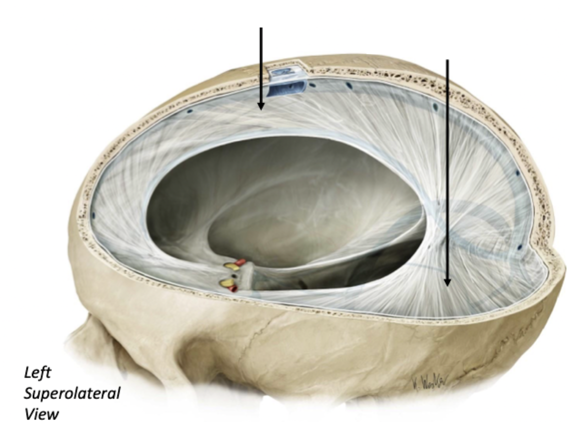
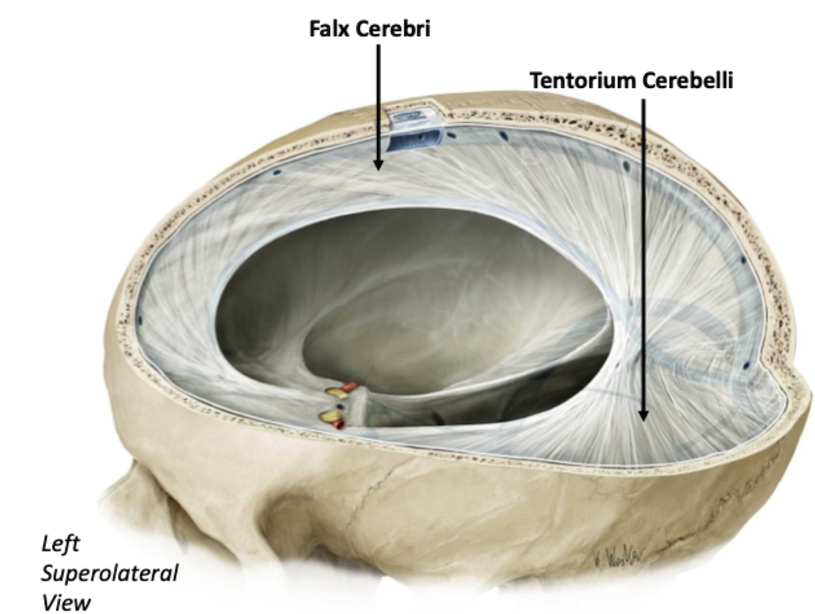
Dura Mater Invaginations (Folds)
Forms three invaginations (folds) within the cranial vault:
Falx Cerebri
Sits on mid-sagittal plane
Tentorium Cerebelli
Lies along transverse plane
Falx Cerebelli
Helps split the other two
Dura Mater Layers
2 Layers:
Periosteal Layer
Meningeal Layer (deeper)
These layers split to help form the dural venous sinuses (but are often together otherwise)
At the edge of the skull, at the Foramen Magnum, this meninx splits
It continues around the edge of the skull (Periosteal Layer) AND around the spinal cord
(Meningeal Layer)
Dura Mater Spaces
2 Spaces:
Epidural Space (between skull and dura)
Subdural Space (between dura and arachnoid)
These spaces are potential spaces... they are not found unless trauma/disease cause separation of these layers
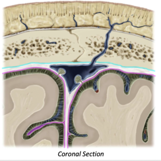
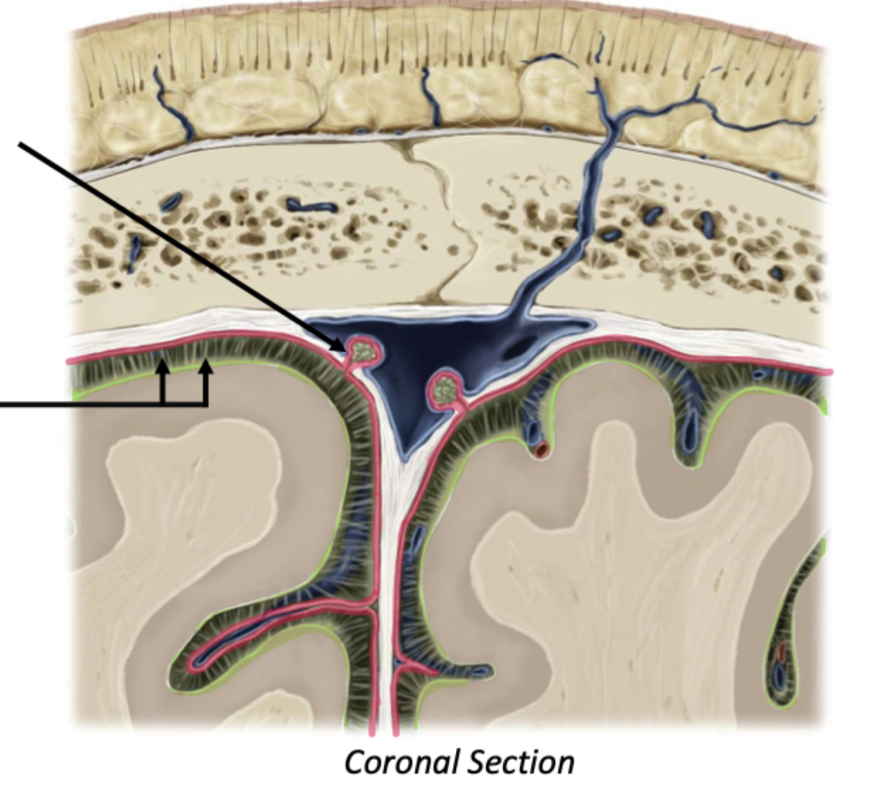
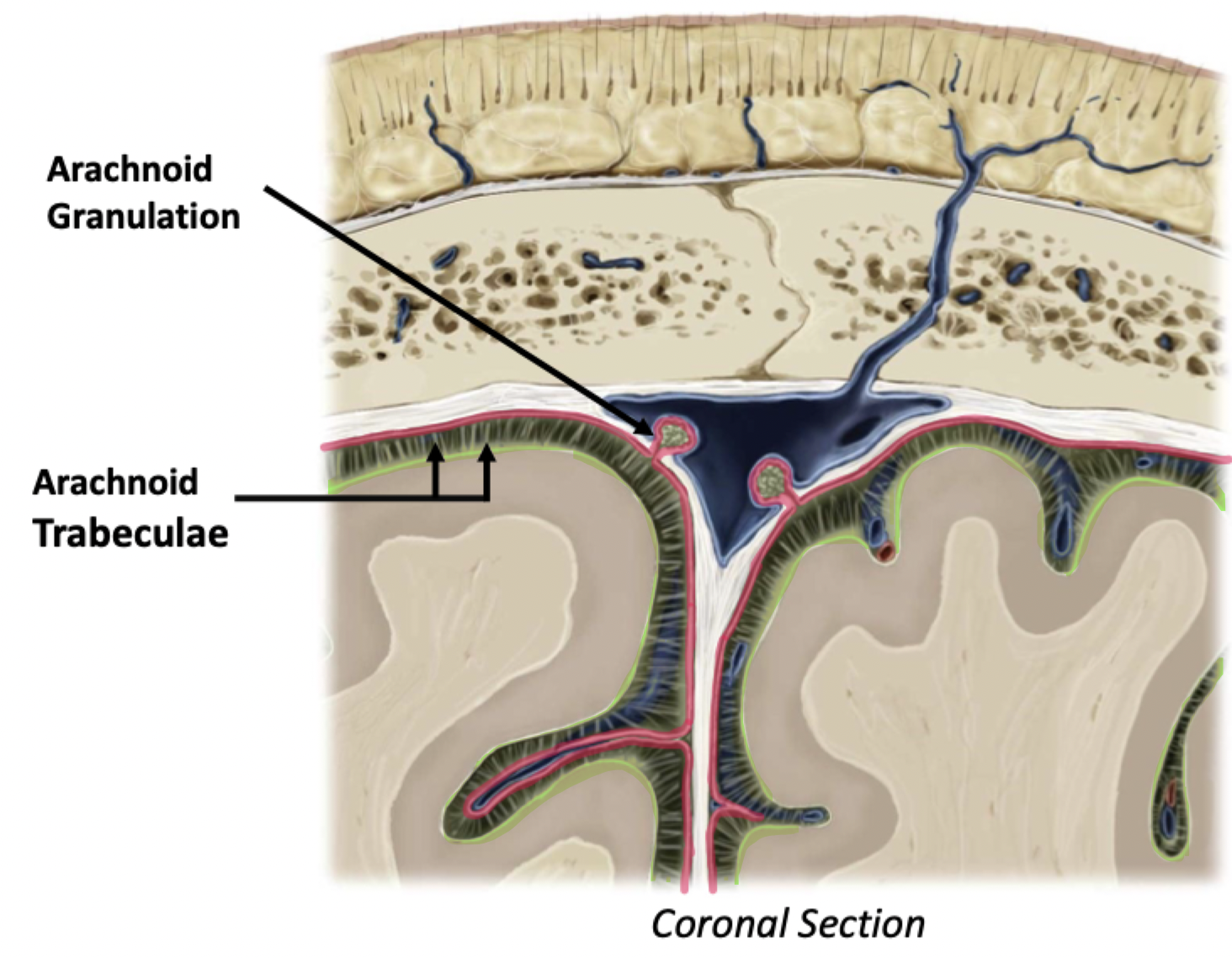
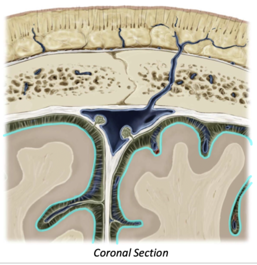
Arachnoid Mater
Sits deep to the dura
Subarachnoid Space, between arachnoid and pia, contains cerebrospinal fluid (CSF)
REAL space
Arachnoid granulations
CSF exits the subarachnoid space through arachnoid granulations
These pierce through the dura to drain CSF into the dural venous sinuses
Arachnoid trabeculae support the arachnoid mater
Cerebrospinal fluid (CSF)
CSF helps to metabolically and physically support the brain
Metabolically -Exchange medium (electrolytes and metabolite exchange)
Physically - Buoyancy and Cushion
Pia Mater
Closely covers cortical sulci and gyri
Subpial Space exists between Pia and Cortex
This is another potential space not normally present
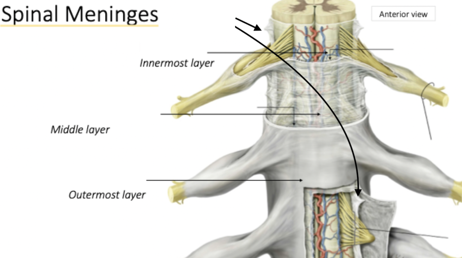
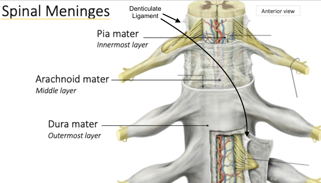
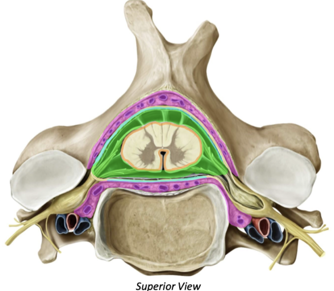
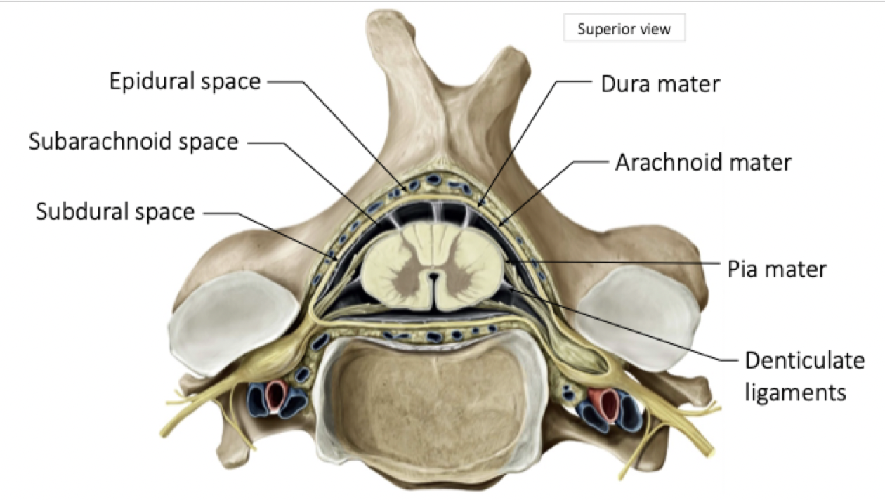
Spinal Meninges
Pia Mater
Thin, transparent connective tissue layer that adheres directly with the surface of the spinal cord and brain
Many blood vessels that supply cord with oxygen and nutrients.
Denticulate Ligaments
Lateral Extensions/thickenings of pia mater
Extend length of spinal cord to increase stability & protect it from sudden displacement (injury)
Arachnoid Mater
Thin, avascular covering , that's composed of epithelial cells and very thin, loose strands of collagen
Cover the Spinal Nerve Roots
Arachnoid - spiderweb arrangement of fibres and collagen
Dura Mater
Strong, composed of dense irregular connective tissue
Cover the Spinal Nerve Roots
Continuous with meningeal layer of cranial dura mater
Extends to S2 vertebrae
Spinal Meninges Spaces
Epidural Space
Real space
Contains fat & venous plexuses & has a cushion effect
Between vertebral canal & dura
Subarachnoid Space
Contains CSF
Between arachnoid & pia
Subdural and Subpial Spaces
Potential spaces
Subdural is between dura & arachnoid
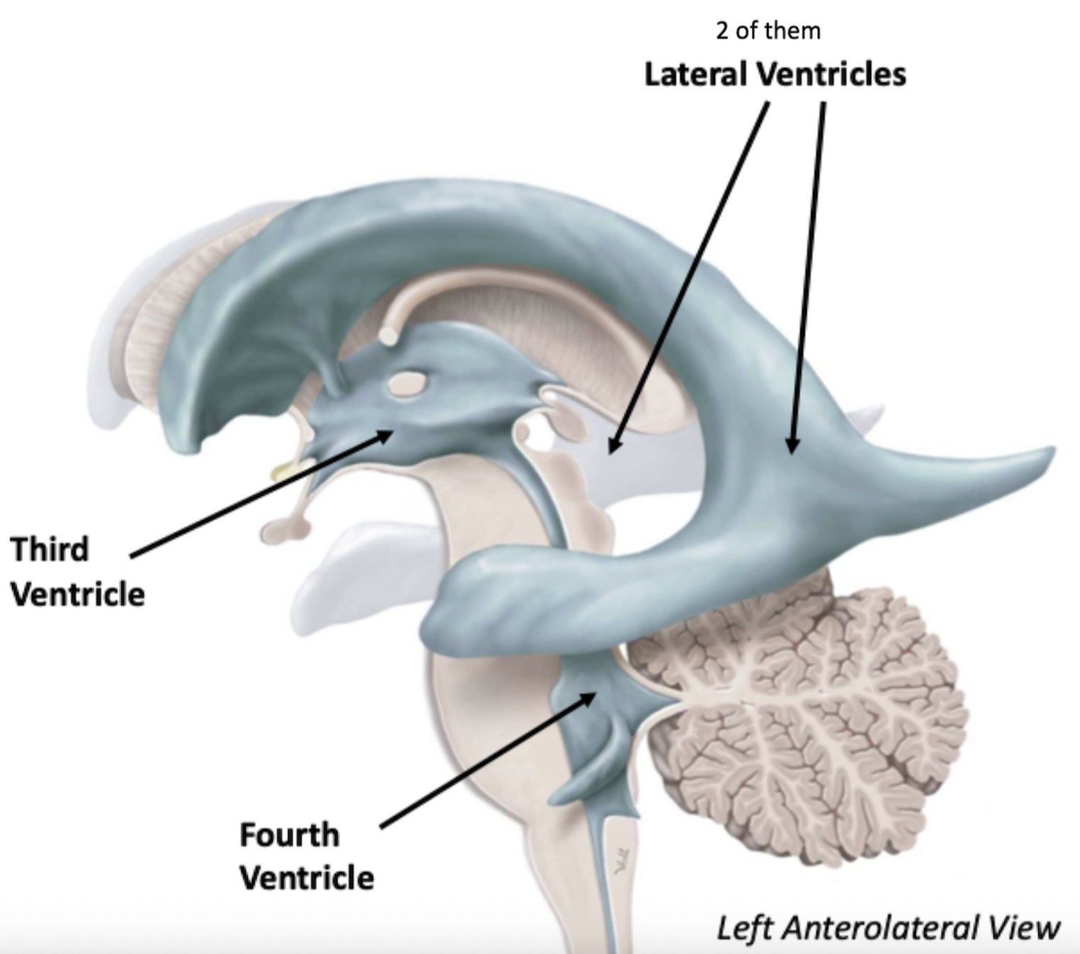
What are the Ventricles?
4 Cavities within the brain responsible for deep CSF flow
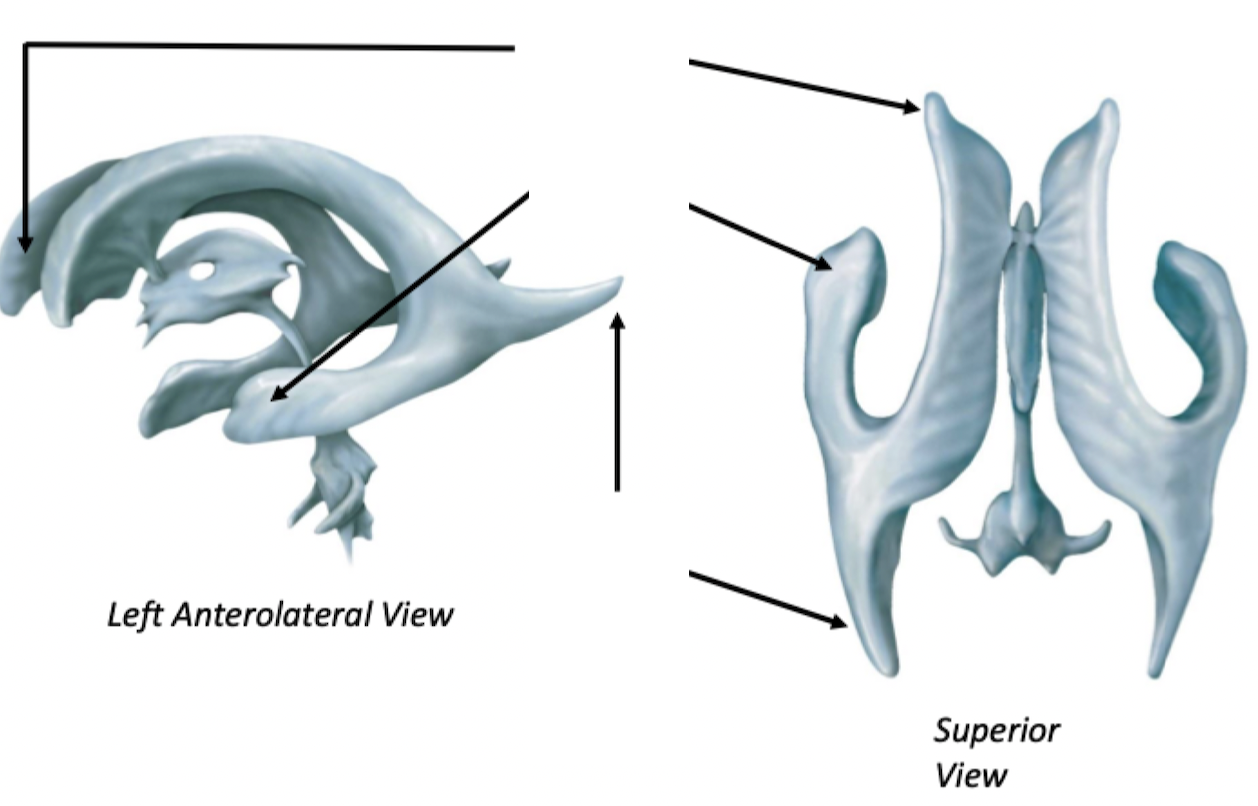
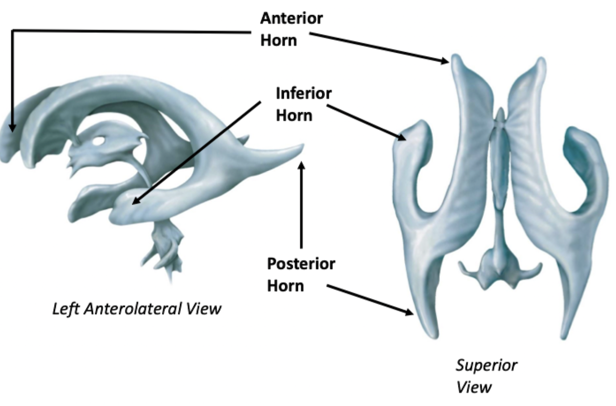
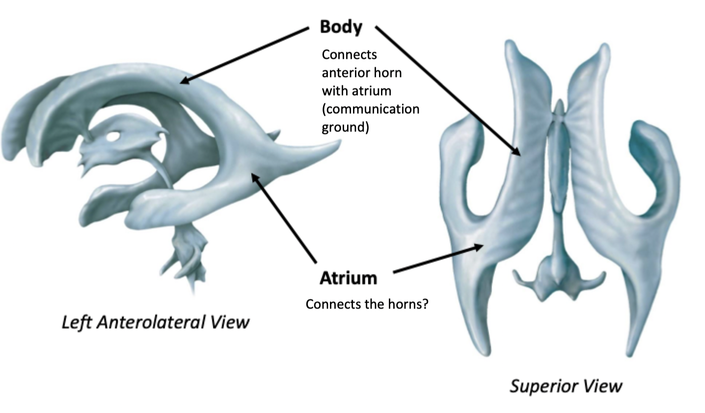
Lateral Ventricles
Explain
Associated with various lobes of the cerebrum (the telencephalon)
Lateral Ventricles
Lateral Ventricles
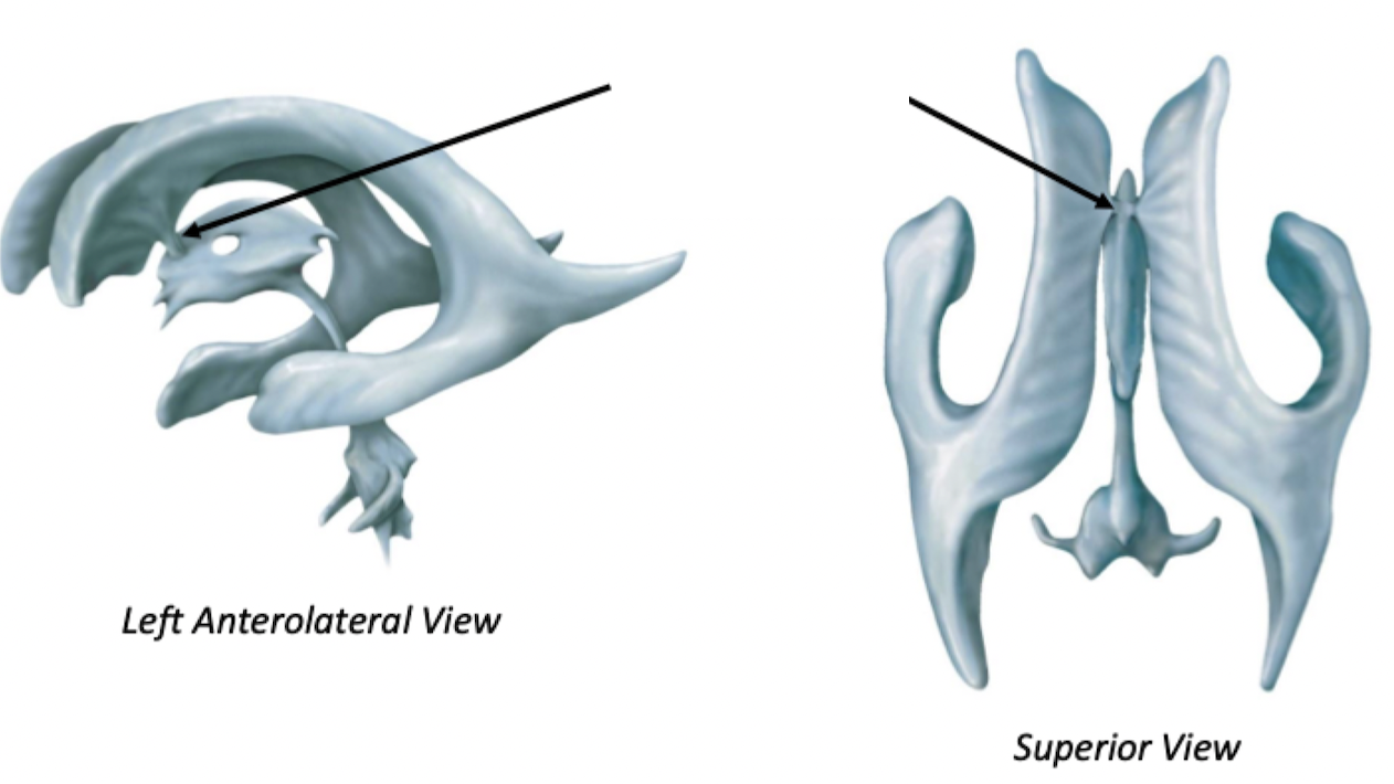
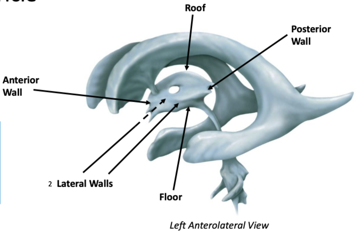
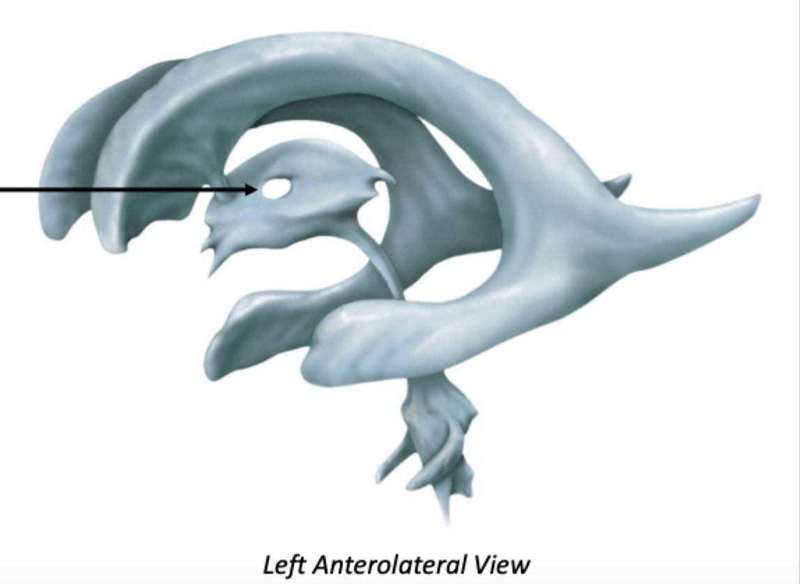
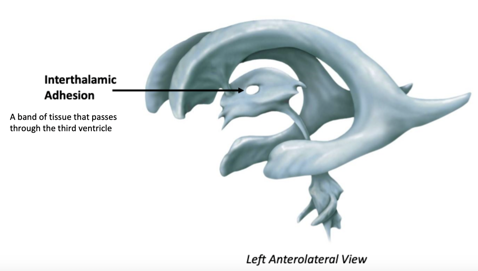
Third Ventricle
Associated with the thalamus and hypothalamus (the diencephalon)
Third Ventricle
Label + Explain
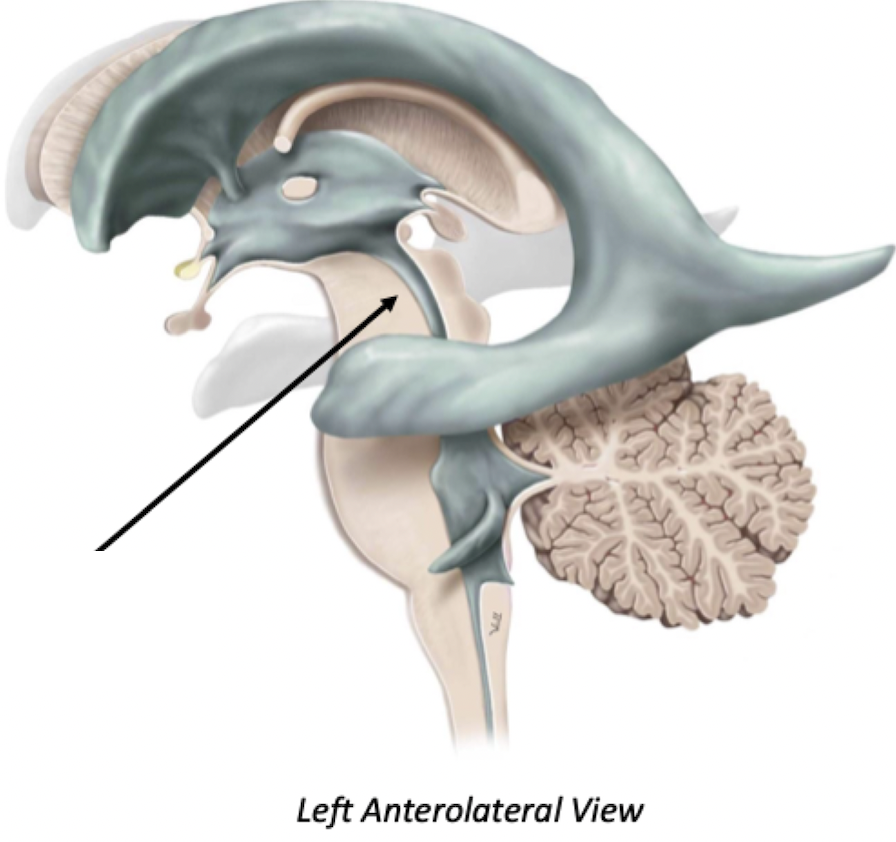
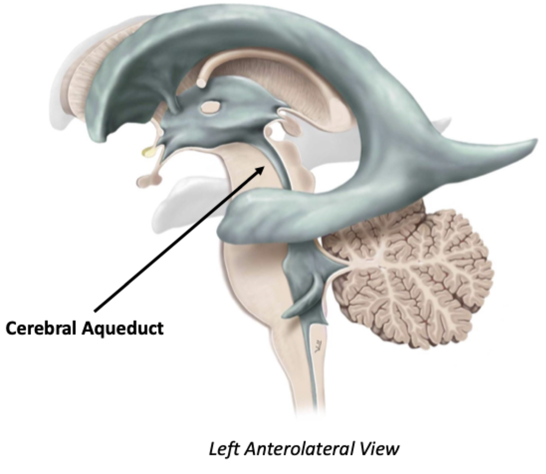
Cerebral Aqueduct
The cerebral aqueduct connects the third and fourth ventricles, passing through the midbrain into the pons
Fourth Ventricle
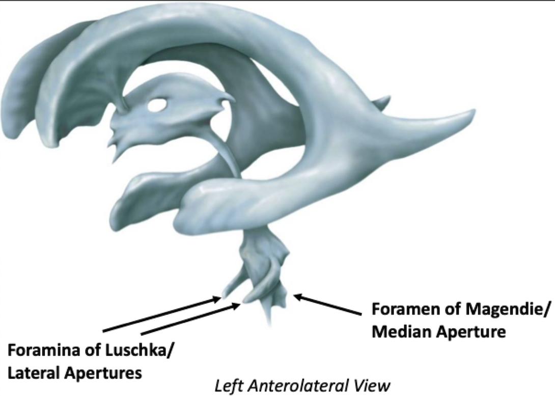
There are 3 apertures in the fourth ventricle:
2 Lateral and 1 Median
Associated with the pons (the myelencephalon)
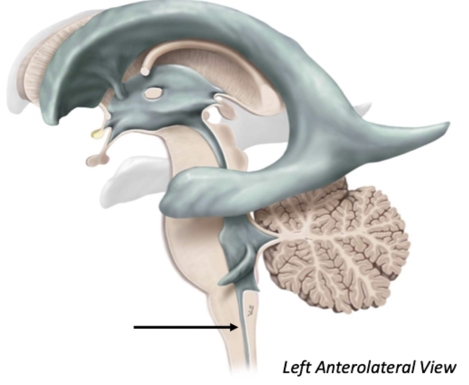
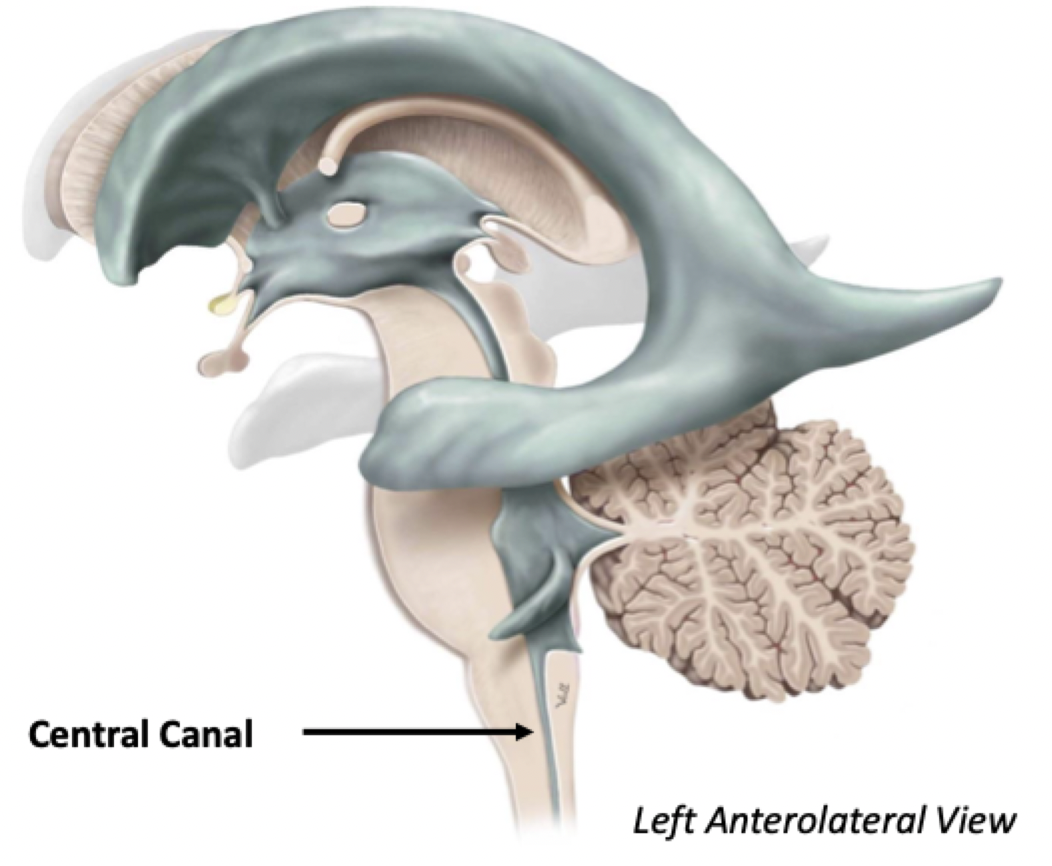
Central Canal
The central canal continues all the way down the spinal cord
Provides metabolic support to the deep spinal cord
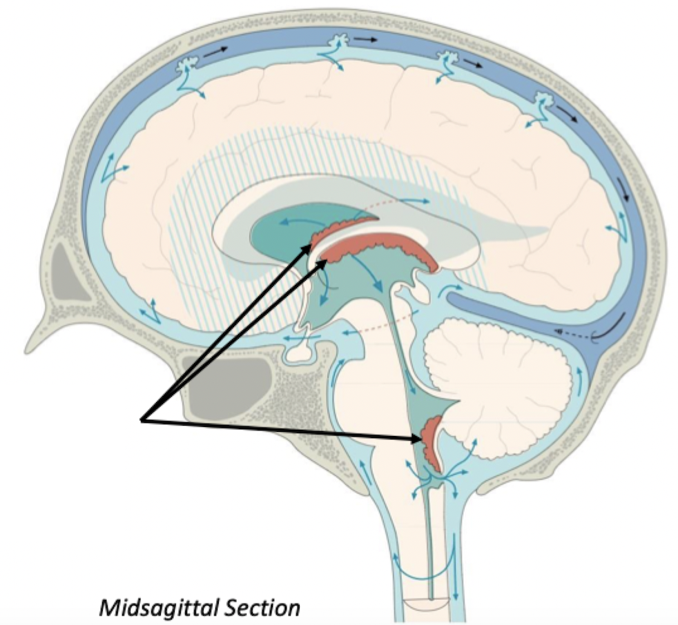
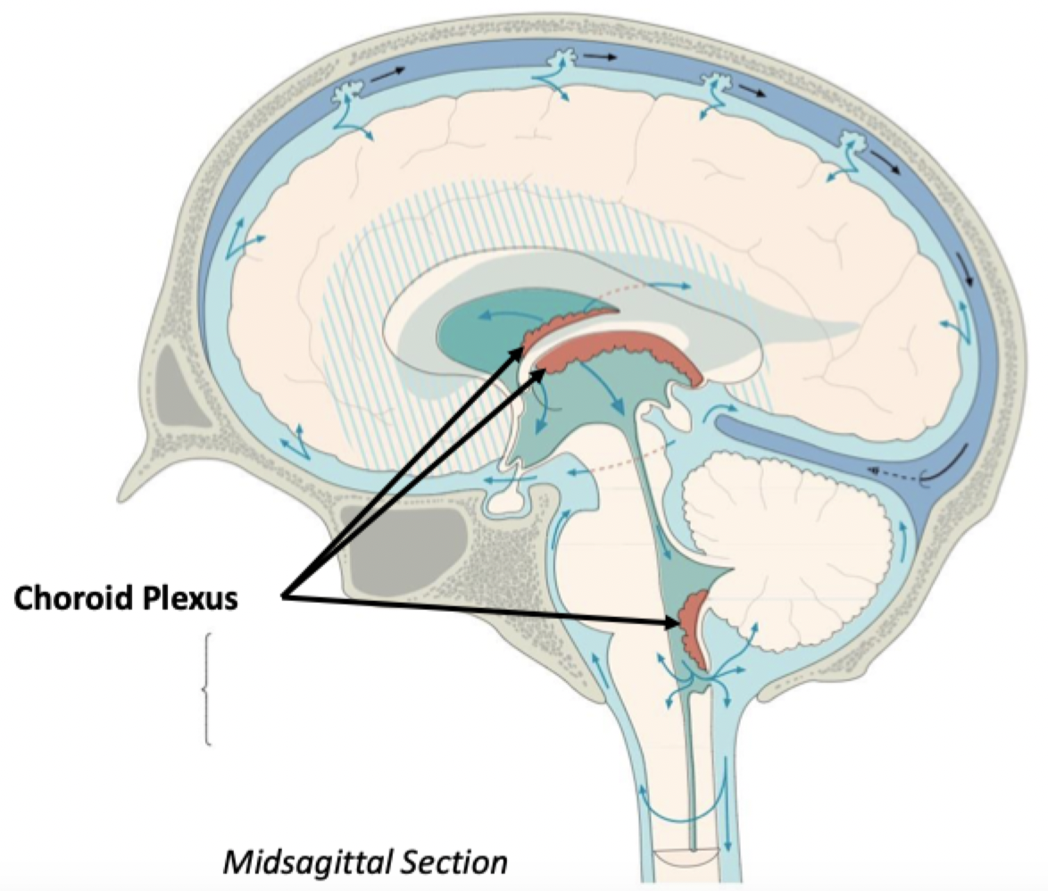
Choroid Plexus
Collection of ependymal cells found on the walls of all 4 ventricles, in specific areas
Produces cerebrospinal fluid, which flows into the ventricles
Ventricular Flow
Lateral Ventricle
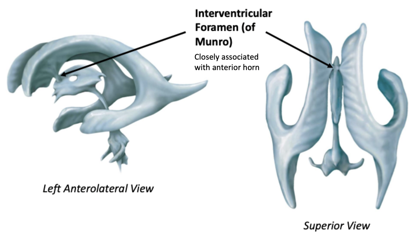
Interventricular Foramen
Third ventricle
Cerebral Aqueduct
Fourth Ventricle
Central Canal/Foramina of Luschka/Foramen of
Magendie
Exiting the Ventricles - CSF Flow
CSF exits into small subarachnoid spaces called cisterns (small cavities)
From these cisterns, CSF flows along the outer cortices of the cerebrum and cerebellum before draining into the dural venous sinuses via arachnoid granulations
Overview of Cranial CSF Flow
Choroid Plexus
Lateral Ventricle
Interventricular Foramen
Third Ventricle
Cerebral Aqueduct
Fourth Ventricle
Median/Lateral Apertures
Subarachnoid Cisterns
Bathes Superficial Brain
Arachnoid Granulations
Dural Venous Sinuses
Overview of Spinal CSF Flow
Choroid Plexus
Lateral Ventricle
Interventricular Foramen
Third Ventricle
Cerebral Aqueduct
Fourth Ventricle
Central Canal
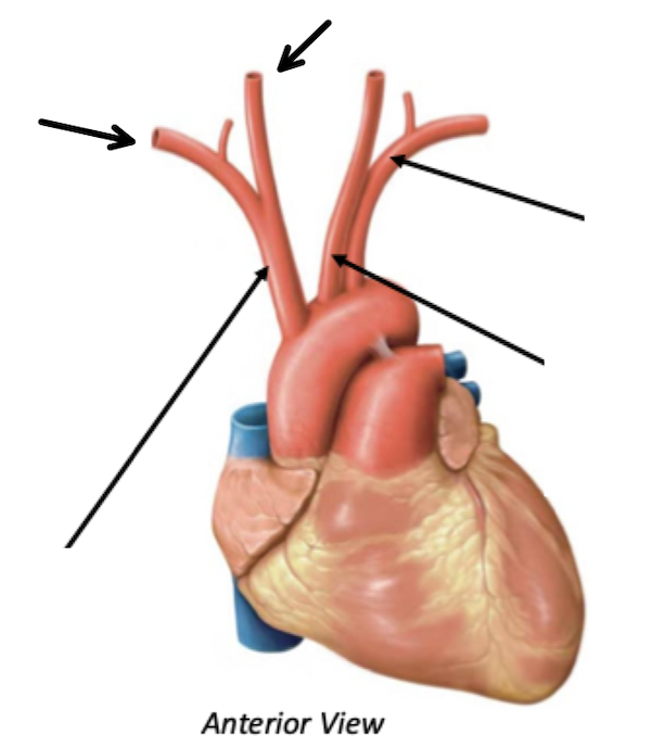
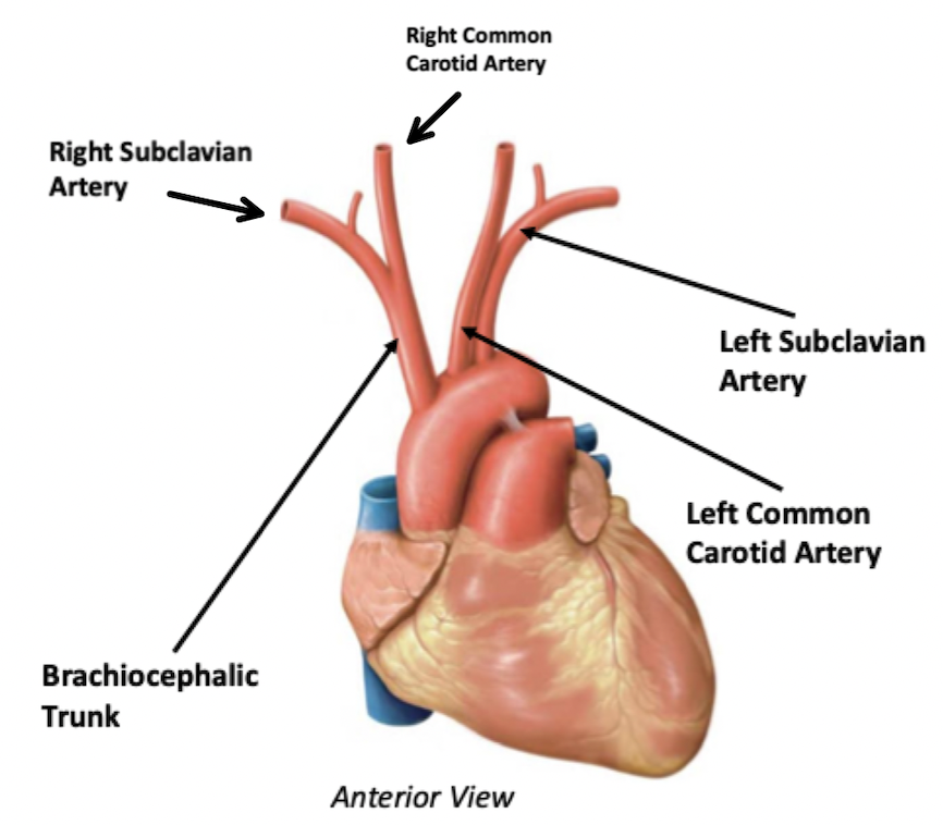
Great Vessels of the Heart
Three major branches off the aorta
Brachiocephalic Trunk
The brachiocephalic trunk bifurcates (ends and splits)
into the right common carotid artery and the right subclavian artery
Left Common Carotid Artery
Left Subclavian Artery
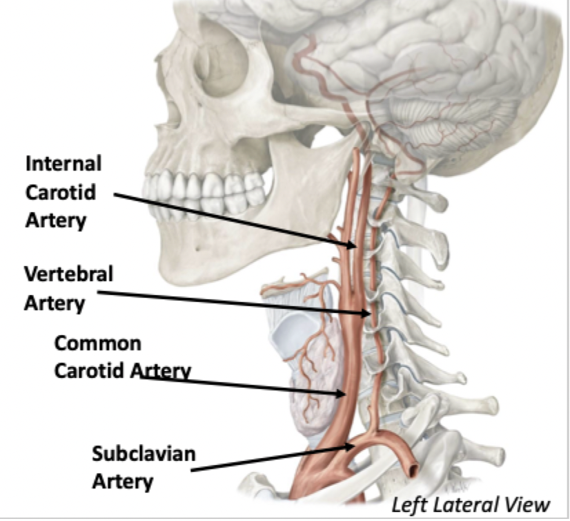
Four Major Arteries to the Brain
Common Carotid Artery → Internal Carotid Artery
Internal Carotid Artery goes through the Carotid Canal
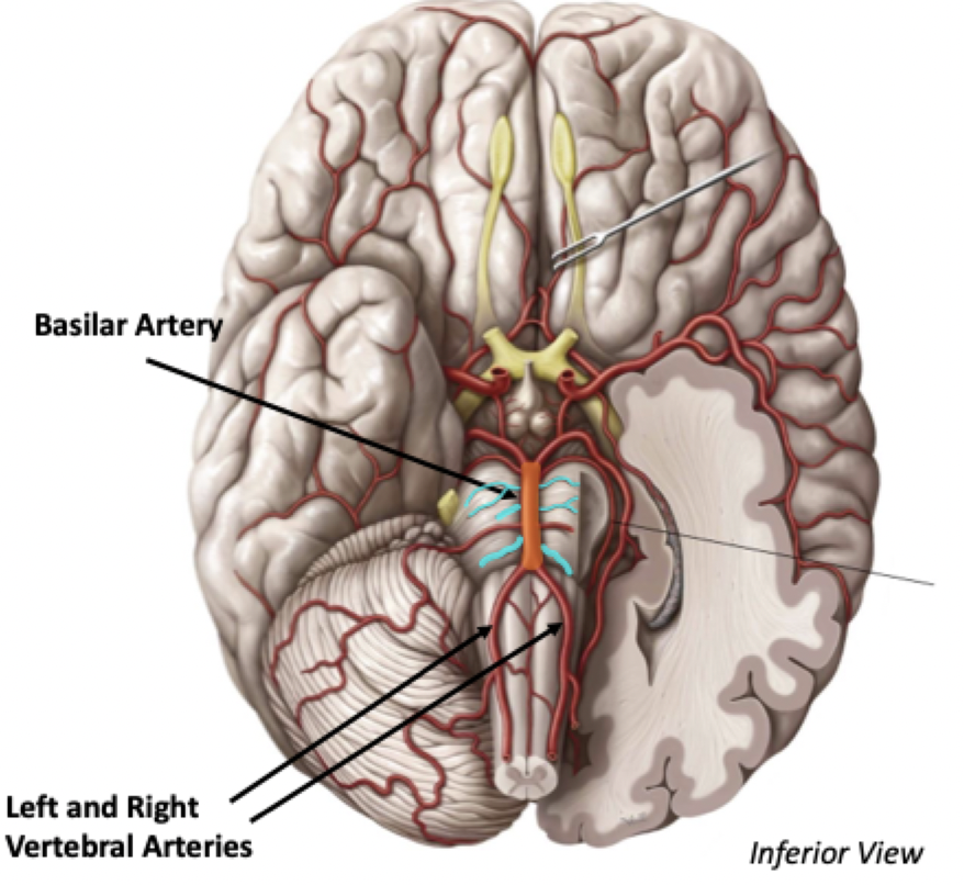
Subclavian Artery → Vertebral Artery
The vertebral artery runs through the vertebral foramina of the cervical vertebrae and goes through the anterior portion of Foramen Magnum
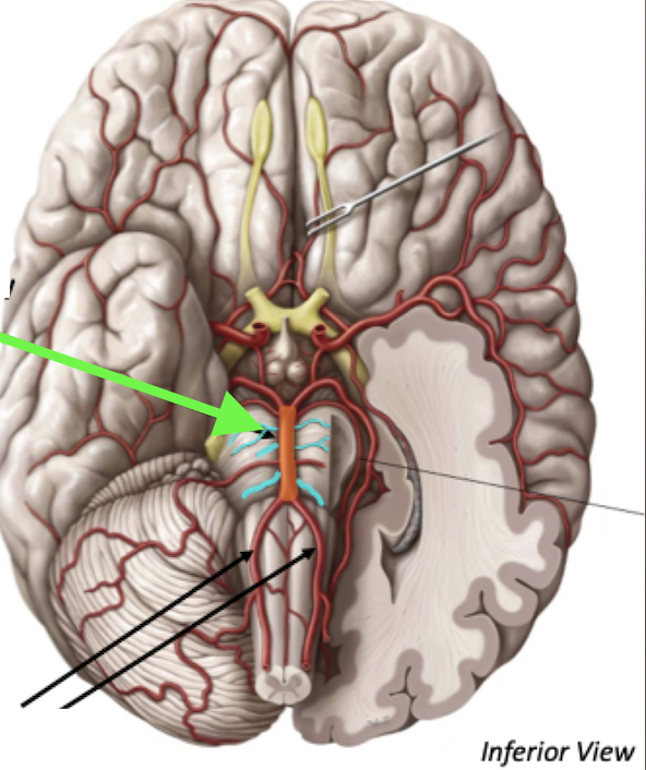
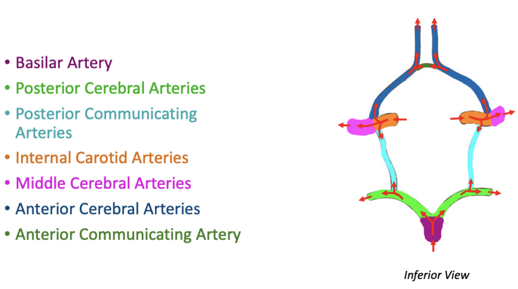
Basilar Artery
Left and Right Vertebral arteries anastomose (join) to form the Basilar artery
Sits in basilar groove of pons
Gives off Pontine arteries to supply the pons
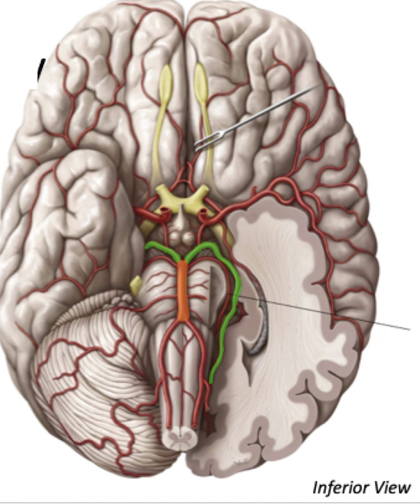
Bifurcation of Basilar Artery
Basilar Artery bifurcates into the 2 Posterior Cerebral arteries
These arteries supply the posterior cerebral cortex
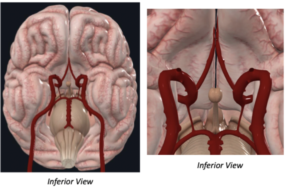
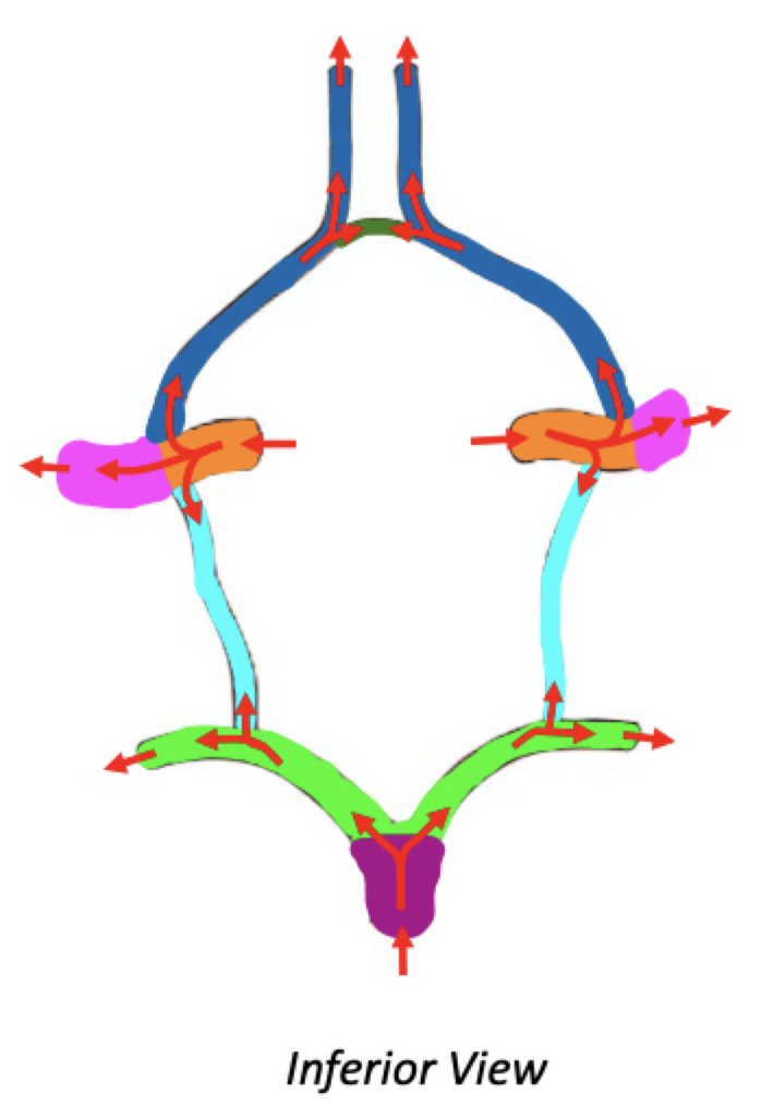
Circle of Willis
Anastomosis of arteries providing major cerebral blood supply
Compensatory mechanism: In the case of a clot in a vertebral or internal carotid artery, maintains cerebral blood flow throughout the brain
Circle of Willis
Label
Basilar artery bifurcates into Posterior Cerebral Arteries
Internal Carotid Artery gives off the Anterior and Middle Cerebral arteries
Internal Carotid Artery also gives off the Posterior Communicating Artery
Posterior Communicating Artery connects Internal Carotid Artery with the Posterior Cerebral Artery
Communication arteries provide connections
Anterior cerebral arteries are connected by the anterior communicating artery
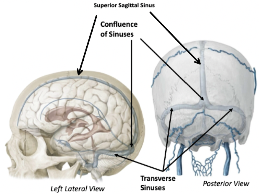
Brain Venous Drainage
Consists of dural venous sinuses that drain the inner structures of the cranial vault
Superior Sagittal Sinus
Lies along mid-sagittal plane, in falx cerebri
Confluence of Sinuses
Transverse Sinuses
The transverse sinuses sit in the tentorium cerebelli