Tendons, ligaments, bursa - #3
1/32
There's no tags or description
Looks like no tags are added yet.
Name | Mastery | Learn | Test | Matching | Spaced |
|---|
No study sessions yet.
33 Terms
Components of extremity MSK
• Bone Surface
articular and non-articular
• Muscle
pennate
• Tendon
fibrillar sonographic signature
musculotendinous junction
• Meniscus/ Discoid (abnormally thickened) tissue
• Ligament
• Bursa
• Nerves
• Synovium
• Articular cartilage
Pennate muscle pattern
fan shaped (feather like)
parallel to bone
Tendons are the attachment of ______ to _________
muscle to bone
tendons have _______ fibers which are _____ - cord like
collagenous
rounded
collagenous fibers of the tendon have flat sheets that are called _________ and have no
aponeuroses
blood flow
synovial sheath may or may not surround the
tendon
synovial sheath has a ______ layer of fluid which makes it easier to visualize
double
Tendons connect ______ to ______ and have _______ sheath in areas of _______ such as in the wrist
muscle to bone
synovial sheath
friction
3 tendons without synovial sheath
achilles
patellar
prox gastrocenmius
paratenon
outer layer of tendon made of loose connective tissue
epitendineum
dense connective tissue surrounding the tendon
MSKUS allows dynamic imaging and ____ ___ ______ comparison s
side to side
a normal tendon is _____ in thickness and echogenicities
normal
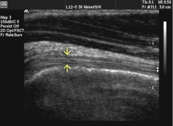
when looking at a tendon you should identify echogenic _______ that respresent the _________ fibers to confirm imaging of tendon
fascicles
collagen
the echogenic facicles /collagen fibers run _____ to long axis of tendon
parallel
modality of choice for tendons
MRI
bicep tendon
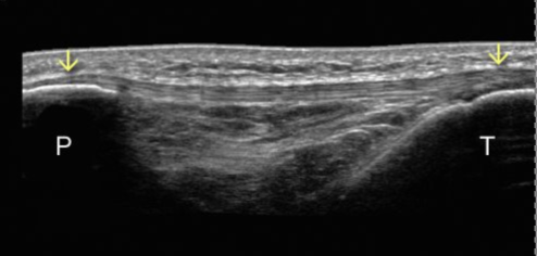
P = patella
T = tibia
Patellar tendon in middle connect both patella and tibia
also helps connect quad muscles to knee and allow leg to straighten
tendon insertion site is called the
enthesis
injury can ________ insertion site/enthesis
thicken
Fibrocartilage at enthesis looks
Avascular
Hypoechoic triangular area
1 cm
Mistaken for pathology
ligaments connect
bone to bone
ligaments are ____ and ________
thin
superficial
measurement and echogenicity of ligament
2-3mm
hypoechoic band
similar appearance to tendons
sonography techniques fro ligaments
stand off pad
long imaging only
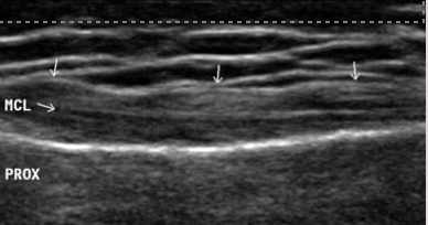
Ligament
synovial sheets are seen in the ____ space
bursa
what is a bursa
Small ‘sacs’ filled with viscous, synovial fluid surrounding muscles, tendons, and ligaments
Bursa fluid allows fro
movement
communication bursa interacts with ______ _______
joint space
Bakers cyst in seen in what bursa
communicating
How is non communication bursa different from communicating
it DOES NOT interact with joint space
ex: prepatellar bursa