NPTE- MSK
1/1055
There's no tags or description
Looks like no tags are added yet.
Name | Mastery | Learn | Test | Matching | Spaced |
|---|
No study sessions yet.
1056 Terms
Biceps Brachii - position of active insufficiency
Shoulder and elbow flex
forearm supination
Biceps Brachii - position of passive insufficiency
Shoulder and elbow ext
forearm pronation
Triceps Brachii - position of active insufficiency
Shoulder and elbow ext
Triceps Brachii - position of passive insufficiency
Shoulder and elbow flex
Wrist extensors - active insufficiency
Elbow flex, wrist ext
Wrist extensors - passive insufficiency
Elbow ext, wrist flex
Wrist flexors - active insufficiency
Elbow flex, wrist flex
Wrist flexors - passive insufficiency
Elbow ext, wrist ext
Hamstrings - active insufficiency
Hip ext, knee flex
Hamstrings - passive insufficiency
hip flex, knee ext
Gastrocnemius - active insufficiency
Knee flex, ankle PF
Gastrocnemius - passive insufficiency
Knee ext, Ankle DF
Insufficiency - when does it come into play?
With two joint muscles.
Active Insufficiency
When a muscle cannot shorten anymore, occurs with the agonist.
Passive Insufficiency
When a muscle cannot stretch anymore, occurs with the antagonist.
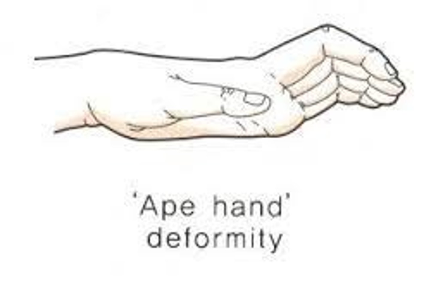
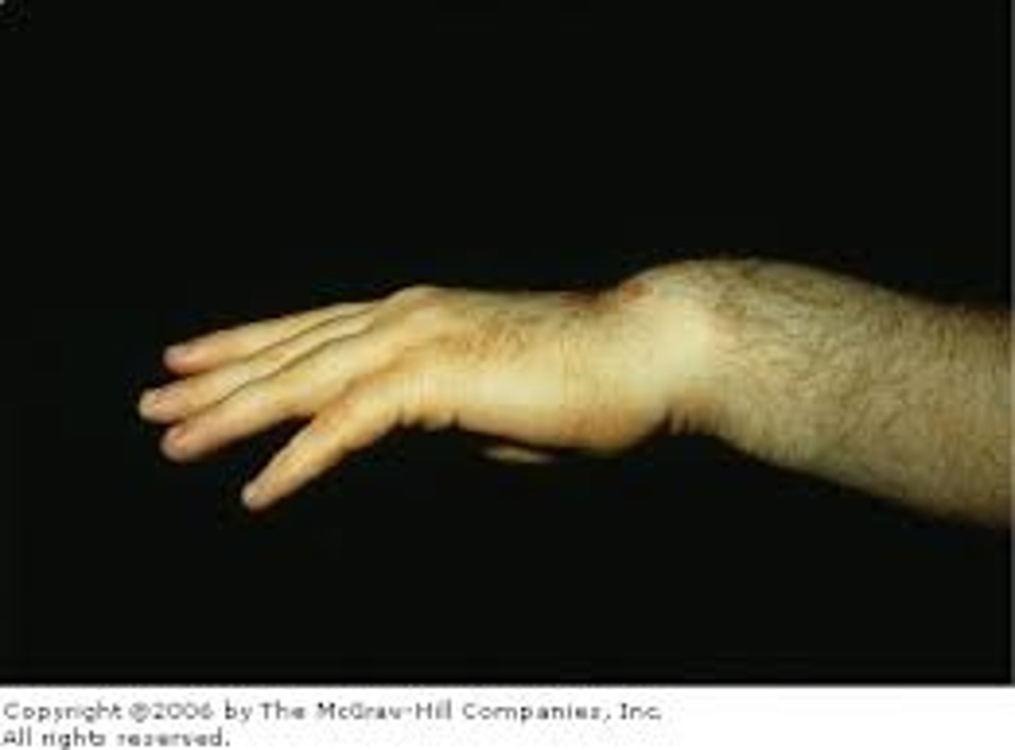
ape hand deformity
wasting of the thenar eminence of the hand occurs as a result of a median nerve palsy
thumb falls back in line with fingers as a result of pull of extensor muscles
**patient is unable to flex or oppose thumb
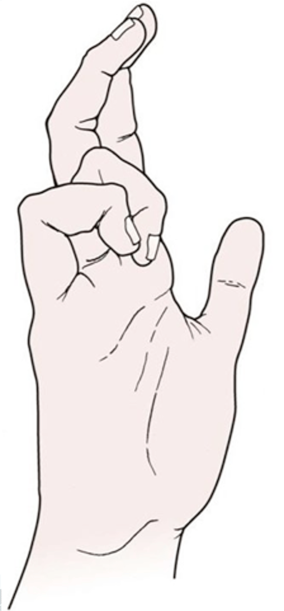
bishop's hand or benediction
wasting of the hypothenar muscles of the hand, interossei ms and 2 medial lumbricals due to ULNAR nerve palsy
**hyperextension of MCP joint and flexion of IP joint
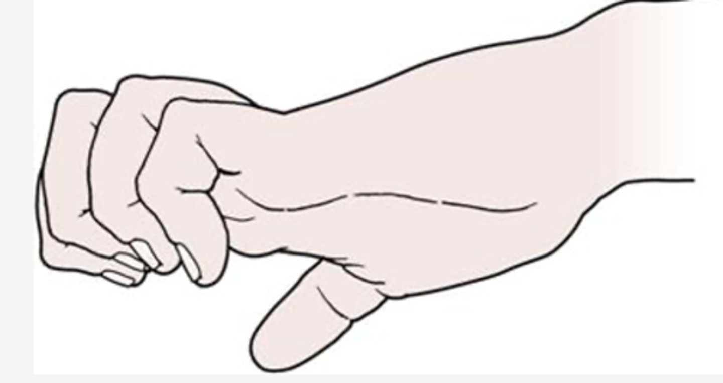
claw fingers
intrinsic minus hand, caused by combination of median & ulnar nerve
results from the loss of intrinsic muscle action and the overaction of the extrinsic extensor muscles on the proximal phalanx of the fingers
MCP joints are hyperextended, and PIP and DIP joints are flexed.
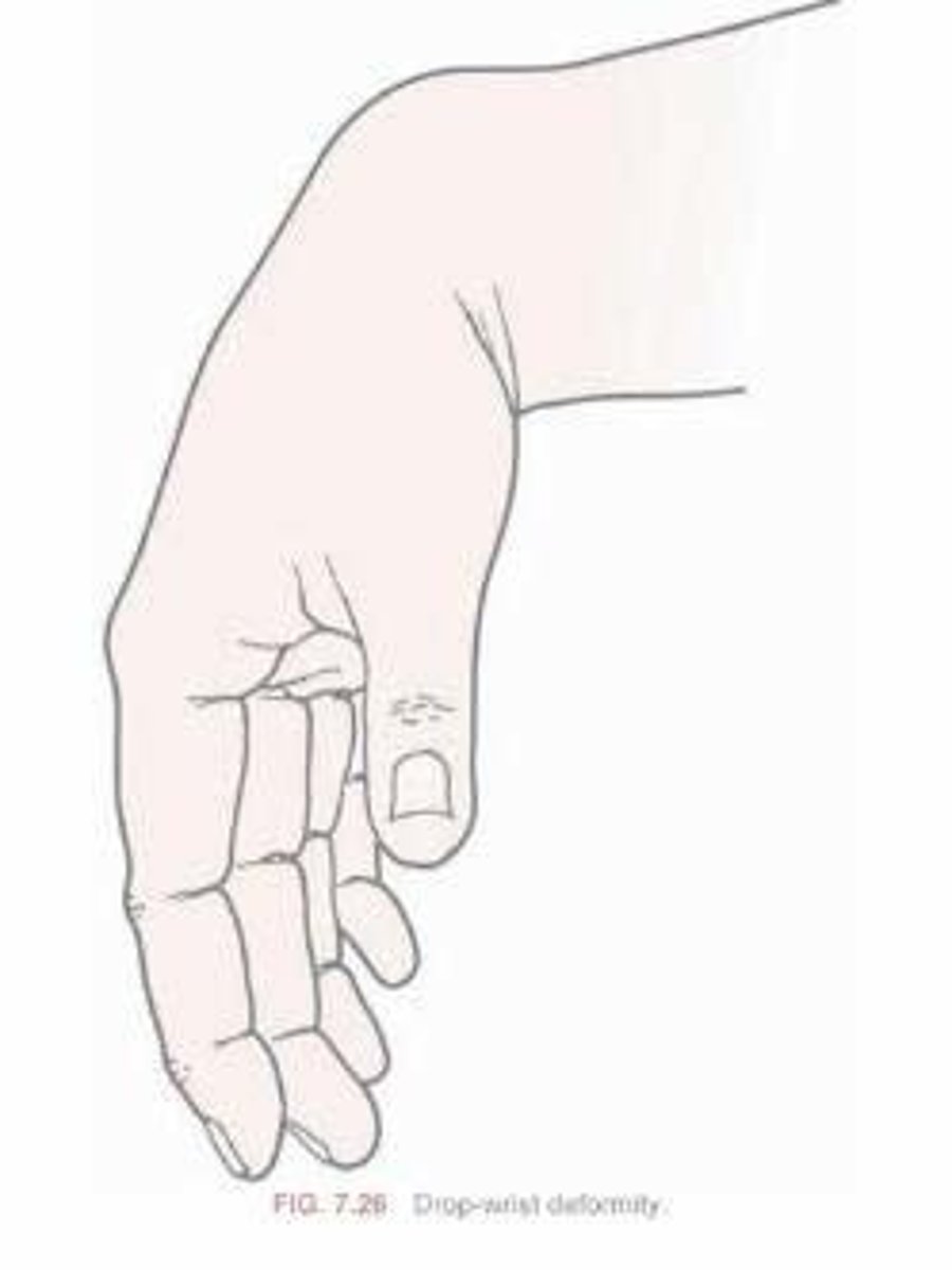
drop-wrist deformity
paralyzed extensor of wrist due to radial nerve palsy.
dupuytren contracture
contracture of palmar fascia, fixed flexion deformity of MCP & PIP
usually seen in ring or little finger
affects men more than women.

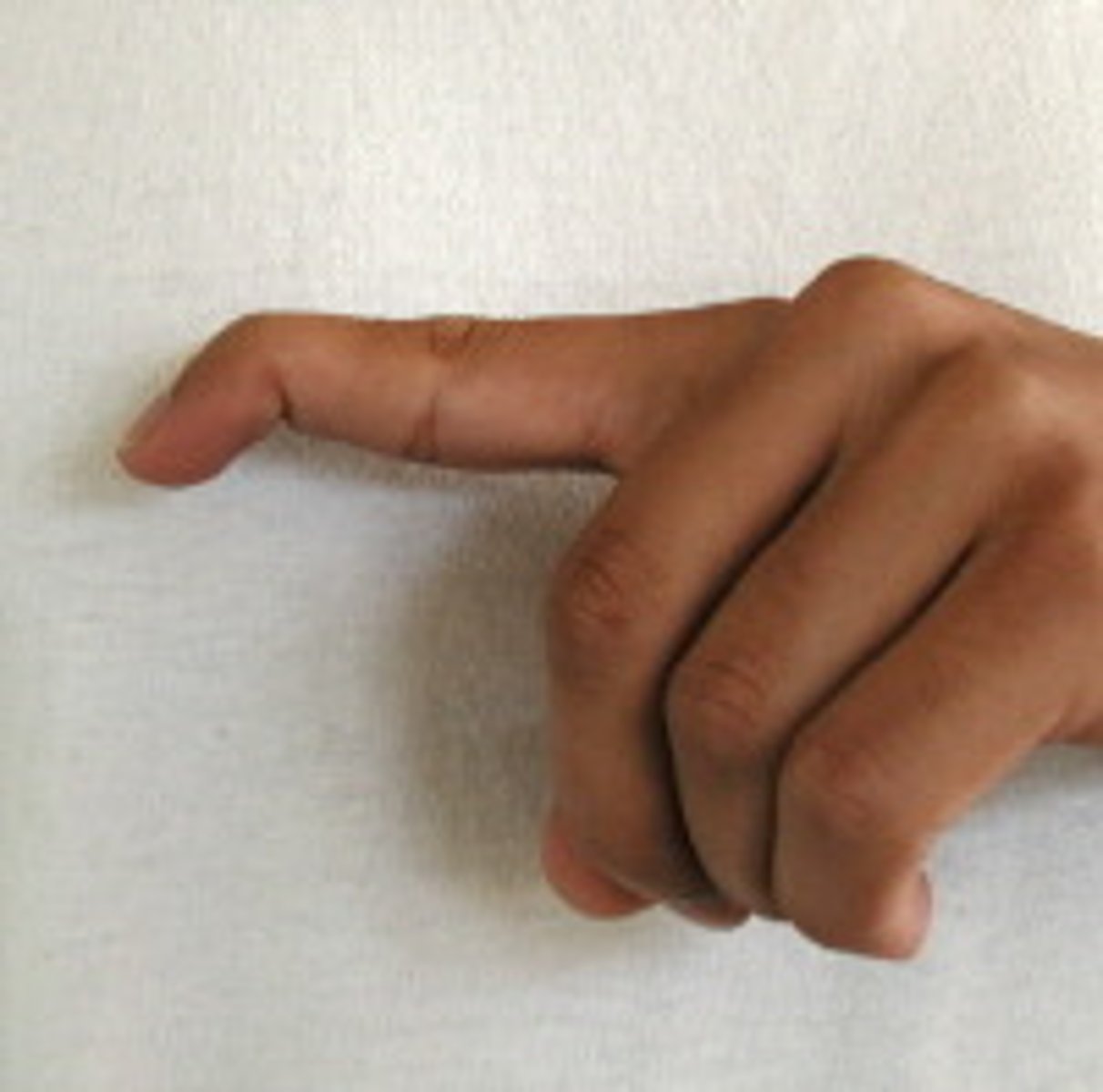
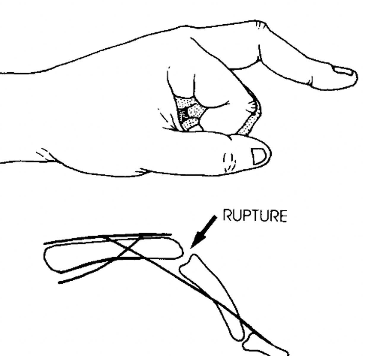
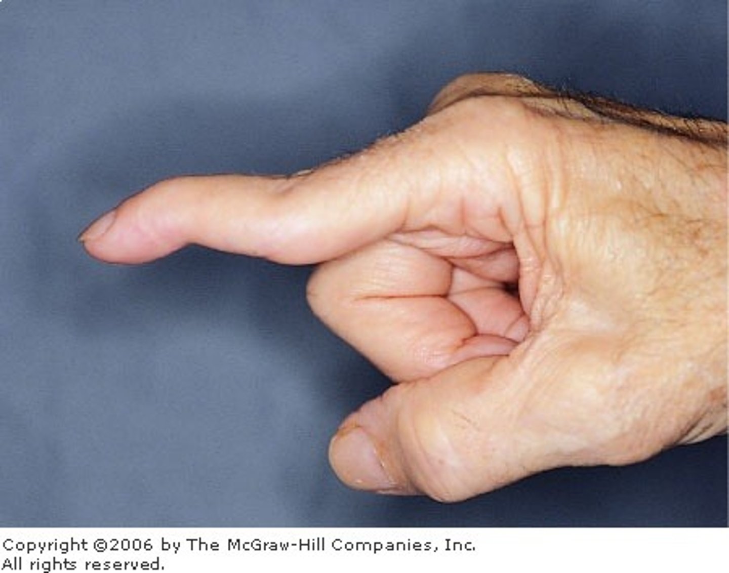
mallet finger
due to rupture or avulsion of the extensor tendon at distal phalanx of finger, distal phalanx rest in flexed
position.
boutonneire deformity
extension of MCP and DIP joint
flexion of PIP joint
rupture of central tendinous slip of the extensor hood and is more common after trauma or in RA
swan neck deformity
extension of PIP
flexion of MCP & DIP due to contracture of intrinsic ma or tearing of volar plate (common in RA)
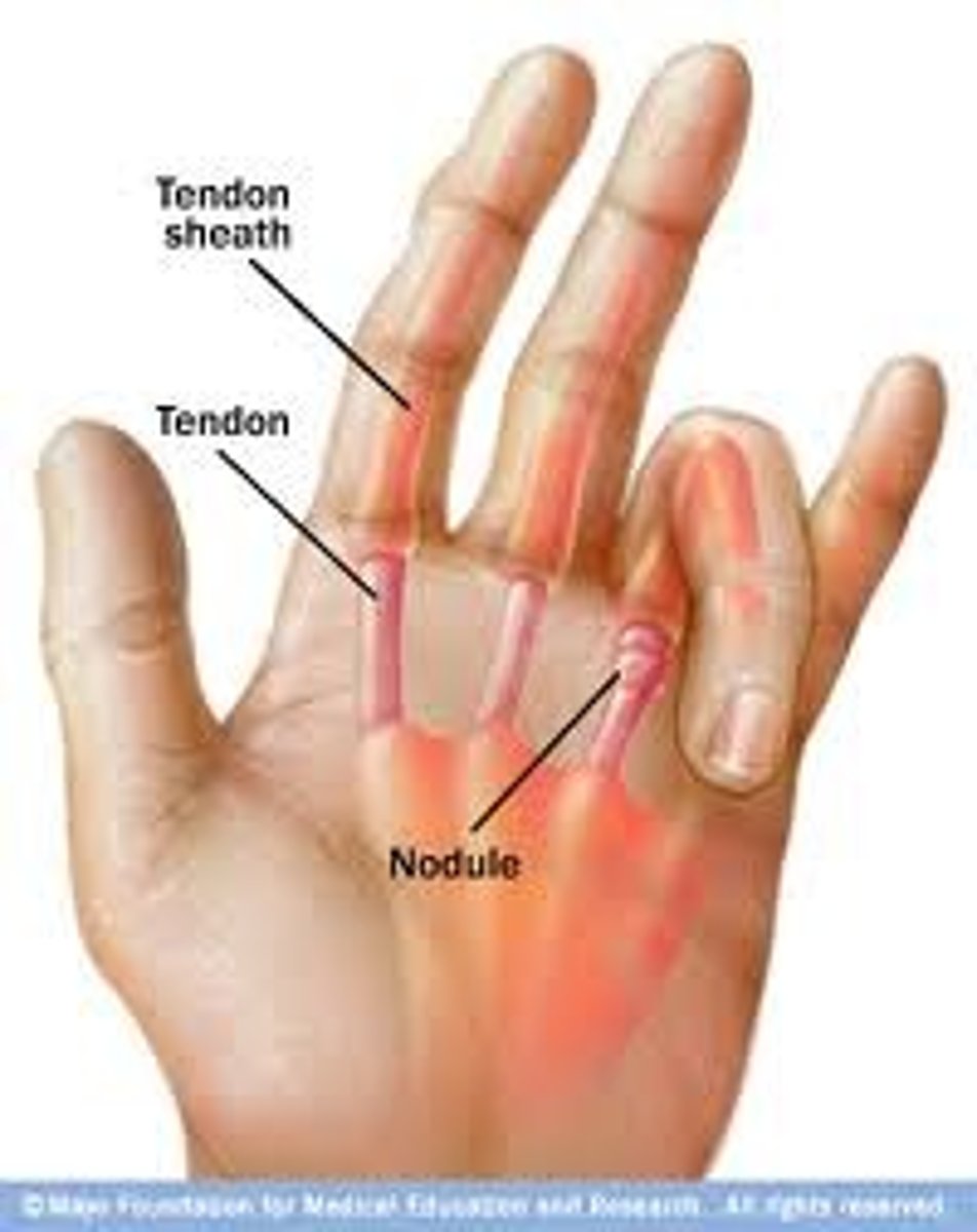
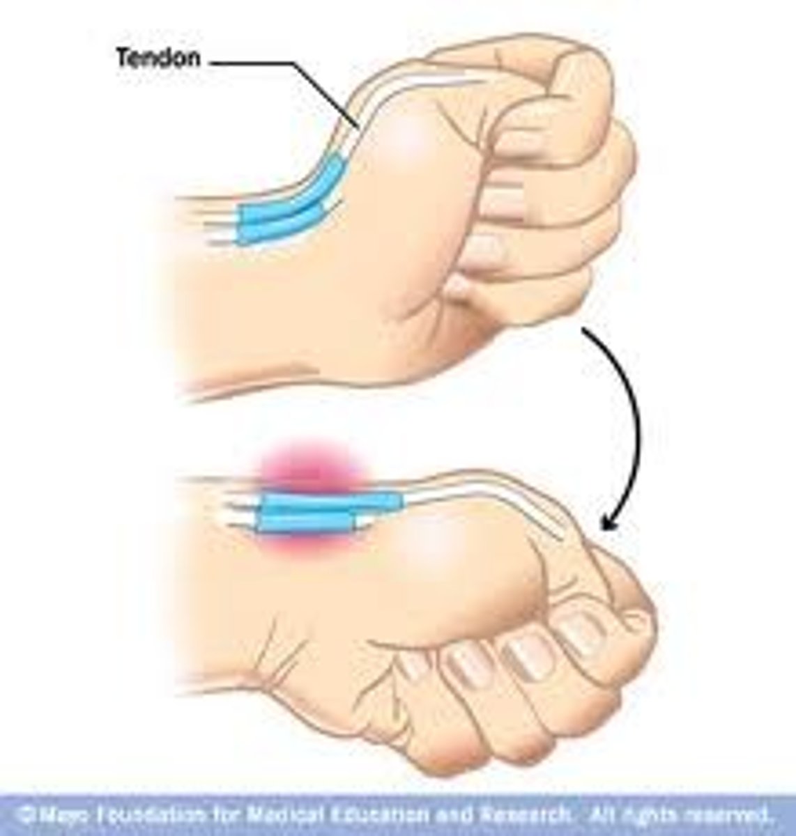
trigger finger
- results of a thickening of the flexor tendon sheath (notta's nodule), which causes sticking of the tendon when the patient attempts to flex the finger
- a low-grade inflammation of the proximal fold of flexor tendon leads to swelling and constriction in digital flexor tendon
-when patient attempts to flex the finger, the tendon sticks, and the finger "let's go," often with a snap.
- as condition worsens, eventually finger will flex but not let go, and it will have to passively extended until finally a fixed flexion deformity occurs.
***usually occurs in 3rd or 4th finger
****most often associated with RA and tends to be worse in the morning.
de quervain's tenosynovitis
inflammation the extensor pollicis brevis and the abductor pollicis longus tendons at 1st dorsal compartment **common in pregnancy
pain at anatomical snuffbox, swelling, decreased grip and pinch strength
Finkelstein's test is positive
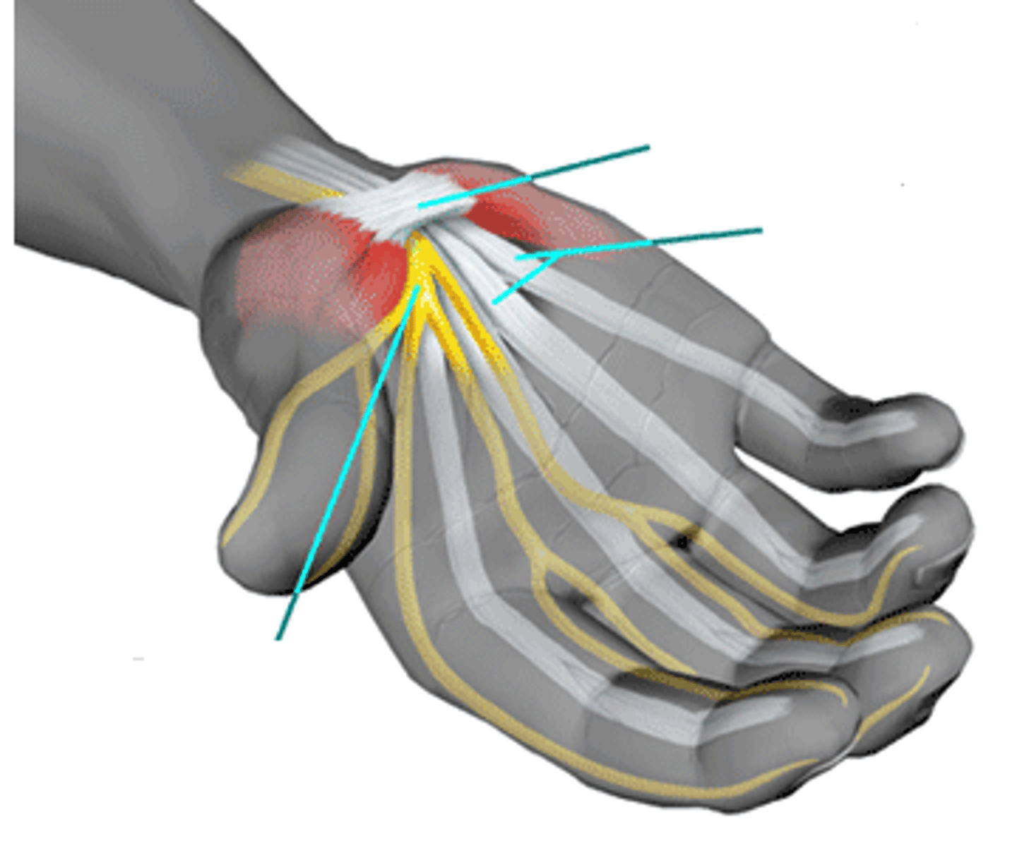
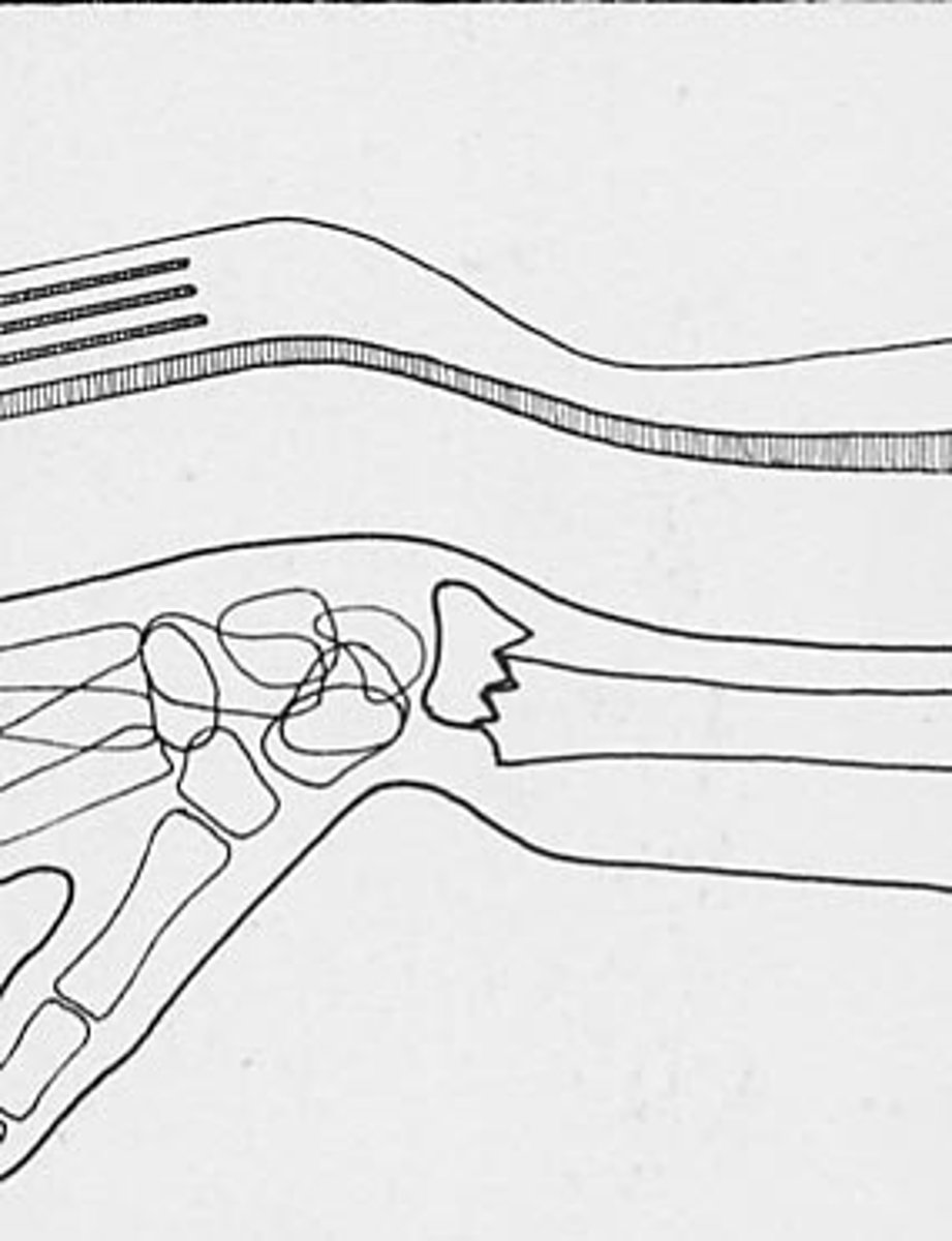
carpal tunnel
compression of median nerve
common in pregnancy, diabetes, RA
burning, tingling, pins and needles and numbness at night
positive tinels sign / phalens
colles fracture
dorsal displacement of distal fragment of radius with radial shift of wrist and hand results in dinner fork deformity
smiths fracture
distal fragment of radius dislocates in a volar direction result in garden spade deformity
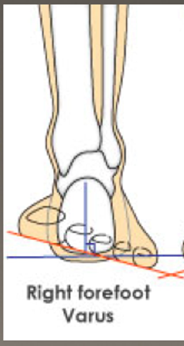
forefoot varus
- inversion of forefoot
- subtalar joint in neutral
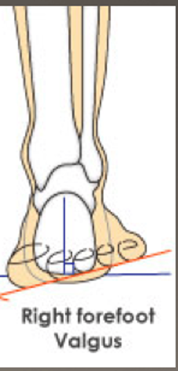
forefoot valgus
- eversion of forefoot
- subtalar joint in neutral
Sever's Disease
age 8-13 yrs
MOI: pronation, tight gastroc-soleus, jumping or landing from height
- limited DF
- pain over posterior-inferior heel which increases with weight bearing
- pain resolves with rest
tests: squeeze test, xray
midfoot sprain
- Age 15-40
- MOI - high impact landing, foot twisted when in fixed position
- walking on toes increases symptoms on midfoot
- generalized tenderness of midfoot
- pain during passive midfoot pronation and supination while the hindfoot is stabilized
- weightbearing lateral and anterior posterior radiographs
Metatarsal Stress Fracture
age 15-45
MOI: overuse
symptoms increase with WB activities on forefoot
tenderness over the fracture site
tests: palpation, ultrasound, tuning fork, bone scan, MRI, SCT
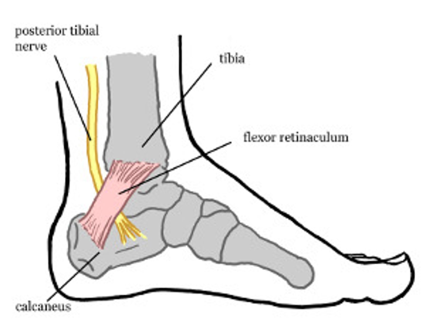
tarsal tunnel syndrome
age 25-50
MOI: posttraumatic, rapid weight gain, fluid retention, inflammatory, abnormal foot/ankle biomechanics or a valgus foot deformity
- symptoms on medial malleolus, distribution of posterior tibuial nerve down into the medial arch and plantar furface of foot and toes
- symptoms increase with excessive pronation in walking or runnin g
- pronated foot, pes planus can be observed
- passive plantarflexion and eversion painful
- resisted toe flexion painful
test: tinels sign positive
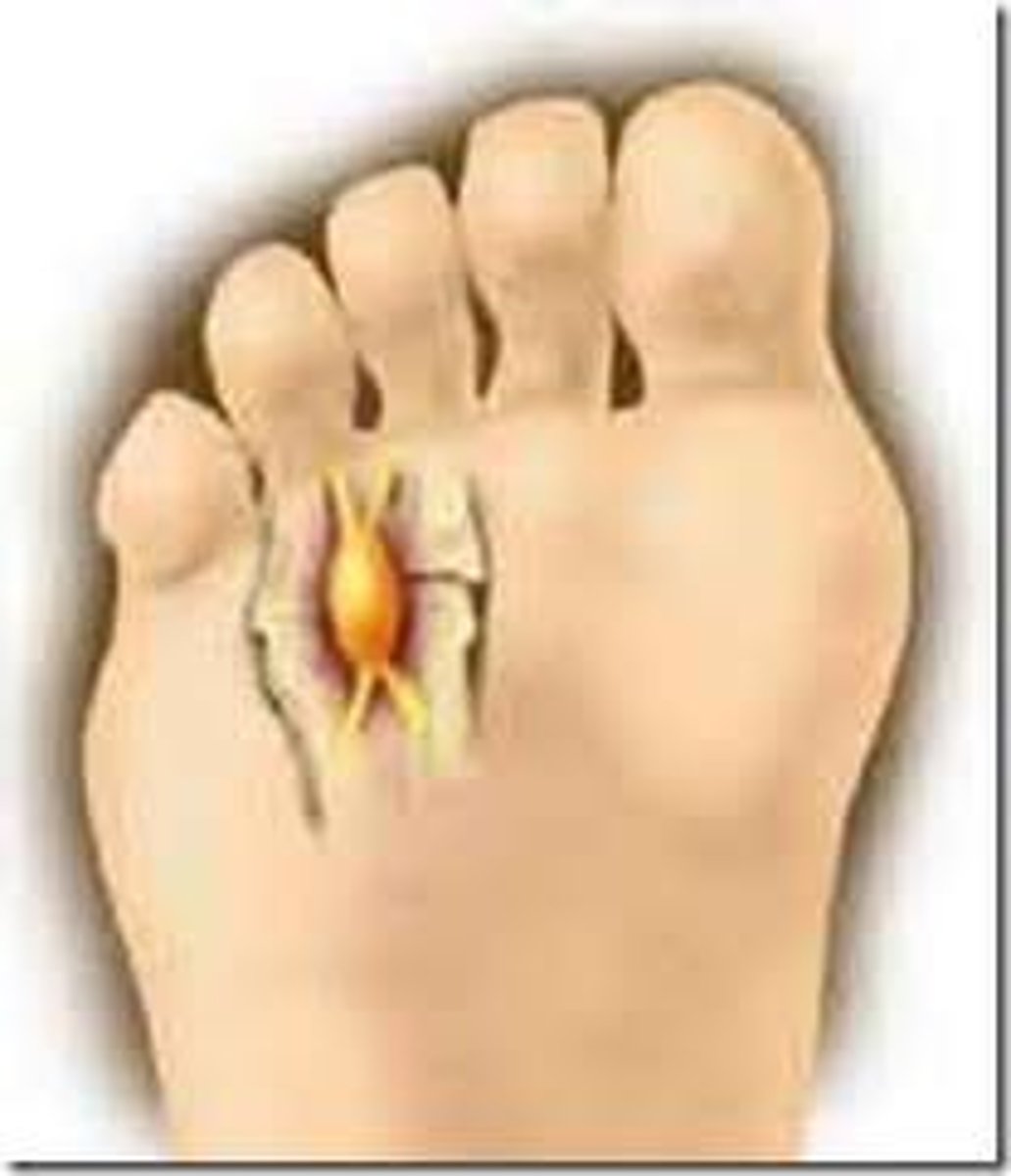
morton's neuroma
age: 40-60
MOI: gradual
weight bearing increases symptoms on sole of foot
pronated foot or flattened arch can be observed
passive toe extension painful
tenderness with palpation of web space or toes
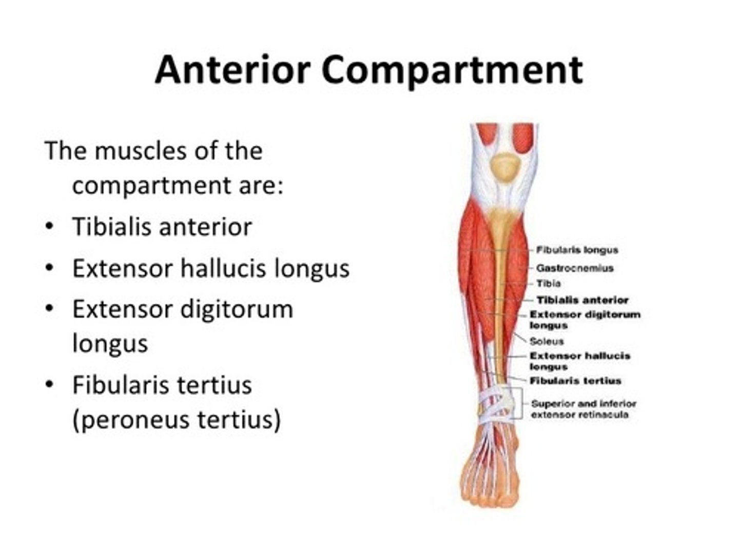
anterior compartment syndrome
- Increased pressure in anterior compartment of leg causing an ischemic condition of the leg
- MOI - direct trauma, muscle hypertrophy, fracture
severe cramping, diffuse pain and tightness
pain decrease with rest and increases with activity
pain worsens with stretching
tender and tight compartment on palpation
**6's: pain, pallor, paresis, paresthesia, pulselessness, palpable tenderness
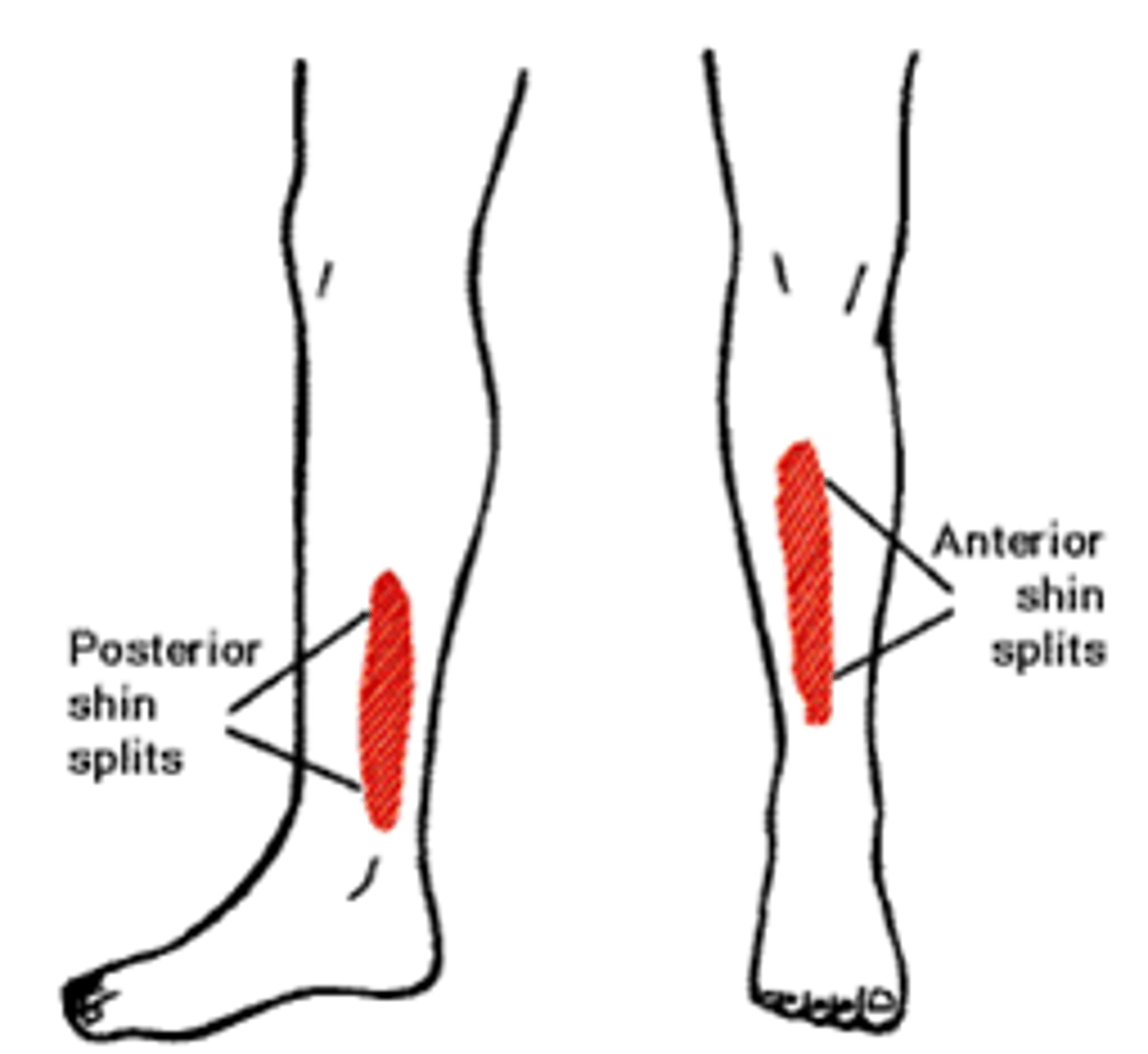
medial tibial stress syndrome (shin splints)
age 15-30
moi: overuse
pain on anterior lower leg, posterior-medial leg
active combined PF and IV painful
resisted PF and EV painful
tenderness on posteromedial calf
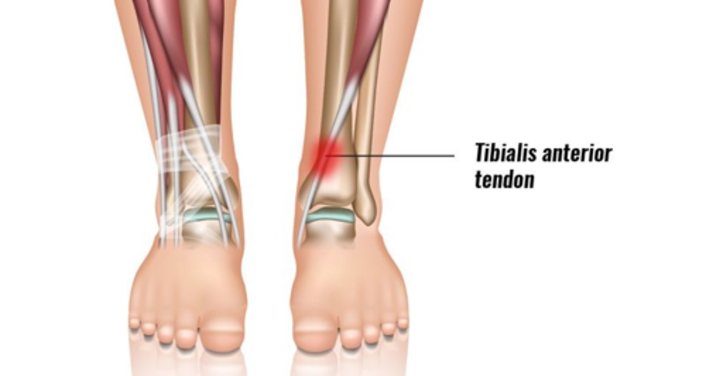
anterior tibialis tendinitis
age: 15-45
moi: overuse
symptoms increase with repetitive DF on anterior lower leg
combined active PF and IV movements painful
passive PF painful
resisted DF painful
tenderness over anterolateral lower leg
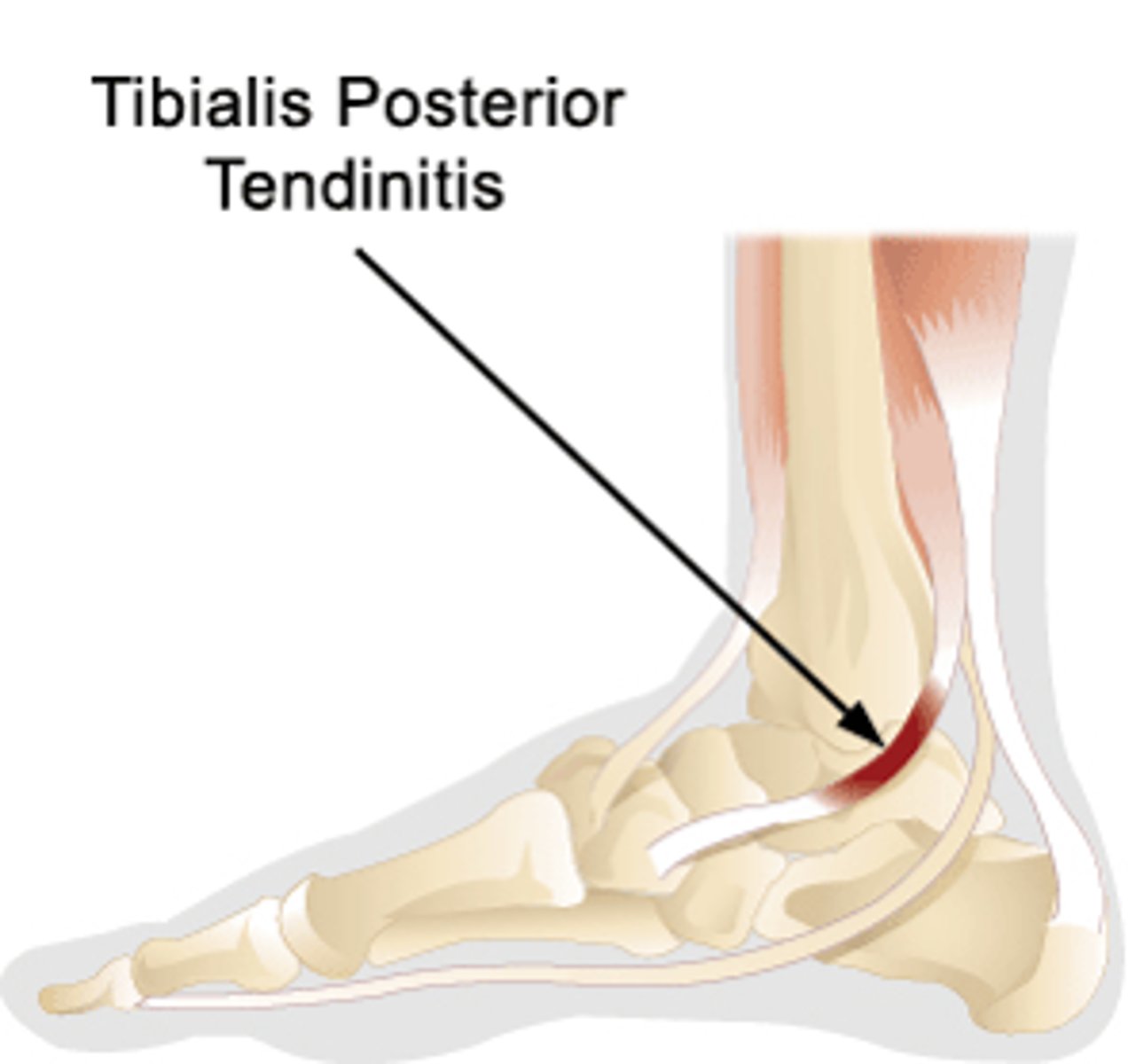
posterior tibialis tendinitis
- Age 20-40
- MOI- overuse with a flat pronated foot
- pain on medial ankle
- swelling and tenderness of medial ankle
- active and passive PF and EV painful
- resisted IV with PF painful
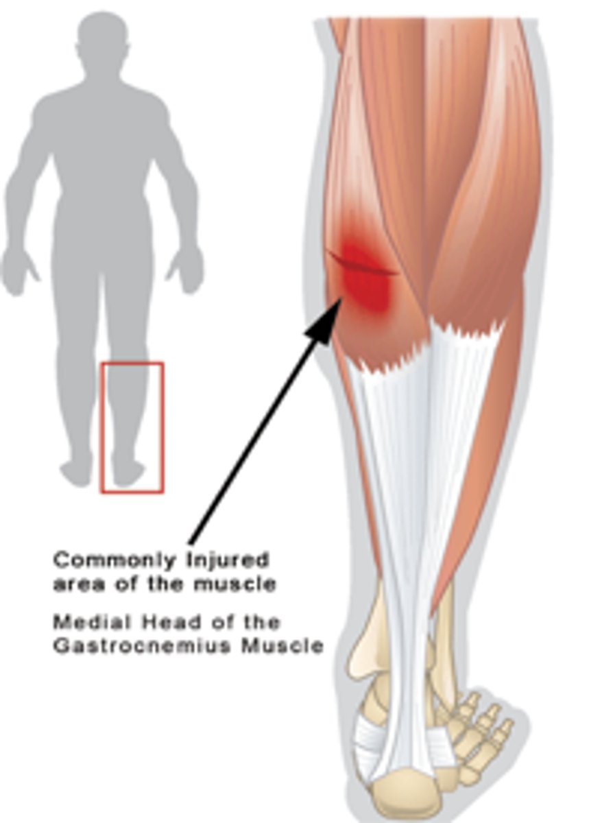
gastrocnemius strain
- Age 20-40
- MOI- sudden overload
pain in upper calf
heel raise increases symptoms
antalgic gait
active and passive DF painful and limited with knee extended
pain on resisted PF
tender mid to upper calf on palpation
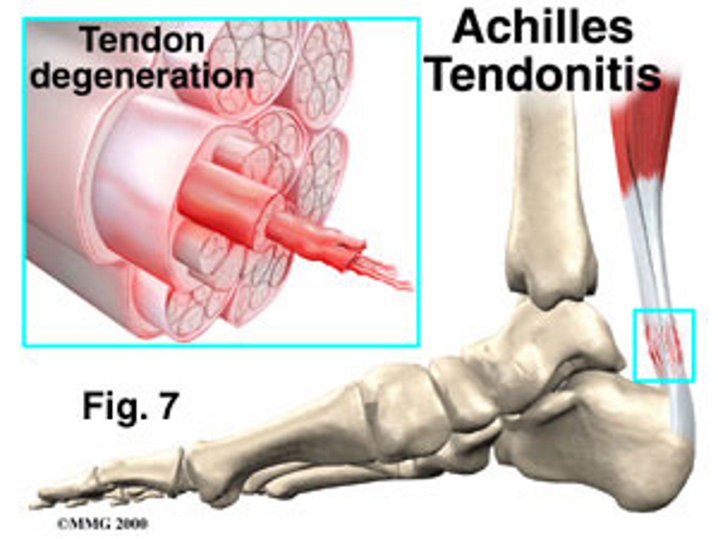
achilles tendinitis
age: 20-40
MOI: overuse
jumping and running increases symptoms on posterior ankle
slight swelling in posterior ankle
active and passive DF painful and limited
passive DF limited with knee in extended position
resisted PF painful
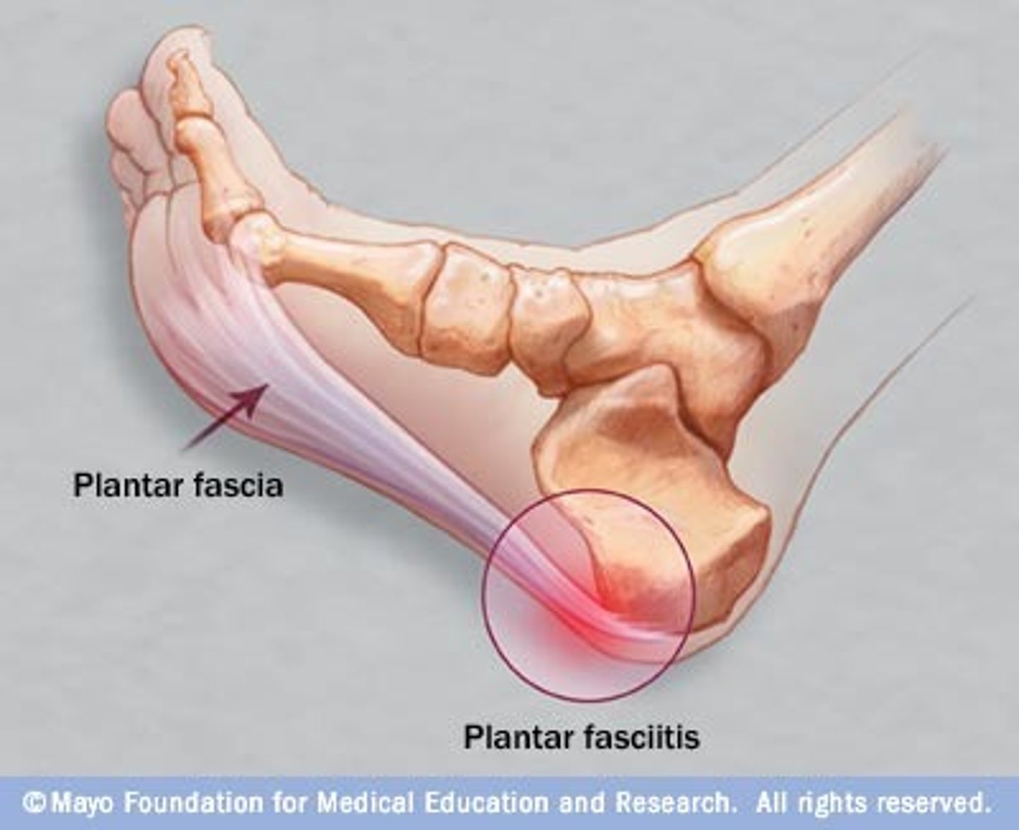
plantar fascitis
age: 20-60
moi: gradual w/ unknown cause
painful sole of foot under heel when WB
pain in morning
pronated foot or flattened arch
passive great toe extension painful
plantar aspect of heel is tender on palpation
test: windlass
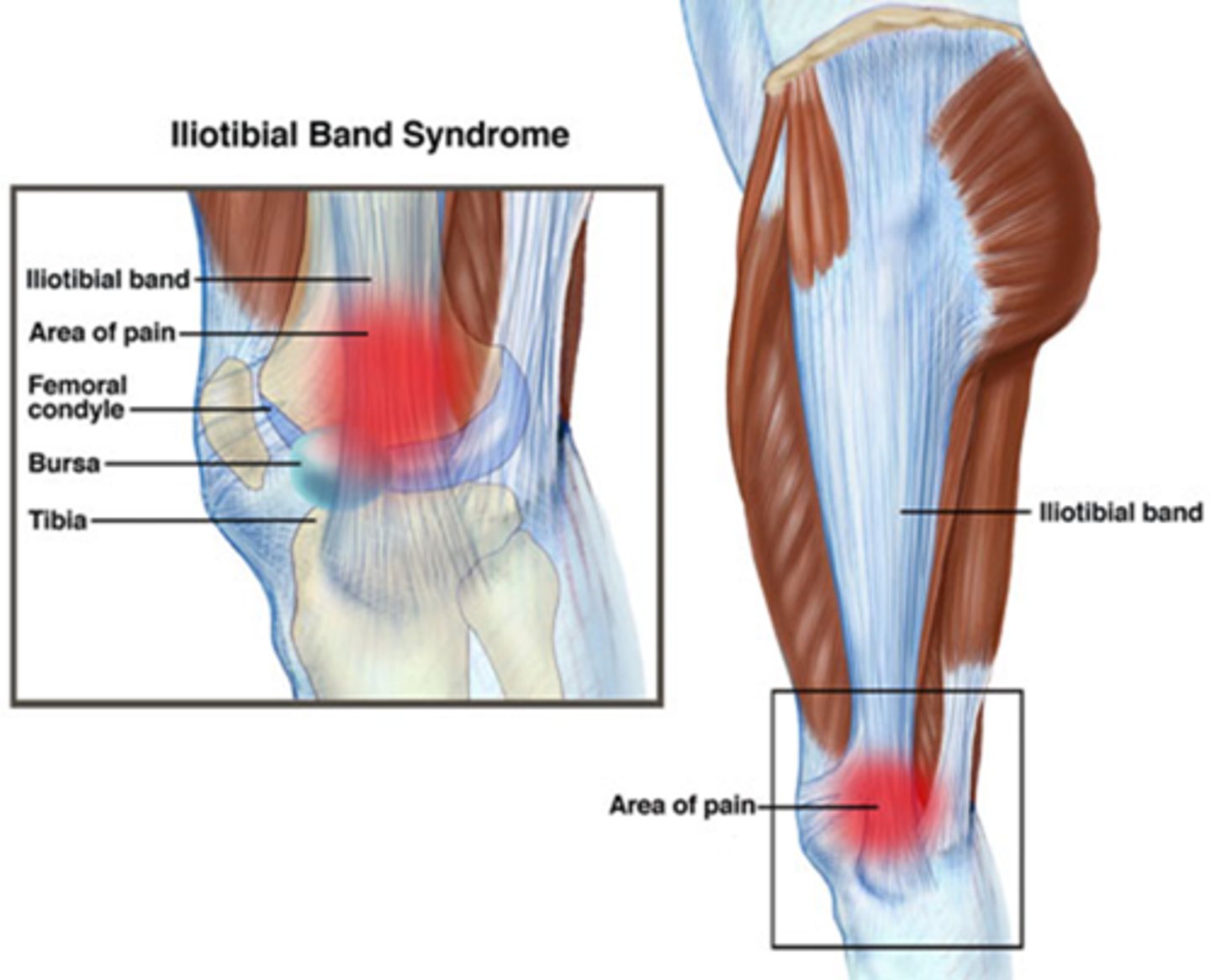
iliotibial band friction syndrome
Age 25-55; MOI- overuse
Pain with repetitive movements, climbing or descending stairs
Lateral knee pain diffuse & hard to localize
Localized tenderness at lateral femoral condyle.
posterior cruciate ligament injury
forced blow to anterior tibia in knee flexed, dashboard or falling into the flexed knee
- hyperflexion
- popping sound
- pain posterior aspect of knee
- aggravated with kneeling
test: posterior sag, posterior drawer, reverse lachman
MCL and LCL injury
valgus forces across medial joint line of knee
lateral collateral ligament- traumatic varus force across knee
- symptoms increase with varus stress (LCL) and valgus stress (MCL)
anterior cruciate ligament injury
age: 15-45
contact and noncontact MOI
contact: glow to the lateral side of knee (valgus force)
- ACL, MCL, and medial meniscus injury (terrible/unhappy triad)
noncontact: tibia ER on planted foot, forceful hyperextension
immediate swelling of knee and popping sound
feeling of instability
symptoms worsen with WB
pain at end ranges
pain with resisted knee rotation
test: anterior drawer, pivot shift, lachmans
sinding-larsen-johansson syndrome
traction apophysitis of the inferior pole of the patella
pain with activities such as cycling, athletics, resisted knee extension
test: xray fragmentation of tibial tubercle or irregular calcification
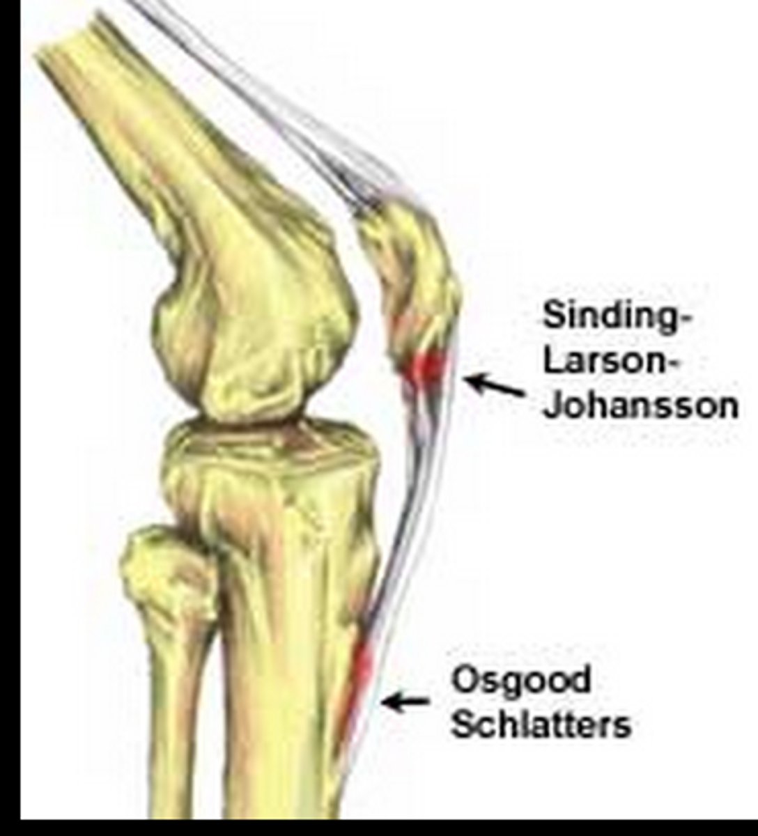
osgood schlatter disease
age 8-13 years in female
age 10-15 in male
onset- sudden
traction apophysistis of tibial tubercle
visible lump over the site
pain with activities such as running, jumping, kneeling, stairs)
knee extension resistance or stressing quads increased symptoms
anterior knee pain
quadriceps muscle tear
age 20-40
moi: sudden eccentric overload
squatting increases symptoms
bruising and/or swelling can occur over anterior thigh/knee
knee flexion is limited
combined hip ext and hip flexion painful
tenderness over anterior thigh
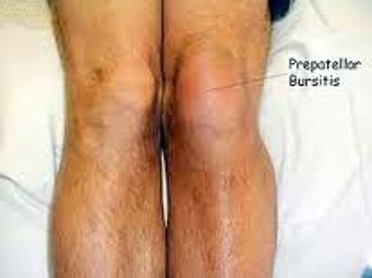
prepatellar bursitis
age 15-50
inflammation of bursa due to recurrent micro trauma of anterior knee
MOI: direct trauma to the anterior aspect of the knee
pain increases with kneeling
local swelling, fluctuation
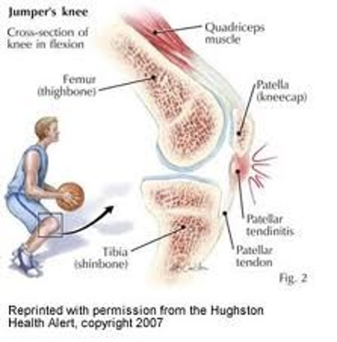
patellar tendinopathy (jumpers knee)
- Age 15-30
- Inflammation of patellar tendon
- Onset- gradual
- MOI- gradual (repeated eccentric overloading during deceleration activities)
pain increases with squatting and jumping
end range knee flexion painful
tender patellar tendon (inferior or superior to patella)
test: patellar grind test, single leg hip test, high step up test
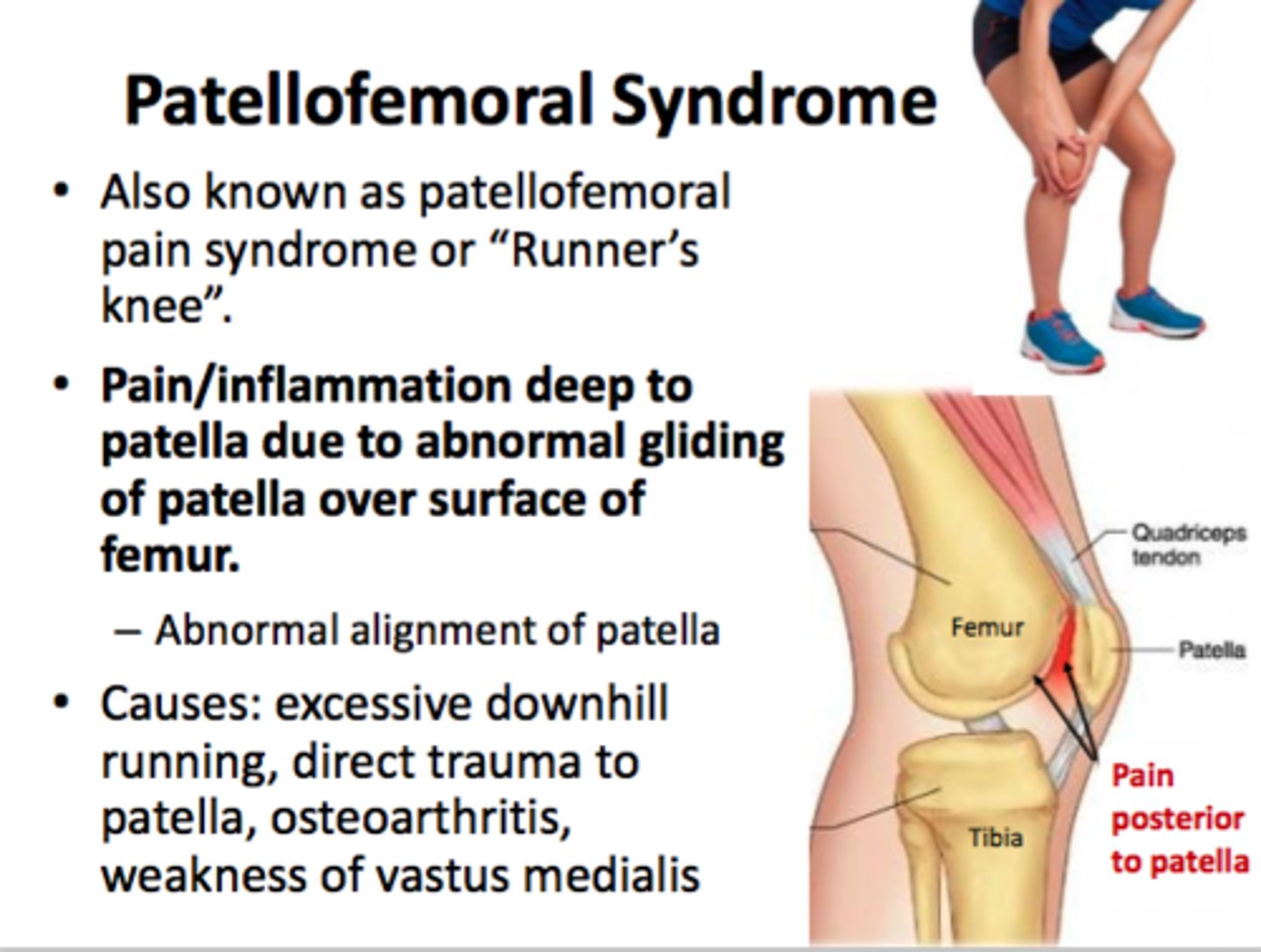
patellofemoral syndrome (runner's knee)
age: 20-50
moi: trauma, tight ITB, gmax, and TFL
worsen symptoms after prolonged sitting (moviegoers sign), stairs, and kneeling
end range knee flexion painful
tenderness on anterior knee especially with patella compression
anterior knee swelling
test: clarke test
keinbocks disease
lunate fracture; blood supply to one of the small bones of the wrist is interrupted
worsened pain with wrist flexion and extension
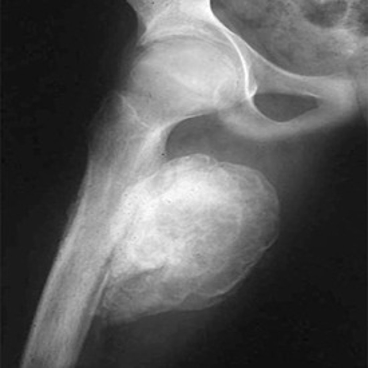
myositis ossificans
-common in brachialis muscle as result of trauma, aggressive stretching
-avoid stretching, massage, resistive exercises, Heat
brachialis strain
-pain on anterior aspect of distal part of the arm, possible tenderness in muscle belly
-painful resisted elbow flexion with forearm pronation
bicep tendon rupture
-Swelling
-Ecchymosis
-palpable gap in biceps tendon
-Weak elbow flexion and supination
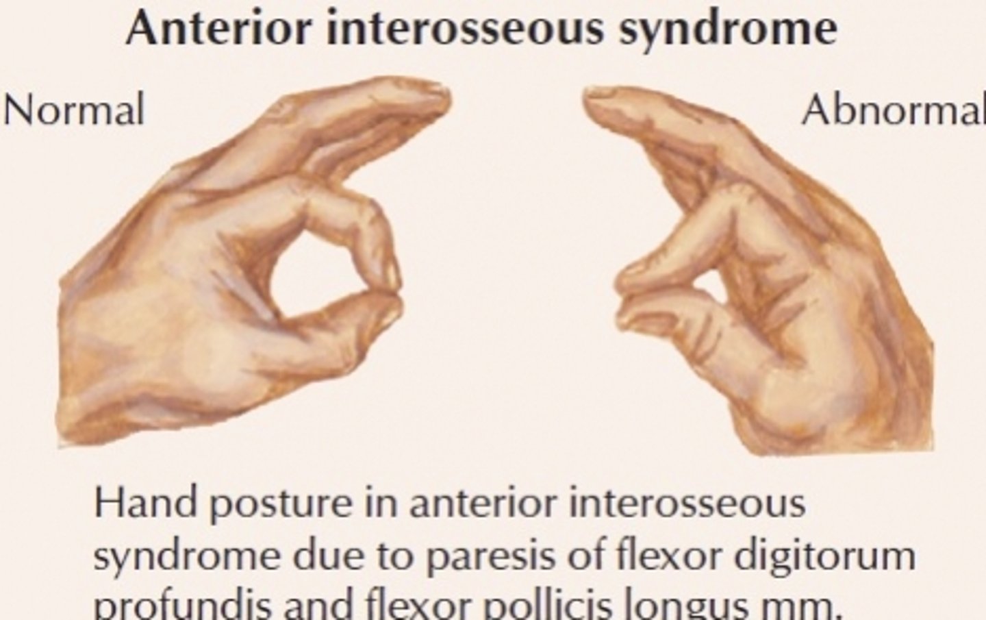
anterior interosseous nerve syndrome
weakness of flexor pollicus longus and flexor digitorum profundus to index finger
weakness of pronator quaradus
median nerve branch
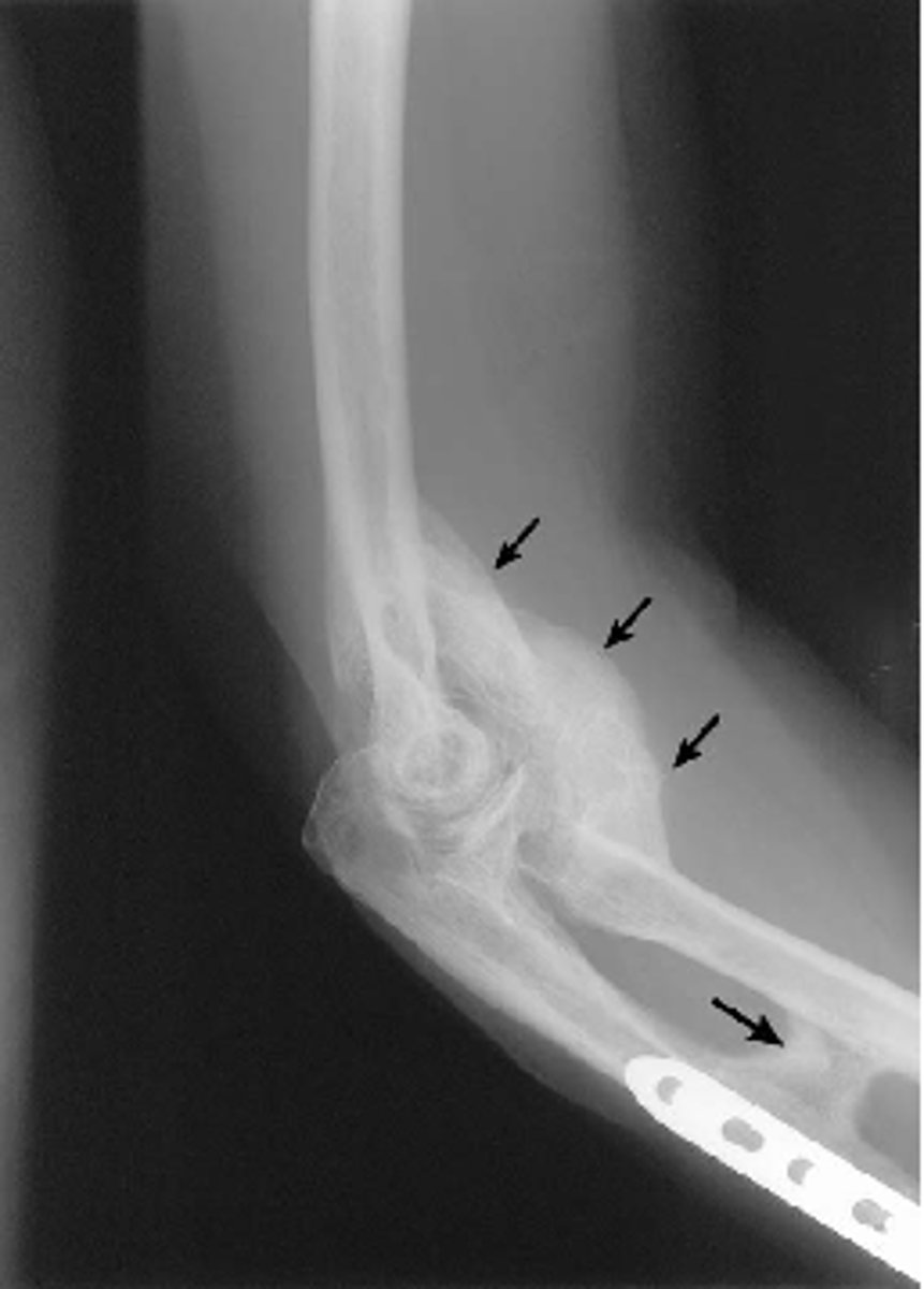
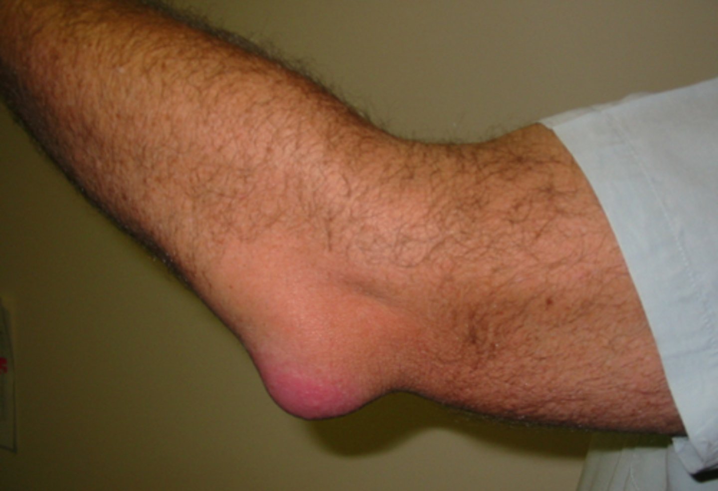
olecranon bursitis
-student's elbow, elbow bursitis
-swelling over posterior elbow
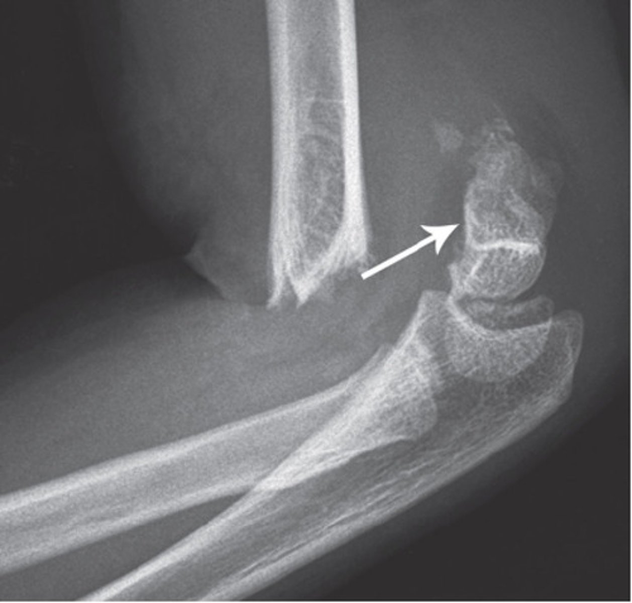
pulled elbow syndrome / nursemaid's elbow
-Age 2-3 years
-Longitudinal traction on an extended elbow --> partial slippage of Annular ligament over the head of radius and radio capitellar jt
-Position of arm - Arm at side, with hand pronated (palm down)
-Radial nerve can get injured
supracondylar fracture
distal humeral fracture
common in kids
typically AIN (branch of median nerve) and brachial artery involvement
complications: volkmanns ischemic contracture, gun stock deformity (reduced carrying angle), high incidence of malunion
requires ORIF
medial epicondylitis
Degenerative condition of pronator teres and flexor carpiradialis at medial epi
overuse in sports or jobs that require strong grip and excessive pronation
pain at passive wrist extension and active wrist flexion
ulnar nerve
lateral epicondylitis
chronic degenerative condition of ECRB at its proximal attachment to the lateral epicondyle of humerus
- repetitive wrist extension or strong grip with wrist extended overloading of ECRB
- painful passive wrist flexion and active wrist extension
SICK scapula
Age 20-40 yrs. Scapular malposition, inferior medial border prominence, coracoid pain and malposition, kinesis abnormalities of scapular movement.
test: scapular assistance test, scapular reposition test
acromioclavicular joint sprain
fall on tip of shoulder or outstretched arm
- step deformity (3rd degree) at point of shoulder
- painful palpation
- horizontal adduction and elevation painful
subacromial bursitis
repetitive overhead activity
painful arc 60-120 of abduction
active abduction and MR is limited
passive MR with 90 degrees shoulder abduction is painful
test: hawkins kennedy test, neer
bicep tendinitis
age 20-45
repetitive overhead activity
longhead gets impinged b/w bicipital groove and anterior acromion
pain with overhead movements
pain with full extension to flexion
resisted elbow flexion painful
test: speed, yergason
rotator cuff tendinitis
Inflammation of RC tendons due to impingement by acromion
painful overhead movements and resisted abduction
limited active abduction
piriformis syndrome
posterior hip and sacral pain
increased pain with sitting, squatting, or ER of hip, radiates to posterior thigh
limited hip IR
hallux rigidus
pain with big toe extension and gait
chronic, osteoarthritis
foot pain due to compensating to decrease toe ext
fused or partially fused first MP joint
toe off phase altered by supination so pressure is on lateral toes
excessive wear on lateral toes
hallux valgus
may be asymptomatic
around first MCP
chronic
secondary lesions: bunions, bursitis, corns, calluses
lateral deviation of big toe
excessive pronation and compensation strategies
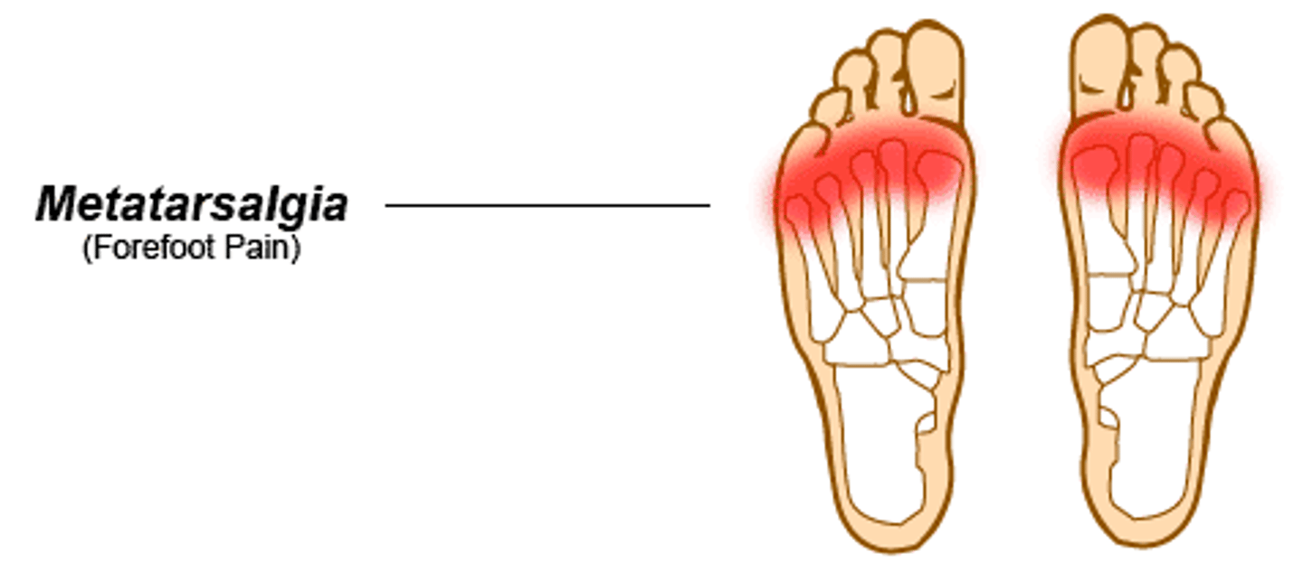
metatarsalgia
soreness of middle metatarsals
abnormal mechanical stress, arthritis
plantar foot pain and fatigue with WB
callus over mid metatarsals
hypermobile or pronated foot
antalgic gait, prontated
special test: metatarsalgia
open / resting position of glenohumeral joint
40-55 degrees abduction
30 degrees horizontal adduction
*scapular plane
close-packed position of glenohumeral joint
maximum abduction and external rotation
open / resting position of the humeroulnar joint
70 degrees flexion
10 degrees supination
close-packed position of the humeroulnar joint
full extension and supination
open / resting position of sternoclavicular joint
arm resting by side
open / resting position of acromioclavicular joint
arm resting by side
close-pack position of sternoclavicular joint
maximum shoulder elevation
close-pack position of acromioclavicular joint
arm abducted to 90 degrees
what is the open / resting position of vertebrae?
midway b/w flexion and extension
what is the close-pack position of vertebrae?
maximal extension
what is open / resting position of the hip?
30 degrees flexion
30 degrees abduction
slight ER
what is the close-pack position of the hip?
full extension, abduction, IR
what is the open / resting position of the knee?
25 degrees flexion
what is the close-pack position of the knee?
full extension and ER
what is the open / resting position of the talocrural joint?
mid inversion / eversion
10 degrees pf
what is the close-pack position of the talocrural joint?
full dorsilexion
what is the open / resting position of the subtalar joint?
midway b/w extreme ranges of position
what is the close-pack position of the subtalar joint?
full inversion
what is the open / resting position of the midtarsal and tarsometatarsal joint?
midway b/w extreme ranges of position
what is the close-pack position of the midtarsal and tarsometatarsal joint?
full supination
what is the open pack / resting position of the proximal radioulnar joint?
70 degrees flexion
35 degrees supination
*distal radioulnar is 10 degrees supination
what is the close-pack position of the proximal radioulnar joint?
5 degrees supination
*same for distal radioulnar
what is the open / resting position of the humeroradial joint?
full extension and supination
what is the close-pack position of the humeroradial joint?
90 degrees flexion
5 degrees supination
what is the open / resting position of the midcarpal joint?
neutral or slight flexion with UD
what is the close pack position of the midcarpal joint?
full extension with UD
glenohumeral arthrokinematics
convex on concave
Abd: superior roll, inferior glide
Flex: anterior roll, posterior glide
glenohumeral abduction arthokinematics
superior roll
inferior glide
glenohumeral flexion arthrokinematics
anterior roll
posterior glide