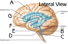
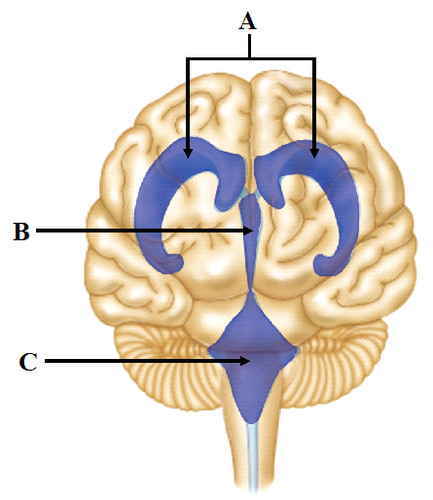
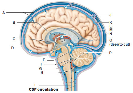
lateral ventricle
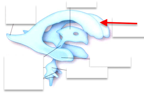
red arrow
interventricular foramen
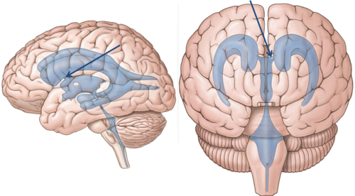
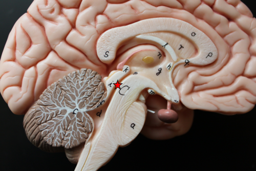
third ventricle sagittal view
F in this picture
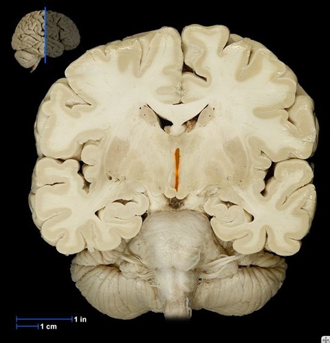
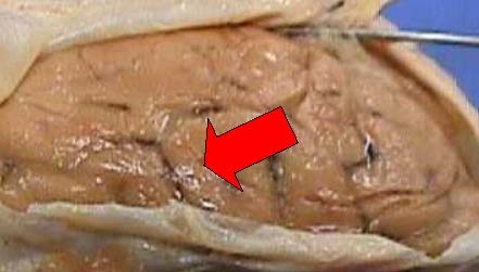
third ventricle frontal section (cadaver)
third ventricle frontal section (diagram)
B in this picture
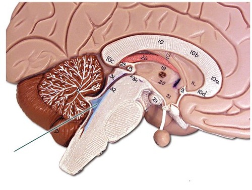
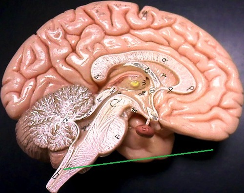
cerebral aqueduct on brain model
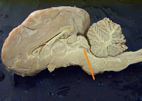
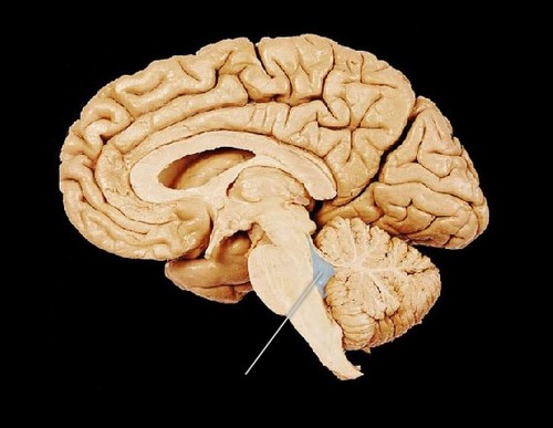
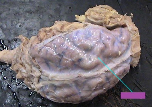
cerebral aqueduct on sheep brain
cerebral aqueduct (sagittal view diagram)
fourth ventricle on brain model
fourth ventricle on cadaver - sagittal view
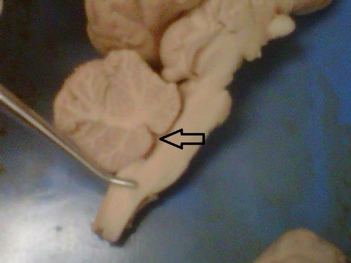
fourth ventricle on sheep brain
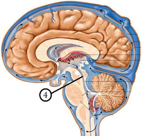
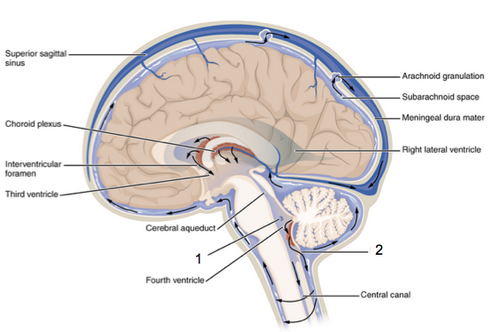
fourth ventricle - diagram, sagittal view
fourth ventricle - frontal view, diagram
lateral apertures and median apertures
openings in the 4th ventricle that allow CSF to exit the ventricles and enter the subarachnoid space to circulate up and around the brain
subarachnoid space
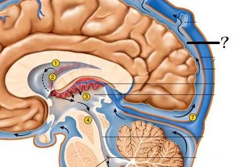
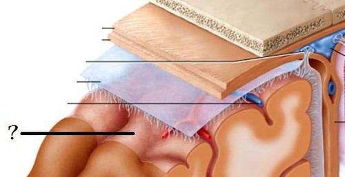
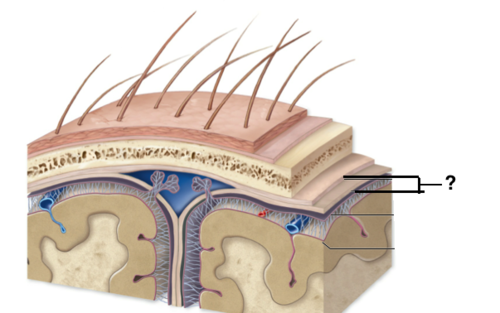
big ? mark
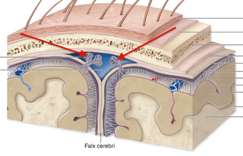
arachnoid villi - frontal view
arachnoid villi - sagittal view
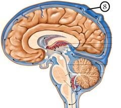
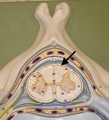
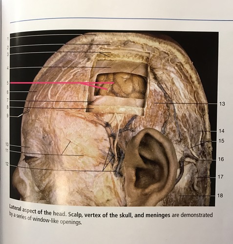
#8 on this picture
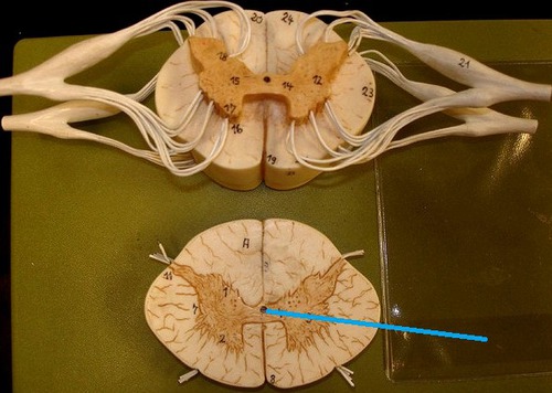
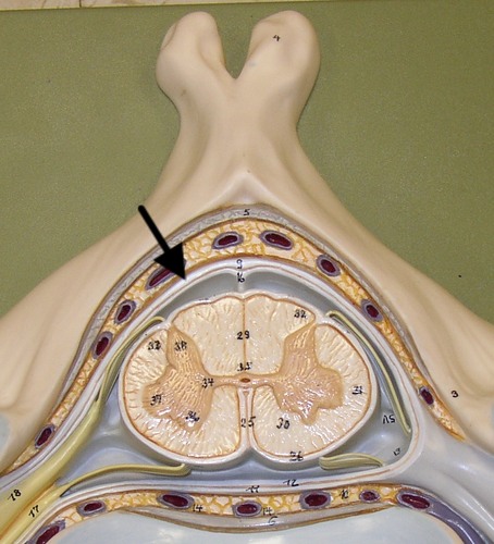
central canal of spinal cord - on model
central canal - sagittal view on brain model
central canal of spinal cord
"I" in this diagram
capillary beds that form cerebrospinal fluid
"B" and "P" in this diagram
Starting point of CSF circulation
subarachnoid space - sagittal view diagram
#2, #3, and "K" in this picture are all this area
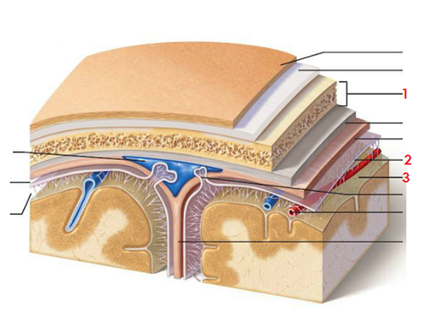
pia mater on brain - diagram
? mark
pia mater on spinal cord - model
pia mater - on cat brain
pia mater - diagram, longitudinal view
#3
pia mater - sagittal view, cadaver
arachnoid mater - diagram, longitudinal view
#4
arachnoid mater - diagram of skull & brain
#2
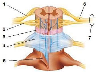
dura mater - spinal cord model
dura mater - sheep brain
dura mater - longitudinal view, diagram
#5
dura mater - cross section, diagram