Week 9+10 - Anatomy Physiology Hearing
1/35
Earn XP
Description and Tags
FINAL EXAM https://create.kahoot.it/share/anatomy-and-physiology-of-hearing/3e1f92e2-e6e4-4d76-aca0-69e3ff640016
Name | Mastery | Learn | Test | Matching | Spaced | Call with Kai |
|---|
No analytics yet
Send a link to your students to track their progress
36 Terms
The hearing organ (“ear”) is composed of series of what structures:
The external ear
The middle ear
The internal ear
The neural impulses registered by the sensory cells are then analyzed by the brain– The external earzed by the brain
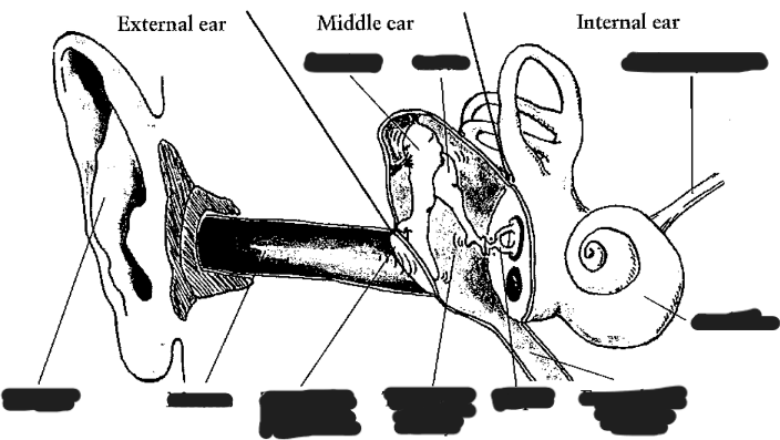
**Label the ear structure**
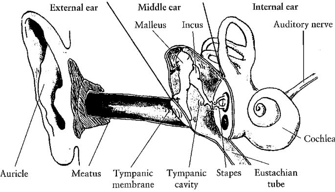
The external ear consists of:
Everyday use of the word “ear” means technically the external ear
External ear consists of:
Auricle (pinna)
Meatus (ear canal)
Tympanic membrane (ear drum)
Task of the auricle: localize the sound source (important: evolution)
Tympanic membrane works like a loudspeaker or microphone membrane
Whats the external auditory meatus
External ear canal
7 mm in diameter and 2.5 cm long
this generates resonance frequencies at 3400 Hz.
From an acoustic point of view: ear canal is a filter that amplifies frequencies between 2kHz and 5 kHz.
Terminates at the tympanic membrane
Two-thirds of ear canal housed in bone (osseous portion)
One-third of ear canal composed of cartilaginous parts
Resonating cavity that contributes to hearing
Determine resonant frequency
Outer third- line with hair cells and cerum (ear wax)-protects by trapping dirt and insects
Whats the tympanic membrane?
Also known as the eardrum
Separates the middle ear from the outer ear
Oval shaped, 10 mm in diameter
Thin three layered sheets of tissue
Landmarks:
Umbo - point of attachment for malleus, middle ear bone- location is cone of light (reflects light from otoscope)
Responsible for initiating mechanical
impedance-matching process of middle ear
First layer: outer (cuticular) layer
Second layer: Intermittent (fibrous) layer
Third layer: inner (mucous) layer
The middle ear consist of:
The middle ear consists of the tympanic cavity
This cavity contains the smallest moving bones of the human body – the ossicles:
Malleus (hammer): touches the tympanic membrane and transmits to
the Incus (anvil) which transmits to the
Stapes (stirrup) which transmits to internal ear (oval window)
Malleus and stapes are attached to muscles (may attenuate to transmission of sound by these bones)
The stapes connect directly to the internal ear through the oval window → transmission of stapes movement to the lymphatic fluid inside the internal ear
What is the malleus?
Largest of the ossicles
Nine mm long and weighs only 25 mg
Provides point of attachment with tympanic membrane
Bulk of bone is the head or caput

What is the incus?
Shaped like an anvil
Weighs 30 mg and is around 7 mm long
Provides intermediate link of ossicular chain
Incus and malleus articulate by means of a saddle join
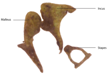
What is the stapes (stirrup)?
Third bone of ossicular chain
Weighs 4 mg with an area of 3.5 mm 2
Helps to transmit sound vibrations from eardrum to oval window
Articulation of the incus and stapes of ball and socket type
Ossicular chain is held in place by ligaments

What are the Tympanic Muscles? (2)
Muscles of middle ear attached to ossicles
Smallest muscles of human body
Stapedius muscle
Imbedded in posterior wall of middle ear
Pulls stapes posteriorly
Tensor tympani
Pulls malleus anterior and medial
What’s the sound attenuation in the middle ear?
The middle ear performs a kind of “volume control” → the muscles of malleus can be tensed, resulting in a low frequency damping
It MUST be activated though by neural impulses to activate these muscles → in order to be activated as noise control, the noise has to be processed by the internal ear → damage could have been occurred already
Additionally, these muscles are activated just before a person starts to speak → damping mechanism to protect against own voice
How does pressure increase in the middle ear form?
Sound waves are mechanically transmitted by ossicles of the middle ear to the internal ear, which is filled with watery liquid
Ossicles perform conversion of pressure changes from an elastic medium (air) to pressure changes of an incompressible liquid (water)
Ossicles function like a cone: from large surface (tympanic membrane) to smaller surface (stapes)
This leads to a pressure increase -> pressure variations at the internal ear are about 20 times stronger than original air pressure variation
This pressure increase is necessary to generate the necessary activation of the liquid (otherwise reflection would occur)
How does pressure equalization in the tympanic cavity form?
The middle ear is not completely airtight, a connection with the Eustachian tube allows for pressure equalization (e.g. meteorological pressure changes)
The Eustachian tube leads from the middle ear to the nasopharynx
Without pressure equalization, the meteorological changes would “push” the ear membrane inwards → feeling of “pressure on the ear” (felt e.g. when going downhill, or in airplane, or fast elevator)
The internal ear consists of:
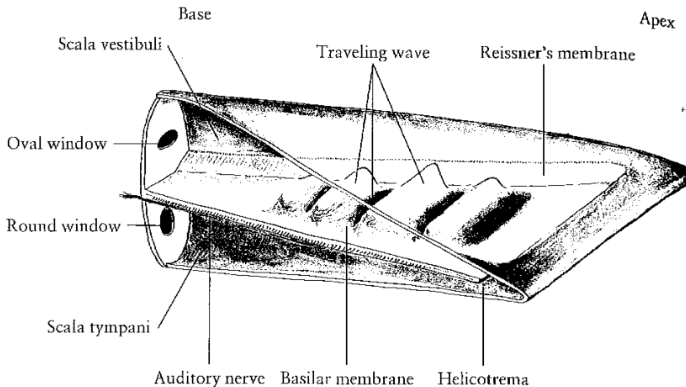
Cochlea: part of the inner ear relevant for hearing-> sound waves are transformed into neural impulses
is shaped like a snail shell
contains two passages, separated by basilar membrane:
Upper: scala vestibul
Lower: scala tympani
These two passages meet at the apex (the tip) in the helicotrema
Scala vestibuli connects to middle ear (stapes) through the oval window
What’s the physiology of hearing
The pressure waves from the middle ear (stapes) reach the cochlea through the oval window -> longitudinal pressure waves are generated in internal ear fluid through scala vestibuli to the apex
These pressure waves return via scala tympani to the round window
Round window serves as pressure release, since the fluids are incompressible
The basilar membrane: How does frequencies occur on different locations?
The travelling (longitudinal) wave meets differing conditions along the basilar membrane:
Cochlea is thinner (i.e. fluid is less deep) near the apex
Basilar membrane is softer near the apex
These two conditions are responsible for a mechanical frequency transformation:
High frequencies reach high displacement (of the transversal wave) near the base (just behind the stapes and oval window)
Low frequencies reach high displacement (of the transversal wave) near the end of the basilar membrane (apex)
What’s the structure of the Basilar membrane? pt1
The basilar membrane contains the organ of Corti, which is linked to the auditory nerve.
The organ of Corti consists of the tectorial membrane
Unlike the basilar membrane, the tectorial membrane does not cover the whole width of the cochlea, but only partly overlaps the basilar membrane the hair cells a number of additional supporting cells
Explain the edges of the tectorial membrane? pt2
One edge of the tectorial membrane is attached to the basilar membrane, close to the point of attachment of Rossner’s membrane
The other edge is supported by about 20000 outer hair cells, which are coded into three rows along the basilar membrane
These outer hair cells rest upon the basilar membrane at their lower end, with, at their upper end, the stereocilia, reach into the tectorial membrane
What are outer hair cells?
Outer hair cells add another mechanical source of frequency differentiation: Stiffer, shorter hair cells at the base are more responsible to high frequencies, while the long, more flexible hair cells at the apex bend more easily to low frequencies
The outer hair cells have only few neural pathways to the brain (and these are slow) → it is assumed that they do not transmit any detailed information about the sound signal to the brain
The actual transmission of information to the brain is done by the inner hair cells
Outer hair cells can be controlled by the brain via a feedback loop (see explanation oto-acoustic emissions), it is assumed that outer hair cells help to control the movement of the basilar membrane, e.g. for damping or frequency amplification purposes
What are inner hair cells?
Around 3500 inner hair cells are responsible for the conversion of the mechanical movement of the basilar membrane into neural impulses
The inner hair cells are grouped in a single row inside the organ of Corti
About 30000 nerve fibers lead from the inner hair cells into the brain → each inner hair cell is linked to about 10 nerve cells
The inner hair cell consist of a main cell body with thin stereocilia on top
The upward movement of the basilar membrane causes the inner hairs cells to fire when touching the tectorial membrane
Where is frequency information encoded along the basilar membrane?
in the tonotopic location along the basilar membrane:
displacements at the base correspond to high frequencies,
displacements at the apex correspond to low frequencies
In other words, different areas of the basilar membrane reach their maximum displacement for different frequencies → causing the nerve cells to fire at different frequencies
What is Auditory Mechanism
Processes the acoustic signals of speech
Has an amazing range of sound pressures
Has a frequency range of around 10 octaves spanning 20 to 20,000 Hz
An octave is a doubling in frequency
Organize the Physiological Principles of the Auditory System
Outer ear: Collects sound; “shapes” frequency components
Middle ear: Matches airborne acoustic signal with fluid medium of cochlea
Inner ear: Performs temporal and spectral analysis on ongoing acoustic signal
Auditory pathway: Conveys and further processes the signal
Cerebral Cortex: Interprets the signal
What’s the function of the Outer Ear?
“Collector” of sounds
Shapes frequency components of sound
Enhances certain midrange frequencies
Pinna of outer ear
Funnels acoustical information to the external auditory meatus; localizes sounds
External auditory meatus
Funnels sound to the tympanic membrane
Acts as a resonator to amplify frequencies between 2kHz and 4kHz
What’s the function of the Middle Ear?
Increases the pressure arriving at the cochlea
Cochlea is a fluid-filled cavity
Acts to overcome impedance
Impedance is resistance to the flow of energy
Designed as an impedance-matching device
Increases pressure of signal arriving at cochlea
Primary function:
Match the impedance of two conductive systems
The outer ear and the cochlea
Three impedance-matching mechanisms
Area ratio provides a 25 dB gain
From tympanic membrane to small oval window
Lever advantage provides a 2 dB gain
Ossicular chain
Buckling effect of tympanic membrane provides a 4-6 dB gain
These three impedence matching devices combined cause a signal gain of around 31 dB.
Disorders that impede middle ear function reduce audition
Otitis media, otosclerosis, glomus tumors
What’s the function of the Inner Ear?
Performs frequency and temporal acoustic analysis of incoming acoustical signal
Inner Ear Function: The cochlea
Establishes first level of auditory processing of incoming acoustic signals
Determines frequency components of signal
Determines amplitude of signal
Identifies temporal aspects of signal
Inner Ear Function: The traveling wave (4)
Wave-like action of basilar membrane
• Determines frequency data going to brain
• Arises from stimulation of perilymph of vestibule
• Moves along basilar membrane until it reaches point of maximum growth
• Wave damps after reaching maximum growth
Inner Ear Function: Basilar membrane
Basilar membrane
Determination of ability to analyze frequency
Characteristics
Thicker, stiffer, and wider at base than at apex
Traveling wave always travels from base to apex due to impedance gradient of basilar membrane and then quickly damps
High frequency sounds are processed at the base whereas low frequency sounds are processed at apex-tonotopic representation
Inner Ear Function: Excitation of hair cells
Hair cells displaced as traveling wave moves along basilar membrane
Excitation of outer hair cells displaced relative to tectorial membrane
Result of shearing effect on cilia
Important for coding intensity in intensity less than 40 dB
Excitation of inner hair cells
Results from fluid flow and endolymph turbulence
Essential for coding frequency
What’s the Auditory loudness scale?
A normal acoustic scale weights all frequencies equally strong, i.e. assumes a linear frequency scale
Psychophysical experiments have shown that humans perceive very low and high frequencies not as well as frequencies in the range of 2-4kHz
The lower frequencies are damped by the middle ear
The higher frequencies are attenuated by the resonance characteristics of the ear canal (or rather the lack of resonances)
What’s the phon-scale (and also dBA- weighting)
Was developed to solve this problem:
0 phon are defined as having the same dBSPL value as the hearing threshold at 1kHz (sine tone)
Loudness at other frequencies is related to this perceived loudness at 1kHz → the hearing threshold at other frequencies
What are Audiograms?
Registration of patient’s hearing threshold over (selected) octave frequencies (i.e. doubling of frequencies)
Sine waves are (traditionally) used between 250 Hz and 8kHz
Determines hearing loss (deviation from “normal” threshold) for certain frequencies, the most common is hearing loss in high frequencies (with age) or mid frequencies (due to noise exposure)
What are Oto-acoustic emissions
It is generally assumed that the nerves first transmit the sound stimulus (the impulse projected into the ear) to the brain → the brain then tells the outer hair cells to react
As a result, the basilar membrane produces oto-acoustic emission → the change of shape of the outer hair cells results in a specific movement of the basilar membrane
Discovered by Kemp (1978) who projected short signal impulses into the ear → with some delay the ear produced an echo of the impulses
The echo occurred much later and much stronger (higher amplitude) than expected by the reflective loop of the ear (external ear → internal ear → external ear) → OAE’s are a reaction of the ear itself (including the brain)
What’s spontaneous OAE’s
Spontaneous OAE’s were discovered (i.e. no stimulus sound needs to be present) → the basilar membrane may move by itself, even without being stimulated by a sound
Spontaneous OAE’s are seen as a kind of “convulsion” of the outer hair cells → OAE’s provide evidence that the basilar membrane is actively controlled (and does not only passively react to external sound input)
Apparently the movements of the basilar membrane can be actively controlled by a feedback process from the brain