anatomy bone practical
5.0(1)
5.0(1)
Card Sorting
1/111
Study Analytics
Name | Mastery | Learn | Test | Matching | Spaced |
|---|
No study sessions yet.
112 Terms
1
New cards
supraorbital foramen
hole above eye socket
2
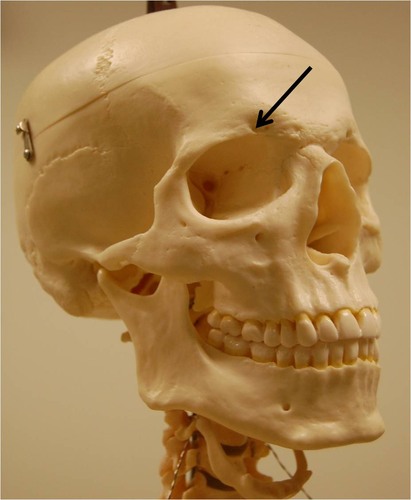
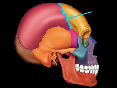
New cards
coronal suture
suture between frontal and parietal
3
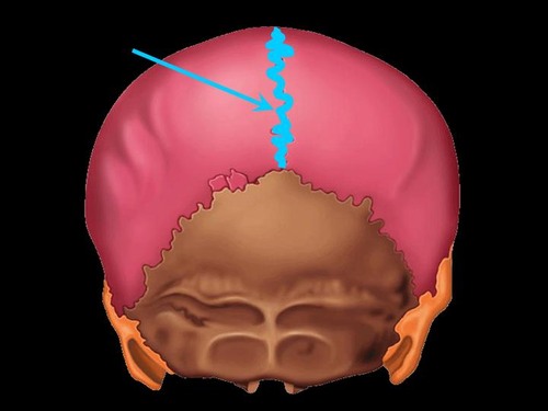
New cards
sagittal suture
suture between parietals
4
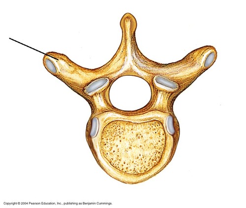
New cards
squamous suture
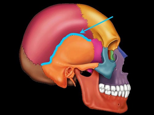
suture between parietals and temporal bones
5
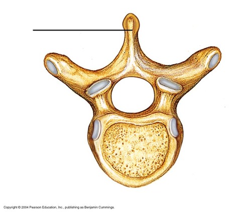
New cards
mandibular condyle
where mandible articulates
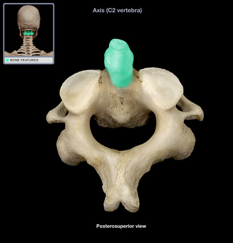
6
New cards
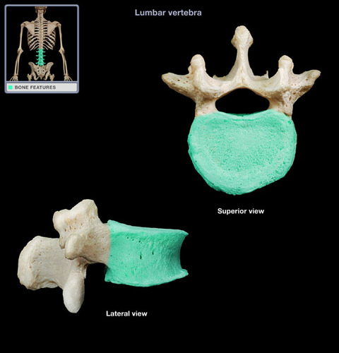
external acoustic meatus
external ear canal

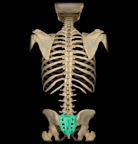
7
New cards
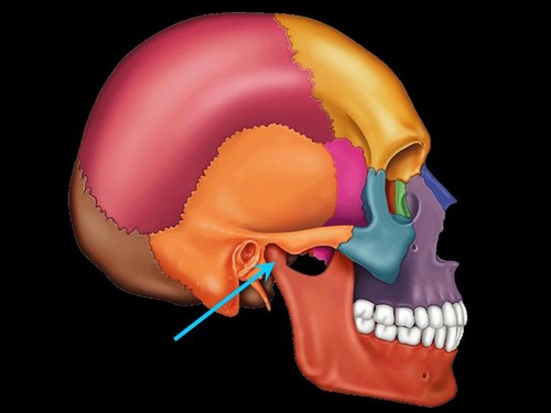
styloid process of temporal bone
long, slender process on temporal bone
8
New cards
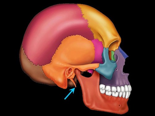
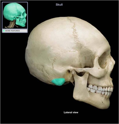
mastoid process
large process posterior and inferior to external acoustic meatus
9
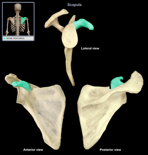
New cards

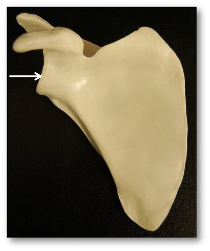
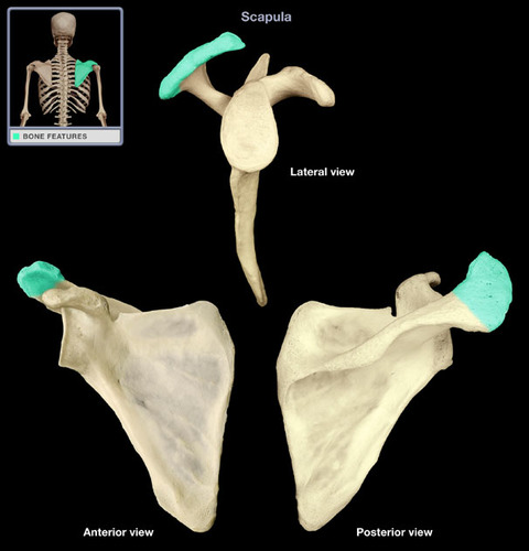
Ethmoid
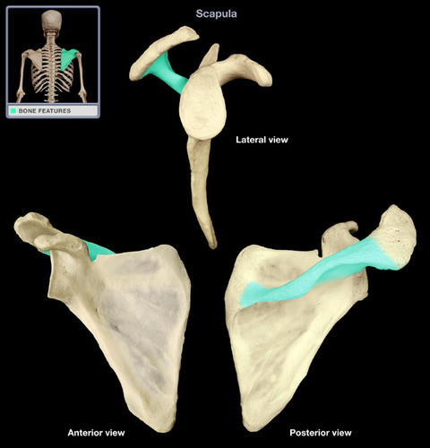
the part next to the highlighted section (until I get new picture)
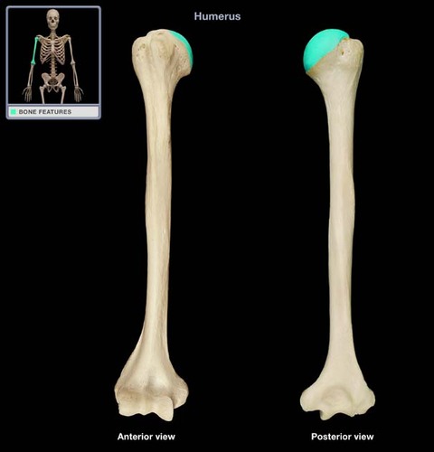
10
New cards
lambdoid suture
suture between occipital and parietal
11
New cards
foramen magnum
largest hole in the skull; spinal cord and brain integrate
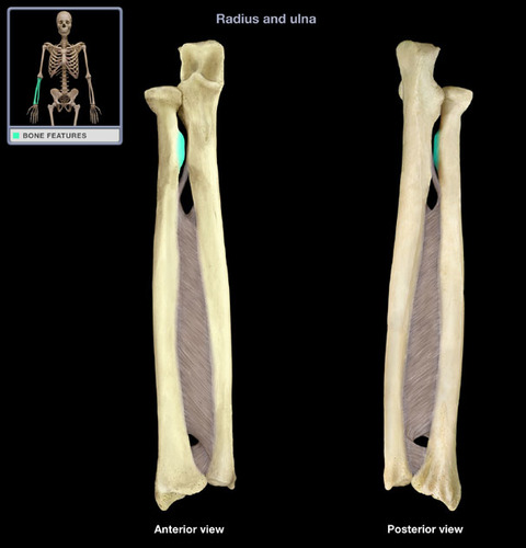
12
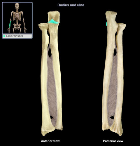
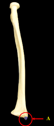
New cards
occipital condyles
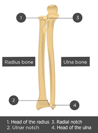
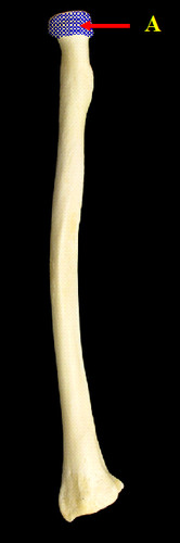
processes adjacent to foramen magnum; articulation for atlas
13
New cards
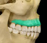
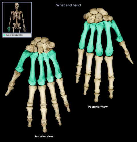
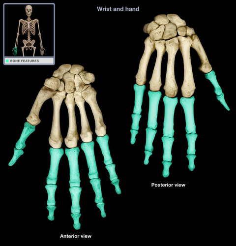
maxilla
above mouth below nose
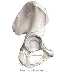
14
New cards
infraorbital foramen
holes below the eyes
15
New cards
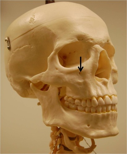
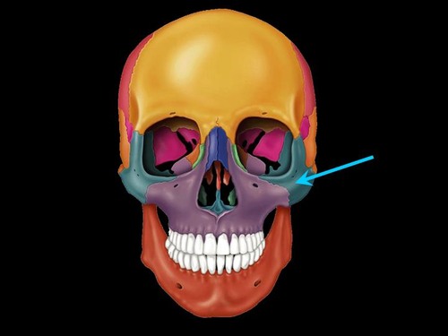
zygomatic bone
lateral side of eye socket and anterior portion of cheek bone
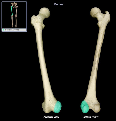
16
New cards
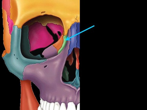
lacrimal bone
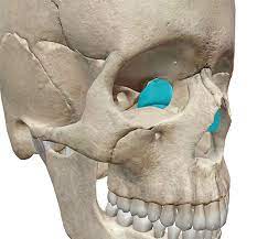
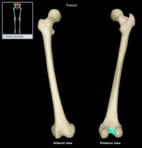
most medial bone located within eye socket
17
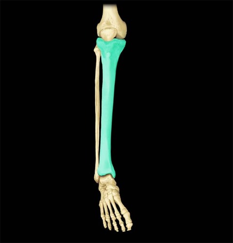
New cards
nasal bone
"bridge" of nose

18
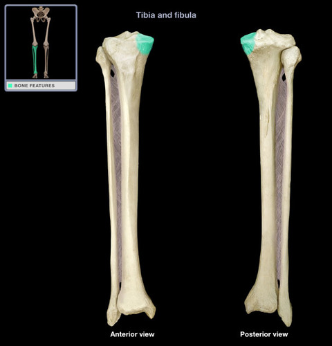
New cards
vomer
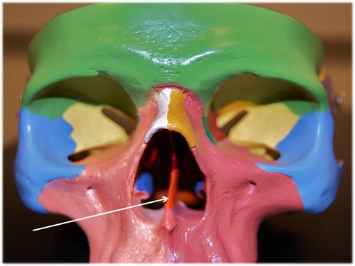
single bone that forms part of the nasal septum
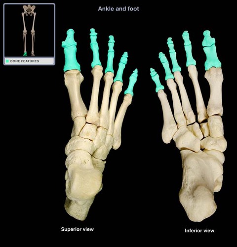
19
New cards
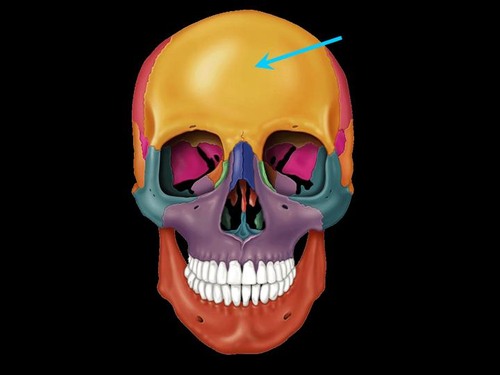
body of mandible
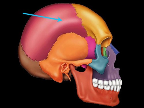
chin/jaw/below the teeth
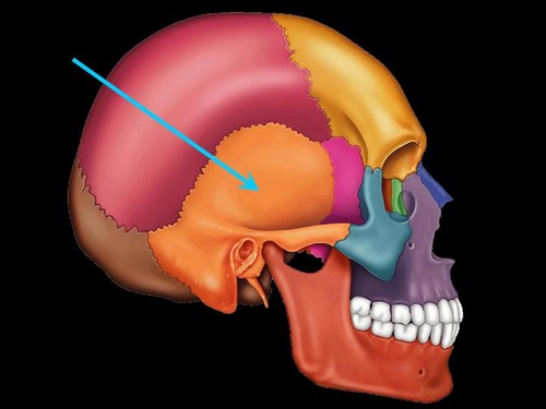
20
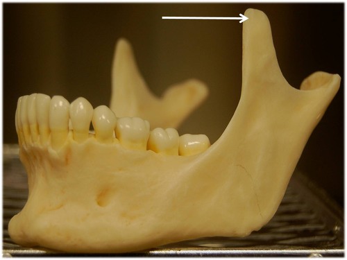
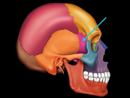
New cards
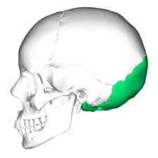
coronoid process of mandible
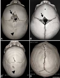
anterior process of mandible
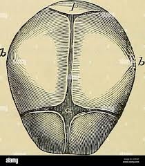
21
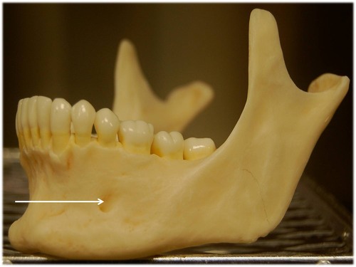
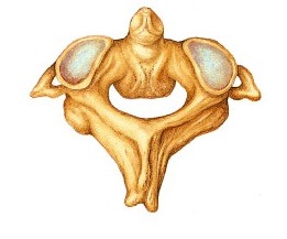
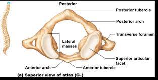
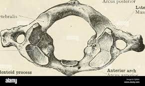
New cards
mental foramen
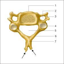
holes on both sided of chin
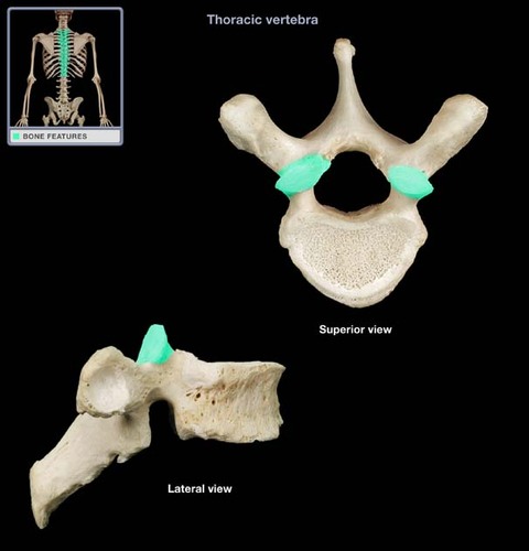
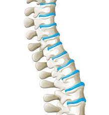
22
New cards
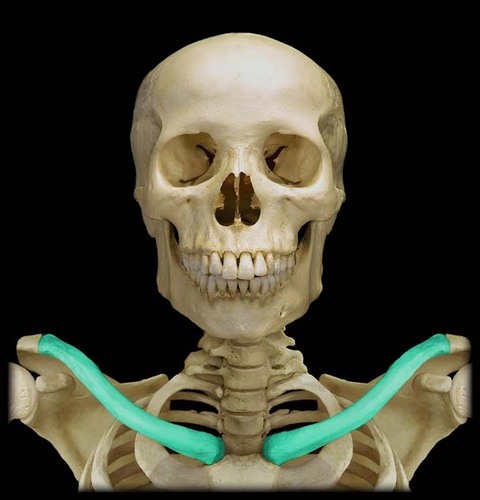
hyoid
floating bone found at junction of floor of mouth and neck
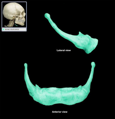
23
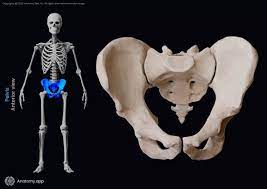
New cards
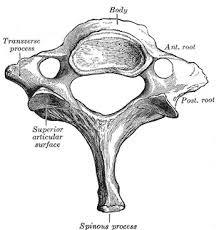
superior articular process
superior; articulates with vertebra above it
24
New cards
transverse process
lateral projections
25
New cards
spinous process
large projection perpendicular to transverse process
26
New cards
body of vertebra
large structure that supports weight of vertebral column
27
New cards
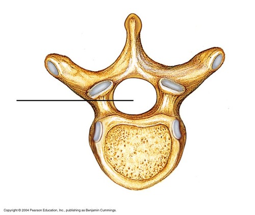
vertebral foramen
large hole for spinal cord
28
New cards
transverse foreman
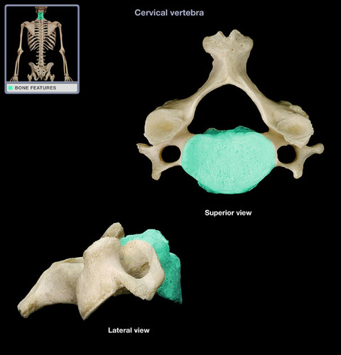
all cervical vertebrae have this
29
New cards
atlas
1st vertebra-no body
30
New cards
axis
2nd vertebra
31
New cards
dens
process that acts as pivot for atlas to allow rotation of the head
32
New cards
lumbar vertebrae
larger bodies with blunt, square spinous process
33
New cards
sacrum
5 fused vertebrae
34
New cards
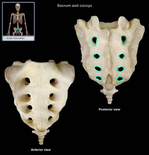
sacral foramen
holes down the sides of median sacral crest
35
New cards
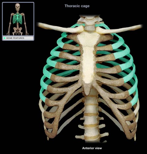
true ribs
1-7
36
New cards
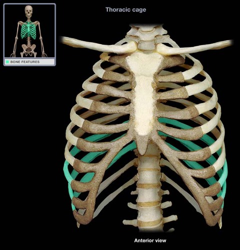
false ribs
8-10
37
New cards
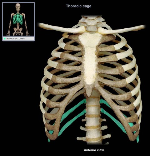
floating ribs
11 and 12
38
New cards
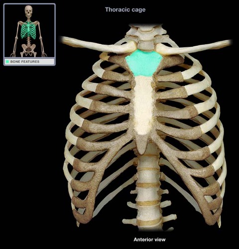
manubrium
superior portion of sternum
39
New cards
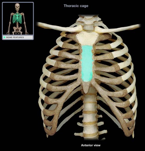
body of sternum
main portion; tie
40
New cards
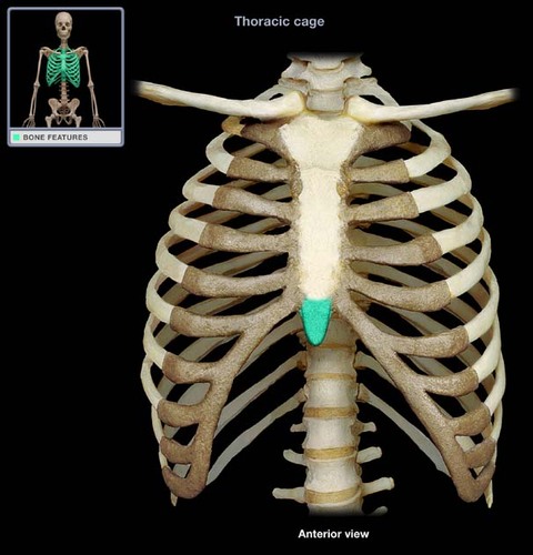
xiphoid process
posterior portion of sternum
41
New cards
clavicle
between scapula and sternum
42
New cards
glenoid cavity
lateral head of humerus fits here
43
New cards
coracoid process
anterior, above glenoid
44
New cards
acromion process
posterior, continuous with scapular spine
45
New cards
scapular spine
posterior
46
New cards
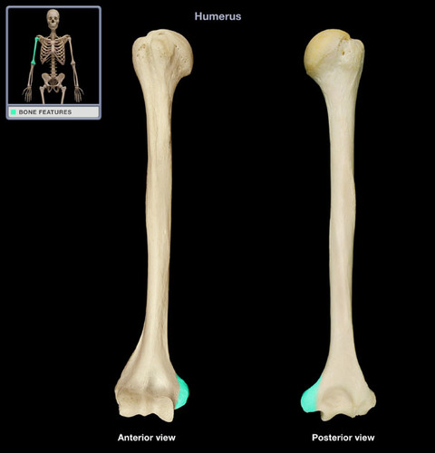
head of humerus
ball; large rounded part
47
New cards
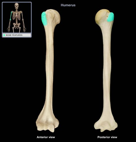
greater tubercle
larger rounded area on lateral side of head
48
New cards
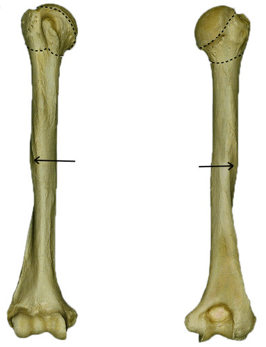
deltoid tuberosity
rough ridge going down humerus bone
49
New cards
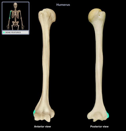
lateral epicondyle
on the humerus; distal end
50
New cards
medial epicondyle
distal end
51
New cards
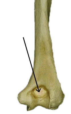
olecranon fossa
large depression posterior on distal end (humerous)
52
New cards
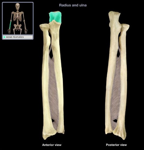
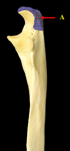
trochlear notch
large c-shaped notch at proximal end
53
New cards
olecranon process
top of trochlear notch
54
New cards
coronoid process of ulna
below trochlear notch
55
New cards
head of ulna
large articular end at distal end of the ulna - roughly at #4
56
New cards
styloid process of ulna
small process on distal end
57
New cards
head of radius
proximal, flat, rounded part
58
New cards
styloid process of radius
distal, sticks out laterally
59
New cards
radial tuberosity
large knob below head; proximal
60
New cards
metacarpals
1-5
61
New cards
phalanges of hands
1-5; proximal, middle, distal
62
New cards
acetabulum
socket for head of femur
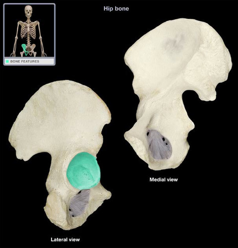
63
New cards
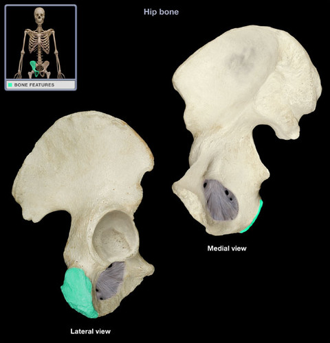
iliac crest
top ridge of bone
64
New cards
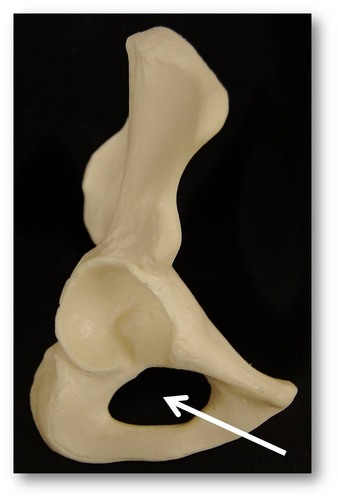
obturator foramen
large opening
65
New cards
ischial tuberosity
most posterior portion
66
New cards
symphysis pubis
67
New cards
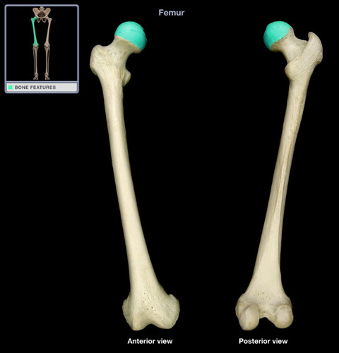
head of femur
large rounded end; proximal
68
New cards
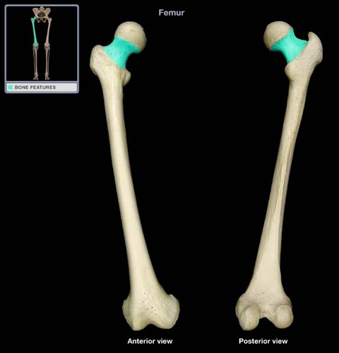
neck of femur
narrowing of bone lateral to the head
69
New cards
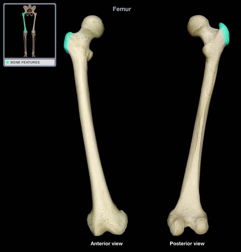
greater trochanter of femur
lateral to head and neck
70
New cards
lateral condyle
distal end of bone; greater trochanter side

71
New cards
medial condyle
distal end of bone; head side of bone
72
New cards
intercondylar fossa
notch between condyles
73
New cards
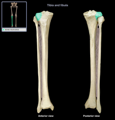
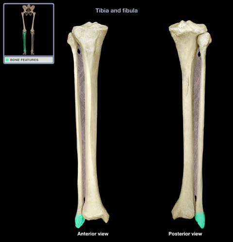
tibia
big toe side
74
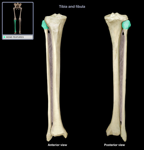
New cards
medial condyle
same side as medial malleolus; medial, flat part
75
New cards
lateral condyle
flat part
76
New cards
medial malleolus
most posterior that sticks out
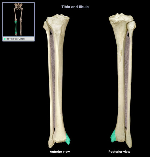
77
New cards
tibial tuberosity
anterior ridge at proximal end
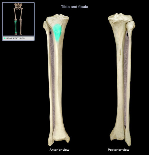
78
New cards
head of fibula
rounded side
79
New cards
lateral malleolus
distal end; point outside of ankle bone
80
New cards
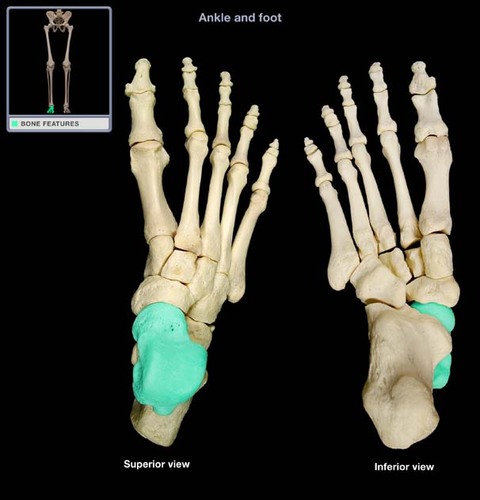
talus tarsal
sits on top of calcaneus
81
New cards
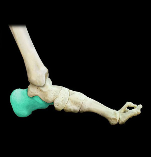
calcaneus
heel bone
82
New cards
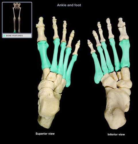
metatarsals
1-5
83
New cards
phalanges of the foot
1-5, proximal, middle, distal
84
New cards
frontal bone
forehead and eyebrow
85
New cards
parietal bones
sides of upper skull
86
New cards
temporal bone
lower sides of skull where ear is
87
New cards
sphenoid bone
bat shaped bone
88
New cards
Occipital Bone
bottom back of skull
89
New cards
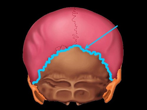
anterior fontanel
90
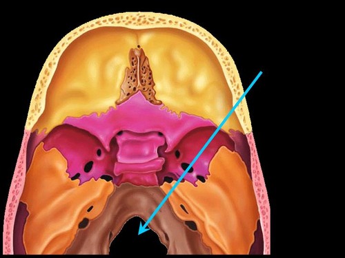
New cards
posterior fontanel
p - towards top of image
91
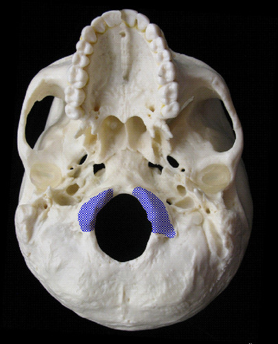
New cards
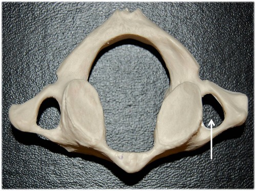
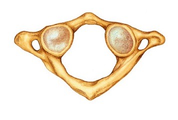
occipital facets
in atlas - two white ovals
92
New cards
den facet
oval indentation on the bottom
93
New cards
bifid spinous process
arrows at the bottom
94
New cards
thoracic vertebrae
8-19 vertebrae
95
New cards
cervical vertebrae
first seven vertebae
96
New cards
intervertebral discs
inbetween each vertebrae
97
New cards
sacral canal
vertical hollow passage through the sacrum
98
New cards
costal cartilage

99
New cards
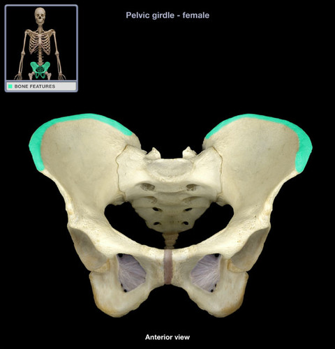
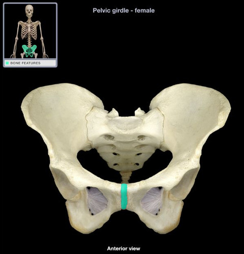
pelvic girdle
100
New cards
os coxa (ossa coxae pl.)
hip bone