Neural Communication II – Chemical synapses

0.0(0)
studied byStudied by 0 people
GameKnowt Play
learnLearn
examPractice Test
spaced repetitionSpaced Repetition
heart puzzleMatch
flashcardsFlashcards
Card Sorting

1/49

flashcard set

Earn XP

Description and Tags

Week 3

Study Analytics
Name
Mastery
Learn
Test
Matching
Spaced

No study sessions yet.

50 Terms

1
New cards

Outline the two types of transmission of information in the brain.

  1. Action Potential Transmission:

    • Action potentials are electrical signals that transmit information within cells.

  2. Chemical Transmission:

    • Occurs at synapses

    • It is the communication between cells mediated by neurotransmitters.

2
New cards

Outline how voltage gated calcium channels work in the synapse.

Process:

  • Open when the membrane depolarises to around -40 to -60 mV.

  • Calcium enters the cell

  • The calcium then allows for vesicle release at the synapse
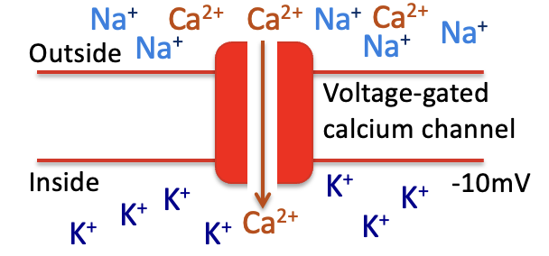
<p><strong>Process:</strong></p><ul><li><p><span>Open when the membrane depolarises to around -40 to -60 mV.</span></p></li><li><p><span>Calcium enters the cell</span></p></li><li><p><span>The calcium then allows for vesicle release at the synapse</span></p></li></ul><p></p>
3
New cards

Describe the process of neurotransmitter release.

  1. Action potential:

    • An action potential transmitted to the presynaptic terminal.

    • Depolarisation spreads over the presynaptic membrane.

  2. Calcium channels:

    • Depolarisation leads to the opening of voltage-gated calcium channels (VGCC) in the membrane

    • Calcium flows into the terminal down the concentration and electrical gradients.

  3. Release of neurotransmitters:

    • The increase in calcium concentration stimulates release of neurotransmitters into the presynaptic cleft

    • The neurotransmitters diffuses to the postsynaptic membrane and bind to receptors
<ol><li><p><strong>Action potential:</strong></p><ul><li><p>An action potential transmitted to the presynaptic terminal.</p></li><li><p>Depolarisation spreads over the presynaptic membrane.</p></li></ul></li><li><p><strong>Calcium channels:</strong></p><ul><li><p>Depolarisation leads to the opening of voltage-gated calcium channels (VGCC) in the membrane</p></li><li><p>Calcium flows into the terminal down the concentration and electrical gradients.</p></li></ul></li><li><p><strong>Release of neurotransmitters:</strong></p><ul><li><p>The increase in calcium concentration stimulates release of neurotransmitters into the presynaptic cleft</p></li><li><p>The neurotransmitters diffuses to the postsynaptic membrane and bind to receptors</p></li></ul></li></ol><p></p>
4
New cards

Outline the types and structure of SNARE proteins

  • SNARE Proteins Types:

    • v-SNAREs: Found on vesicles.

    • t-SNAREs: Found on the target membrane.

  • SNARE Peptide Structure:

    • Lipophilic region inside the membrane.

    • Long tail projecting into the cytosol.
<ul><li><p><strong>SNARE Proteins Types:</strong></p><ul><li><p><strong>v-SNAREs</strong>: Found on vesicles.</p></li><li><p><strong>t-SNAREs</strong>: Found on the target membrane.</p></li></ul></li><li><p><strong>SNARE Peptide Structure:</strong></p><ul><li><p>Lipophilic region inside the membrane.</p></li><li><p>Long tail projecting into the cytosol.</p></li></ul></li></ul><p></p>
5
New cards

State the function and mechanism of SNARE proteins.

  1. Vesicle Release:

    • Vesicles need SNARE proteins to dock and release.

  2. Docking Mechanism:

    • Tails bind to each other in the presence of calcium, allowing the vesicle to dock
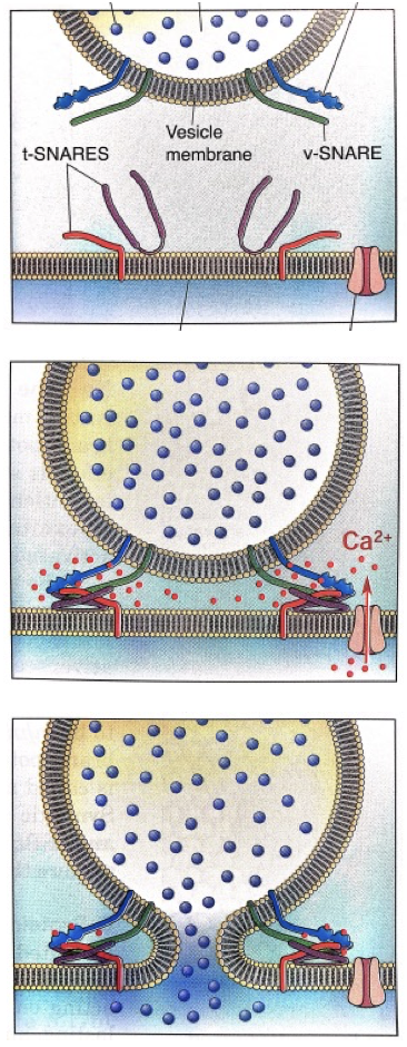
<ol><li><p><strong>Vesicle Release:</strong></p><ul><li><p>Vesicles need SNARE proteins to dock and release.</p></li></ul></li><li><p><strong>Docking Mechanism: </strong></p><ul><li><p>Tails bind to each other in the presence of calcium, allowing the vesicle to dock</p></li></ul></li></ol><p></p>
6
New cards

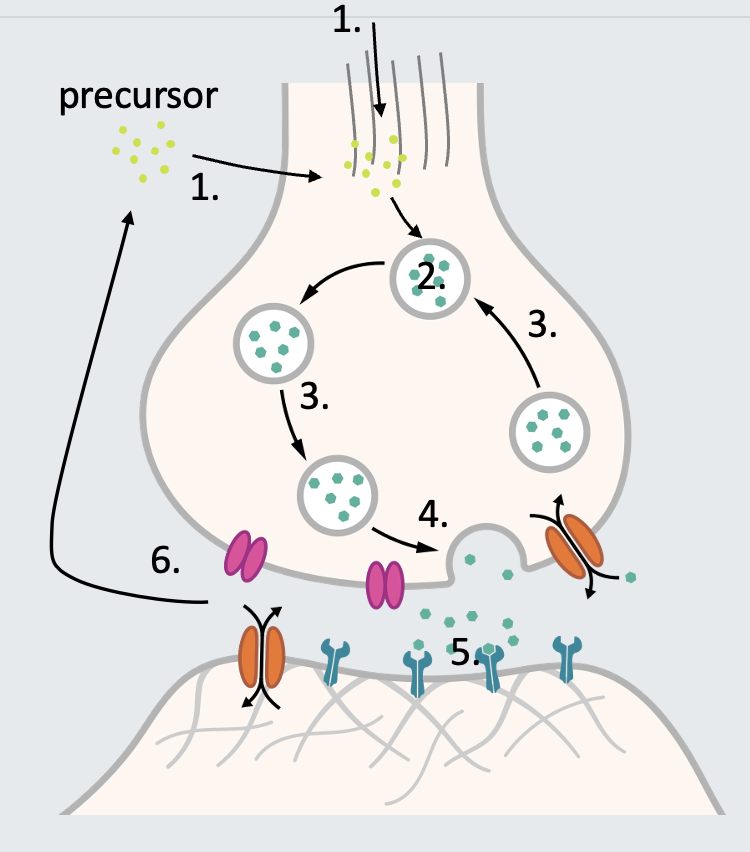
Outline the synthesis step in the lifecycle of neurotransmitters.

  • Synthesis:

    • Process: Neurotransmitters (NTs) are created from precursor molecules.

    • Locations:

      • Soma (Cell Body): Slow synthesis process.

      • Axon Terminal: Local synthesis.

      • External Sources: NTs can be obtained externally.

7
New cards

State the steps of the the neurotransmitter lifecycle.

  1. Synthesis

  2. Packaging and storage

  3. Translocation

  4. Release

  5. Receptor activation

  6. Inactivation

<ol><li><p>Synthesis</p></li><li><p>Packaging and storage</p></li><li><p>Translocation</p></li><li><p>Release</p></li><li><p>Receptor activation</p></li><li><p>Inactivation </p></li></ol><p></p>
8
New cards

Outline what is necessary for repetitive signalling and what cell is used to achieve this.

  • Immediate Clearance for Repetitive Signalling:

    • Neurotransmitters in the synaptic cleft need to be cleared immediately to allow for repetitive signalling between neurons.

  • Role of Astrocytes:

    • Astrocytes play a crucial role in neurotransmitter (NT) clearance.

    • Well-known for clearing glutamate using Excitatory Amino Acid Transporters (EAATs).
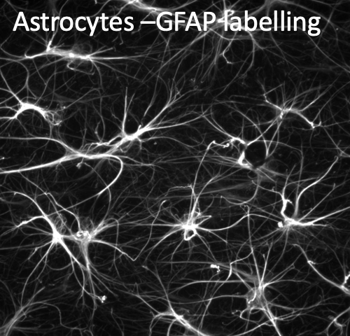
<ul><li><p><strong>Immediate Clearance for Repetitive Signalling:</strong></p><ul><li><p>Neurotransmitters in the synaptic cleft need to be cleared immediately to allow for repetitive signalling between neurons.</p></li></ul></li><li><p><strong>Role of Astrocytes:</strong></p><ul><li><p>Astrocytes play a crucial role in neurotransmitter (NT) clearance.</p></li><li><p>Well-known for clearing glutamate using <strong>Excitatory Amino Acid Transporters (EAATs)</strong>.</p></li></ul></li></ul><p></p>
9
New cards

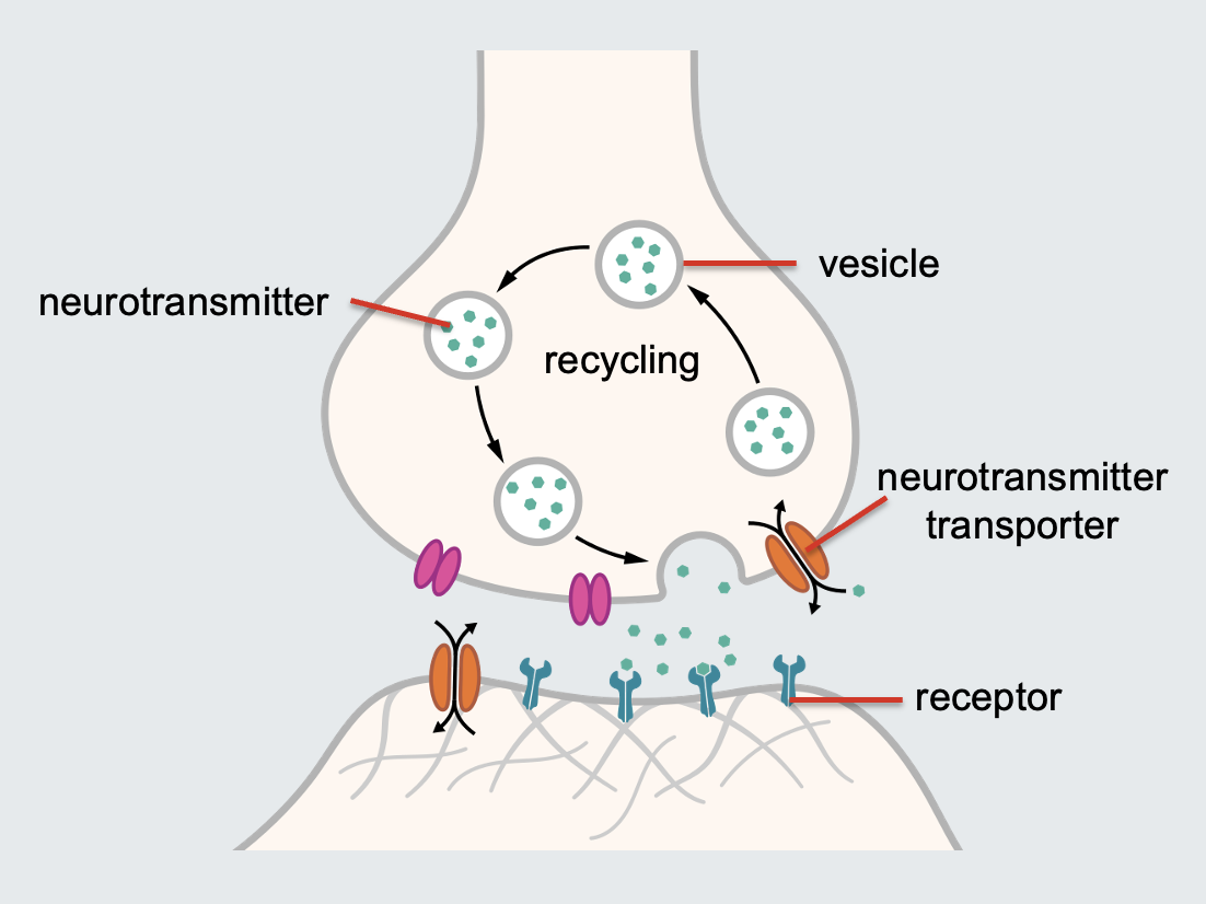
Describe the relationship between neurotransmitters and receptors.

  • Postsynaptic Density:

    • As neurotransmitters reach the postsynaptic membrane, they encounter a dense area of receptors located in the postsynaptic density.

  • Variety of Receptors:

    • The types of receptors present depend on various factors:

      1. Type of Neuron: Different neurons have different receptors.

      2. Signalling Mechanisms: Receptors vary based on the cell's signalling processes.

      3. Location: The location within the nervous system influences receptor types.

      4. Function: The specific function of the neuron also determines receptor variety.
<ul><li><p><strong>Postsynaptic Density:</strong></p><ul><li><p>As neurotransmitters reach the postsynaptic membrane, they encounter a dense area of receptors located in the postsynaptic density.</p></li></ul></li><li><p><strong>Variety of Receptors:</strong></p><ul><li><p>The types of receptors present depend on various factors:</p><ol><li><p><strong>Type of Neuron:</strong> Different neurons have different receptors.</p></li><li><p><strong>Signalling Mechanisms:</strong> Receptors vary based on the cell's signalling processes.</p></li><li><p><strong>Location:</strong> The location within the nervous system influences receptor types.</p></li><li><p><strong>Function:</strong> The specific function of the neuron also determines receptor variety.</p></li></ol></li></ul></li></ul><p></p>
10
New cards

Outline the characteristics and types of Classical neurotransmitters (NTs)

Characteristics:

  • Small (amino acids and amines)

  • Their receptors far outnumber the neurotransmitters.

Types:

  • Excitatory neurotransmitters:

    • Examples: Glutamate (Glu), Acetylcholine (ACh), Histamine, Dopamine (DA)

  • Inhibitory neurotransmitters:

    • Examples: γ-Aminobutyric acid (GABA), Serotonin (5-HT), Dopamine (DA)
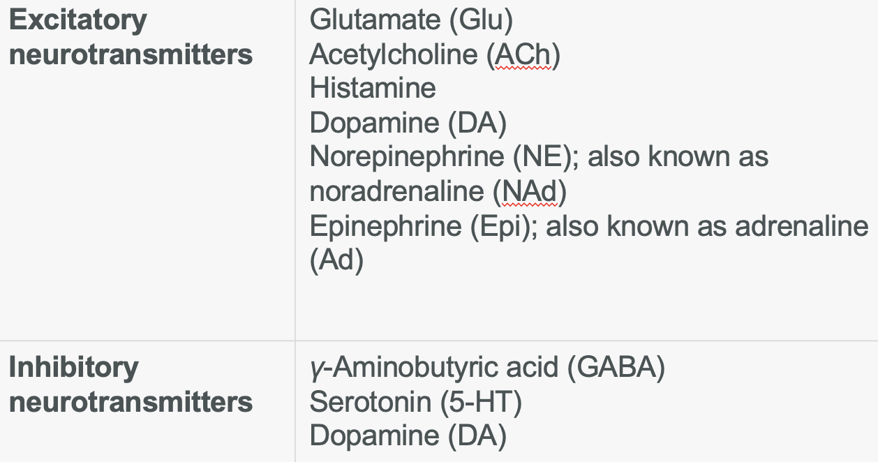
<p><strong>Characteristics: </strong></p><ul><li><p>Small (amino acids and amines)</p></li><li><p>Their receptors far outnumber the neurotransmitters.</p></li></ul><p></p><p><strong>Types:</strong></p><ul><li><p><strong>Excitatory neurotransmitters:</strong></p><ul><li><p><strong>Examples</strong>: Glutamate (Glu), Acetylcholine (ACh), Histamine, Dopamine (DA)</p></li></ul></li><li><p><strong>Inhibitory neurotransmitters:</strong></p><ul><li><p><strong>Examples: </strong><em>γ</em>-Aminobutyric acid (GABA), Serotonin (5-HT), Dopamine (DA)</p></li></ul></li></ul><p></p>
11
New cards

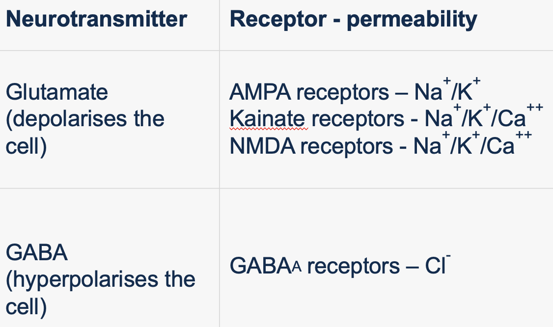
Outline the characteristics and types of ligand gated ion channels.

Characteristics:

  • Activation:

    • Only activated by specific molecules (neurotransmitters, NTs).

    • Ligand binding opens the channel.

  • Permeability:

    • Channels are permeable to different ions.

  • Excitatory Receptors:

    • Allow an influx of Na+ and Ca2+ and efflux of K+.

  • Inhibitory Receptors:

    • Permeable to Cl-.

Types of neurotransmitters with corresponding receptor:

  • Glutamate (Depolarizes the Cell):

    • AMPA Receptors: Na+/K+

    • Kainate Receptors: Na+/K+/Ca++

    • NMDA Receptors: Na+/K+/Ca++

  • GABA (Hyperpolarizes the Cell):

    • GABAA Receptors: Cl-

<p><strong>Characteristics:</strong></p><ul><li><p><strong>Activation:</strong></p><ul><li><p>Only activated by specific molecules (neurotransmitters, NTs).</p></li><li><p>Ligand binding opens the channel.</p></li></ul></li><li><p><strong>Permeability:</strong></p><ul><li><p>Channels are permeable to different ions.</p></li></ul></li><li><p><strong>Excitatory Receptors:</strong></p><ul><li><p>Allow an influx of Na+ and Ca2+ and efflux of K+.</p></li></ul></li><li><p><strong>Inhibitory Receptors:</strong></p><ul><li><p>Permeable to Cl-.</p><p></p></li></ul></li></ul><p><strong>Types of neurotransmitters with corresponding receptor:</strong></p><ul><li><p><strong>Glutamate (Depolarizes the Cell):</strong></p><ul><li><p><strong>AMPA Receptors:</strong> Na+/K+</p></li><li><p><strong>Kainate Receptors:</strong> Na+/K+/Ca++</p></li><li><p><strong>NMDA Receptors:</strong> Na+/K+/Ca++</p></li></ul></li><li><p><strong>GABA (Hyperpolarizes the Cell):</strong></p><ul><li><p><strong>GABAA Receptors:</strong> Cl-</p></li></ul></li></ul><p></p>
12
New cards

Outline the process of generating post-synaptic potentials, with examples.

Process:

  1. Activation of receptors within dendrites causes ion flow.

  2. Channel permeability and electrochemical forces dictate the direction of ion flow.

  3. Ion flow through these channels generates changes in membrane potential (post-synaptic potentials).

Examples:

  • Excitatory post-synaptic potential (EPSP):

    • AMPA receptor: forces sodium ions into the cell and potassium ions out the cell

    • NMDA receptor: forces sodium and calcium ions into cell and potassium ions out the cell

  • Inhibitory post-synaptic potential (IPSP):

    • GABAA/C receptor: forces chloride ions into the cell
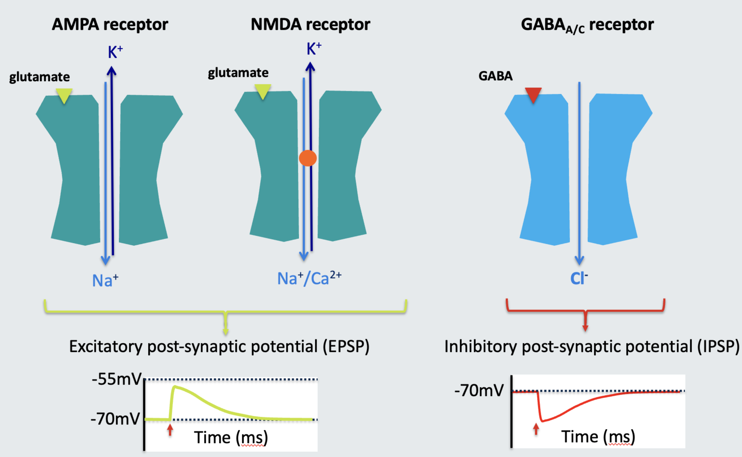
<p><strong>Process:</strong></p><ol><li><p>Activation of receptors within dendrites causes ion flow.</p></li><li><p>Channel <strong>permeability</strong> and <strong>electrochemical</strong> forces dictate the direction of ion flow.</p></li><li><p>Ion flow through these channels generates changes in membrane potential (<strong>post-synaptic potentials</strong>). </p></li></ol><p></p><p><strong>Examples:</strong></p><ul><li><p><span><strong>Excitatory post-synaptic potential (EPSP):</strong></span></p><ul><li><p><strong>AMPA receptor: </strong>forces sodium ions into the cell and potassium ions out the cell</p></li><li><p><strong>NMDA receptor: </strong>forces sodium and calcium ions into cell and potassium ions out the cell</p></li></ul></li><li><p><strong>Inhibitory post-synaptic potential (IPSP):</strong></p><ul><li><p><span><strong>GABA<sub>A/C</sub> receptor: </strong>forces chloride ions into the cell</span></p></li></ul></li></ul><p></p>
13
New cards
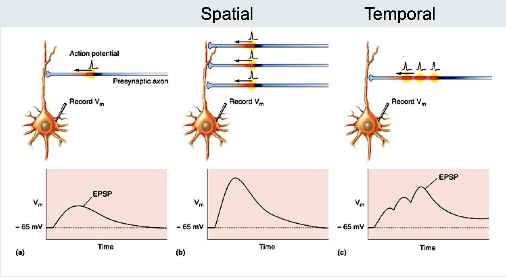
<p>State the event (a) occurring in the image.</p>

State the event (a) occurring in the image.

A presynaptic action potential triggers a small EPSP in a postsynaptic neuron.

14
New cards

State how different information is coded in the within and between neurons.

  • Inside cells = frequency of action potentials (digital)

  • Between cells = amount of neurotransmitter (NT) release
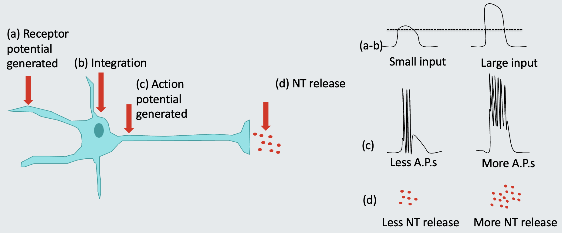
<ul><li><p><strong>Inside cells</strong> = frequency of action potentials (digital)</p></li><li><p style="text-align: left"><strong>Between cells</strong> = amount of neurotransmitter (NT) release</p></li></ul><p></p>
15
New cards

Outline the findings from David Hubel on the structure and function of the visual system.

Structures:

  • Lateral geniculate nucleus (LGN) neurons

  • Functions:

    • Respond to a dot of light in a particular part of the visual field
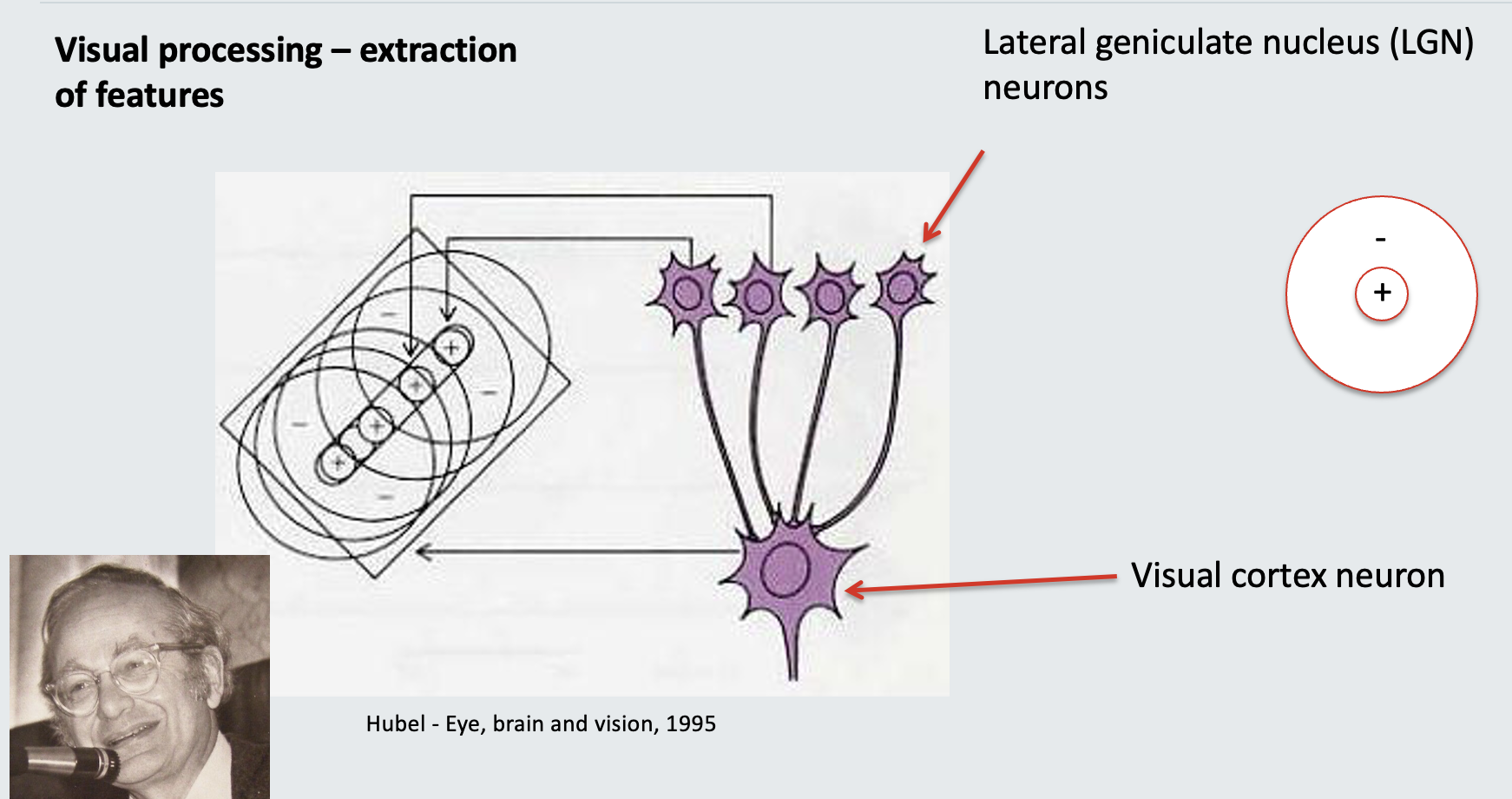
    • No response to general illumination over a wide area

  • Visual cortex neuron:

  • Functions:

    • Gets inputs from several LGN neurons

    • Fires an action potential when there is a line in a particular part of the visual field

<p><strong>Structures: </strong></p><ul><li><p>Lateral geniculate nucleus (LGN) neurons</p></li><li><p><strong>Functions: </strong></p><ul><li><p>Respond to a dot of light in a particular part of the visual field</p></li><li><p>No response to general illumination over a wide area</p></li></ul></li><li><p>Visual cortex neuron:</p></li><li><p><strong>Functions:</strong></p><ul><li><p>Gets inputs from several LGN neurons</p></li><li><p>Fires an action potential when there is a line in a particular part of the visual field</p></li></ul></li></ul><p></p>
16
New cards

State the different neuromodulators and how they interact.

  • Types:

    • Dopamine

    • Serotonin

    • Noradrenaline

  • Interaction: different neurotransmitter systems can modulate each other.

<ul><li><p><strong>Types:</strong></p><ul><li><p>Dopamine</p></li><li><p>Serotonin</p></li><li><p>Noradrenaline</p></li></ul></li><li><p><strong>Interaction: </strong><span>different neurotransmitter systems can modulate each other.</span></p></li></ul><p></p>
17
New cards

Outline the impact of dopamine as a neuromodulator.

  • Dopamine can be excitatory or inhibitory depending on the receptors expressed in the post-synaptic cell

  • Gating of excitatory signals,

    • e.g. by reward expectation or response selection in the striatum
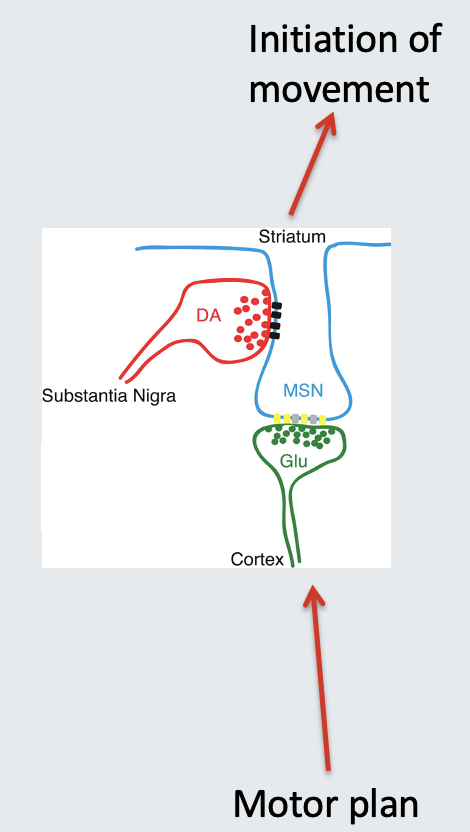
<ul><li><p style="text-align: left">Dopamine can be <strong>excitatory or inhibitory </strong>depending on the receptors expressed in the post-synaptic cell</p></li><li><p style="text-align: left">Gating of excitatory signals, </p><ul><li><p style="text-align: left">e.g. by reward expectation or response selection in the striatum</p></li></ul></li></ul><p></p>
18
New cards

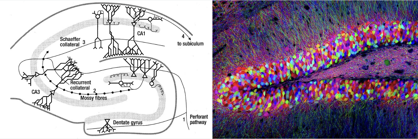
Outline the role of the hippocampus as an association network.

  • Memory Formation:

    • Process: Cells that fire at the same time become associated, leading to memory formation.

  • Memory Recall:

    • Mechanism: When some of these cells are reactivated, they can reactivate the entire network, enabling memory recall.

  • Potential for Epilepsy:

    • The same network that facilitates memory formation and recall can also become epileptic under certain conditions.
<ul><li><p><strong>Memory Formation:</strong></p><ul><li><p><strong>Process:</strong> Cells that fire at the same time become associated, leading to memory formation.</p></li></ul></li><li><p><strong>Memory Recall:</strong></p><ul><li><p><strong>Mechanism:</strong> When some of these cells are reactivated, they can reactivate the entire network, enabling memory recall.</p></li></ul></li><li><p><strong>Potential for Epilepsy:</strong></p><ul><li><p>The same network that facilitates memory formation and recall can also become epileptic under certain conditions.</p></li></ul></li></ul><p></p>
19
New cards

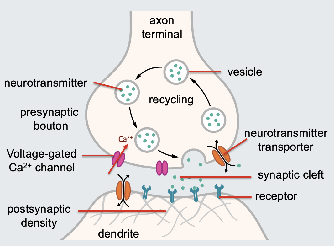
Outline the steps in transmission through the synapse.

  1. Action Potential Arrival:

    • Depolarisation of the membrane activates voltage-gated calcium channels.

  2. Calcium Influx:

    • Calcium enters the cell and is essential for docking neurotransmitter-filled vesicles to the presynaptic membrane.

  3. Neurotransmitter Release:

    • Neurotransmitters spread out into the synaptic cleft.

  4. Receptor Binding:

    • Neurotransmitters bind to receptors within the postsynaptic density on the dendrite.

  5. Surplus Removal:

    • Surplus neurotransmitters are taken up by glial cell transporters or transporters in the presynaptic membrane.

  6. Recycling:

    • Neurotransmitters are often recycled into new vesicles, ready for further release when the axon terminal depolarises again.

20
New cards

Postsynaptic Density

A huge protein complex on the dendrite that contains receptors for neurotransmitters.

21
New cards

Presynaptic bouton

Another name for axon terminals.
22
New cards
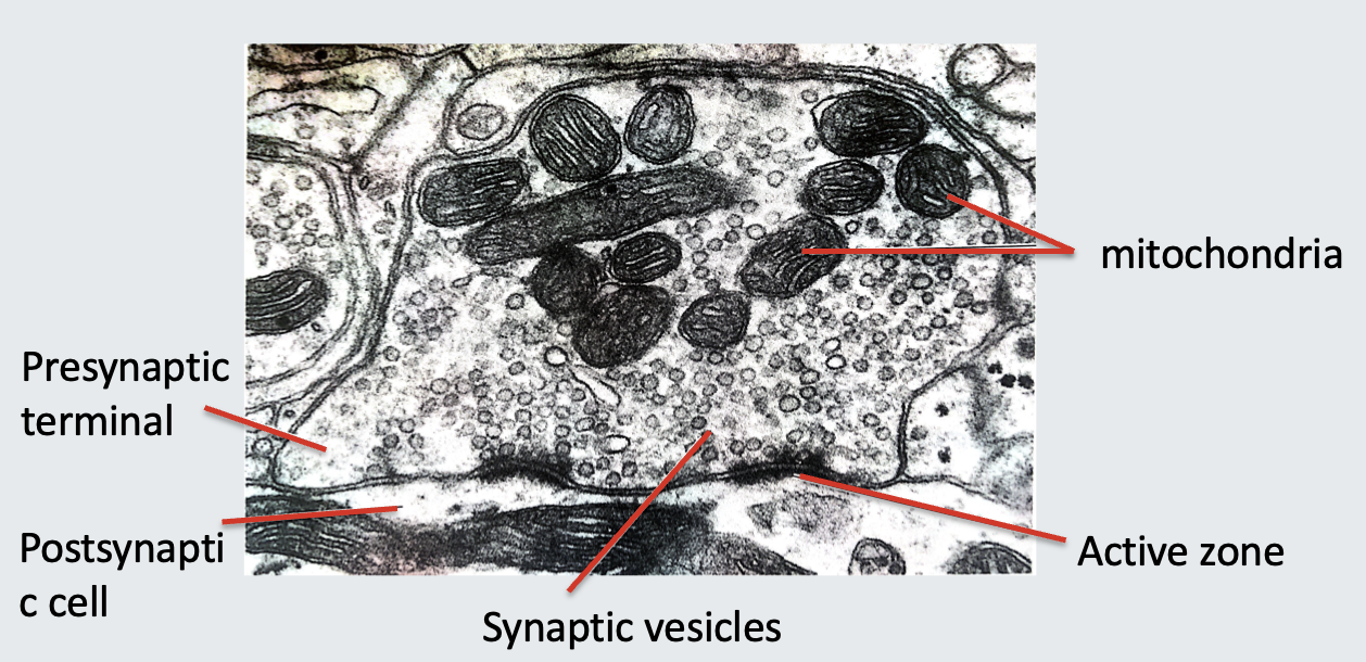
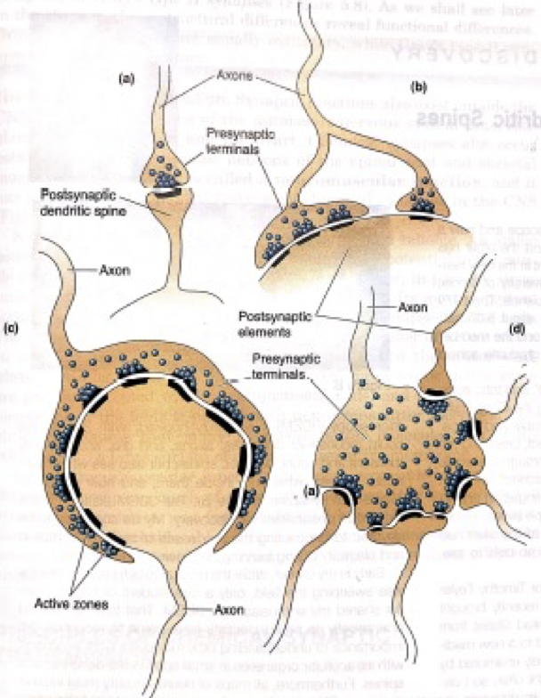
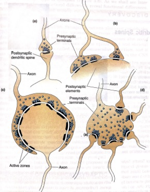
<p>Outline the shape and size of the synapse (a) shown in the image. </p>

Outline the shape and size of the synapse (a) shown in the image.

Axospinous Synapse:

  • Structure: A small presynaptic axon terminal contacts a postsynaptic dendritic spine.
23
New cards
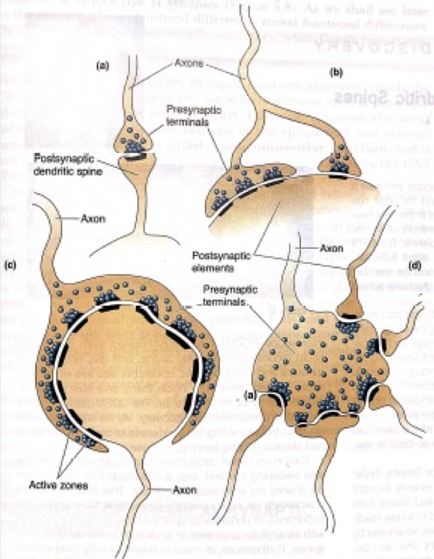
<p>Outline the shape and size of the synapse (b) shown in the image. </p>

Outline the shape and size of the synapse (b) shown in the image.

Axosomatic Synapse:

  • Structure: An axon branches to form two presynaptic terminals, one larger than the other, both contacting the postsynaptic soma (cell body).
24
New cards
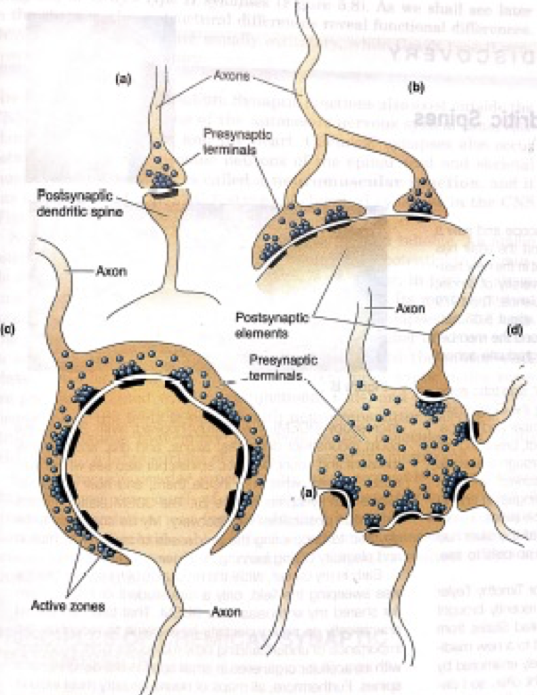
<p>Outline the shape and size of the synapse (c) shown in the image.</p>

Outline the shape and size of the synapse (c) shown in the image.

Calyx of Held:

  • Structure:

    • An unusually large axon terminal that contacts and envelops a postsynaptic soma.

  • Location:

    • Found where auditory sensory neurons connect to neurons in the trapezoid nucleus in the pons.

  • Function:

    • Rapid Transmission: Essential for quick relay of auditory information, ensuring timely processing of sound.

    • Mechanism: The large contact area allows an action potential in the presynaptic neuron to swiftly induce a new action potential in the postsynaptic neuron.
25
New cards
<p>Outline the shape and size of the synapse (d) shown in the image.</p>

Outline the shape and size of the synapse (d) shown in the image.

Large Presynaptic Terminal to Multiple Dendritic Spines:

  • Structure: An unusually large presynaptic axon terminal contacts five postsynaptic dendritic spines.

26
New cards

Outline the packing and storage step in the lifecycle of neurotransmitters.

  • Packaging and Storage:

    • Process: NTs are moved into vesicles from early endosomes.

    • Function: Vesicles store NTs and await the arrival of an action potential.

27
New cards

Outline the translocation step in the lifecycle of neurotransmitters.

  • Translocation:

    • Process: Vesicles move in and out of the synaptic bouton (axon terminal).

    • Role: Prepares vesicles for release upon action potential arrival.

28
New cards

Outline the release step in the lifecycle of neurotransmitters.

  • Release:

    • Trigger: An action potential causes the release of NTs by exocytosis.

    • Mechanism Requirements:

      • Docking: Vesicles dock at the presynaptic membrane.

      • Priming: Vesicles prepare for fusion.

      • Fusion: Vesicles fuse with the membrane to release NTs.

29
New cards

Outline the receptor activation step in the lifecycle of neurotransmitters.

  • Receptor Activation:

    • Process: NTs bind to receptors in the synaptic cleft.

    • Outcome: Initiates a response in the postsynaptic neuron.

30
New cards

Outline the inactivation step in the lifecycle of neurotransmitters.

  • Inactivation:

    • Processes to Terminate Signal:

      • Diffusion Away: NTs drift out of the synaptic cleft.

      • Enzymatic Degradation: NTs are broken down by enzymes.

      • Reuptake: NTs are taken up by:

        • Terminal Neuron (Presynaptic): Recycled for future use.

        • Astrocytes: Glial cells that help maintain neurotransmitter balance.

31
New cards

Define and outline the function of the tripartite synapse.

The Tripartite Synapse:

  • Components:

    1. Presynaptic Neuron

    2. Postsynaptic Neuron

    3. Astrocyte

  • Function:

    • Astrocytes regulate neurotransmitter levels and support synaptic function.

    • Ensure efficient synaptic transmission and prevent excitotoxicity.

32
New cards

Outline the mechanism of glutamate clearance by astrocytes.

Mechanism of Glutamate Clearance:

  • Glutamate Uptake:

    • Astrocytes "hoover up" glutamate from the synaptic cleft via EAATs.

  • Conversion to Glutamine:

    • Inside astrocytes, glutamate is converted into glutamine.

  • Recycling to Neurons:

    • Glutamine is sent back to neurons.

    • Neurons convert glutamine back into glutamate for reuse.

33
New cards

Describe EPSP summation and its two types.

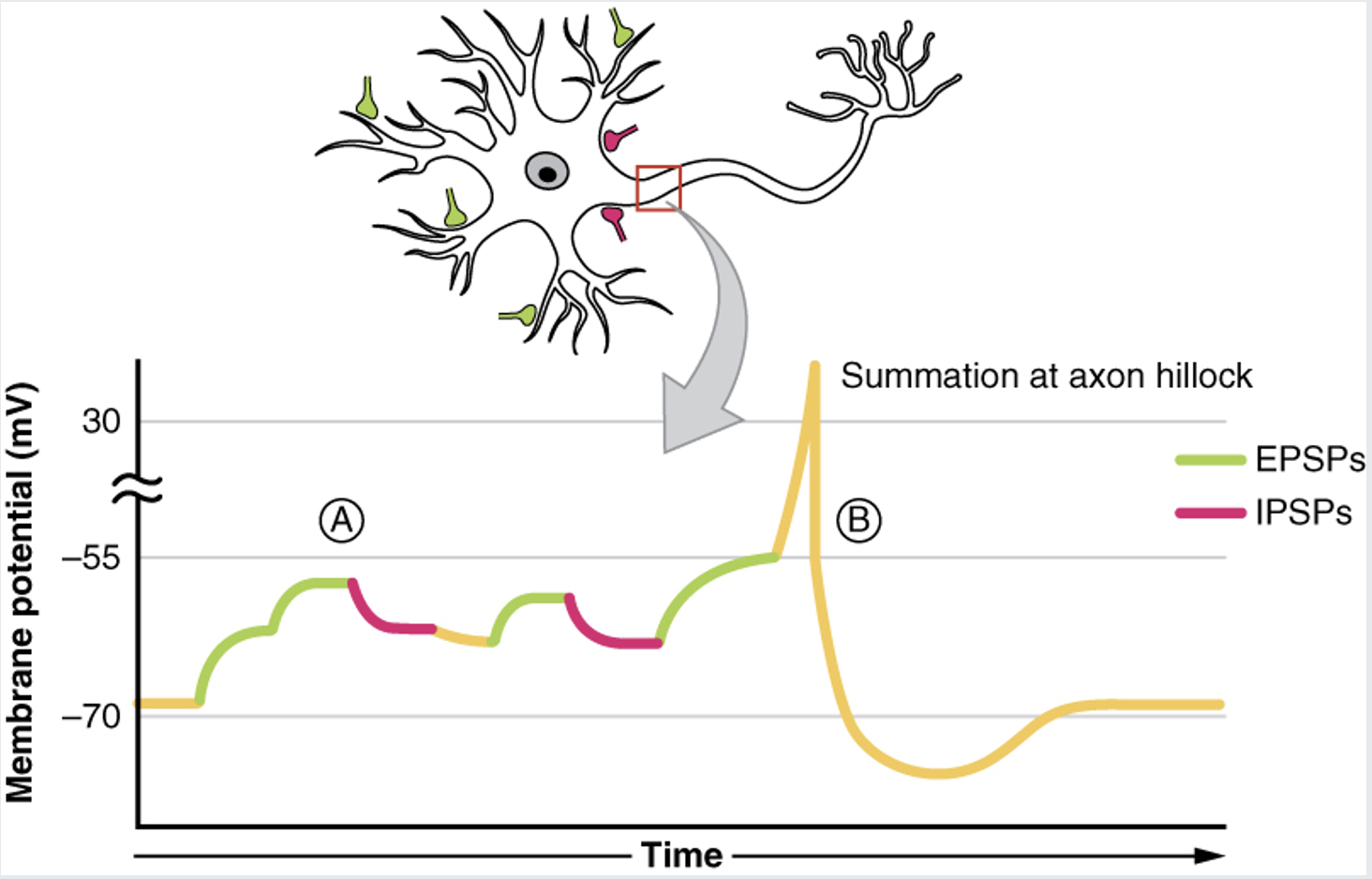
EPSP summation:

  • The combined effect of various potentials

    • The voltage changes from 2+ potentials and adds them

  • Due to a single EPSP not being enough to reach the threshold potential and create an action potential

    • Also, IPSPs reduced the likelihood of this happening.

  • Summation occurs in the axon hillock

  • It decreases in voltage as it travels along the cell body to the axon hillock

    • But, it must be enough where the membrane potential reaches the threshold so the action potential is generated.

Types:

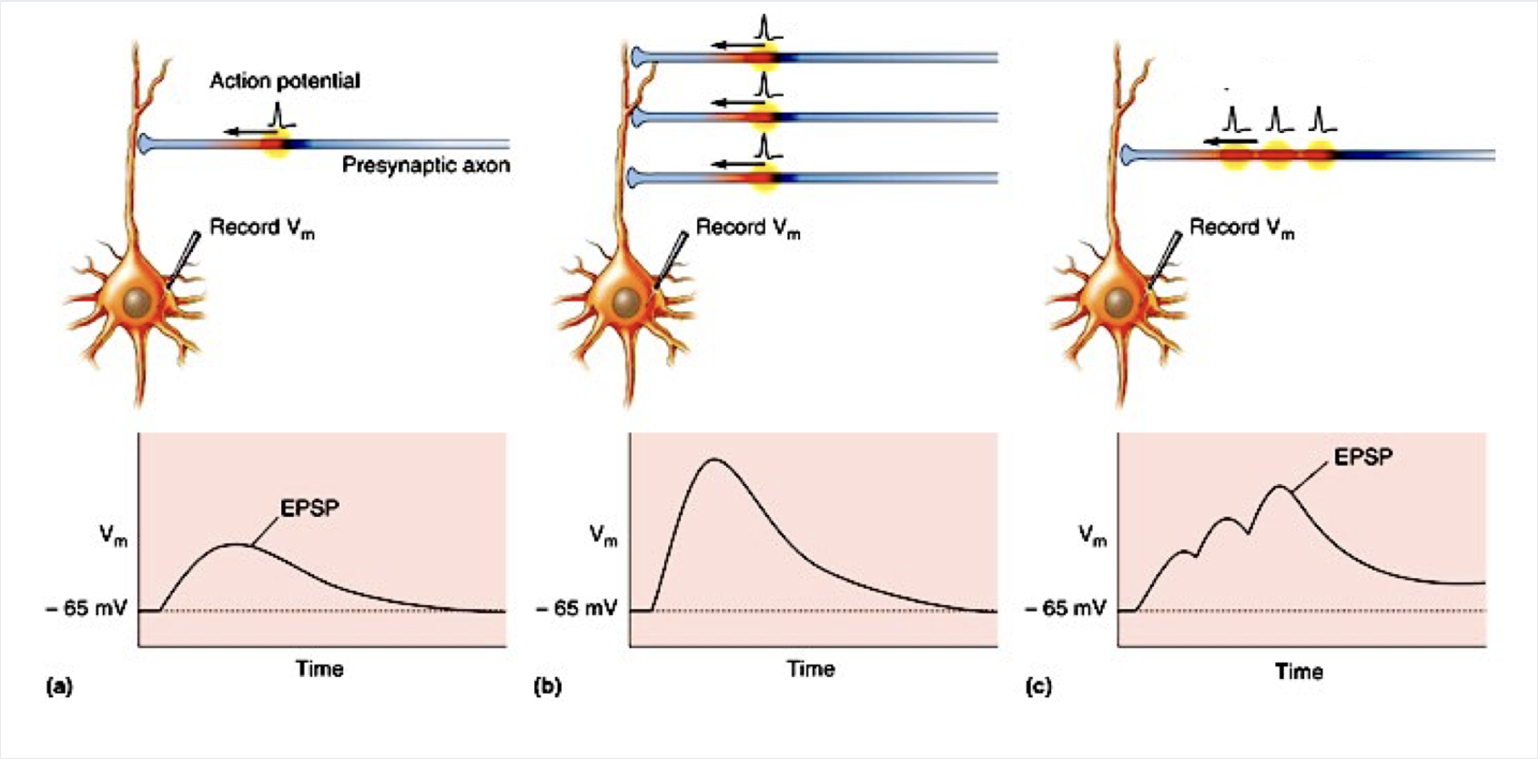
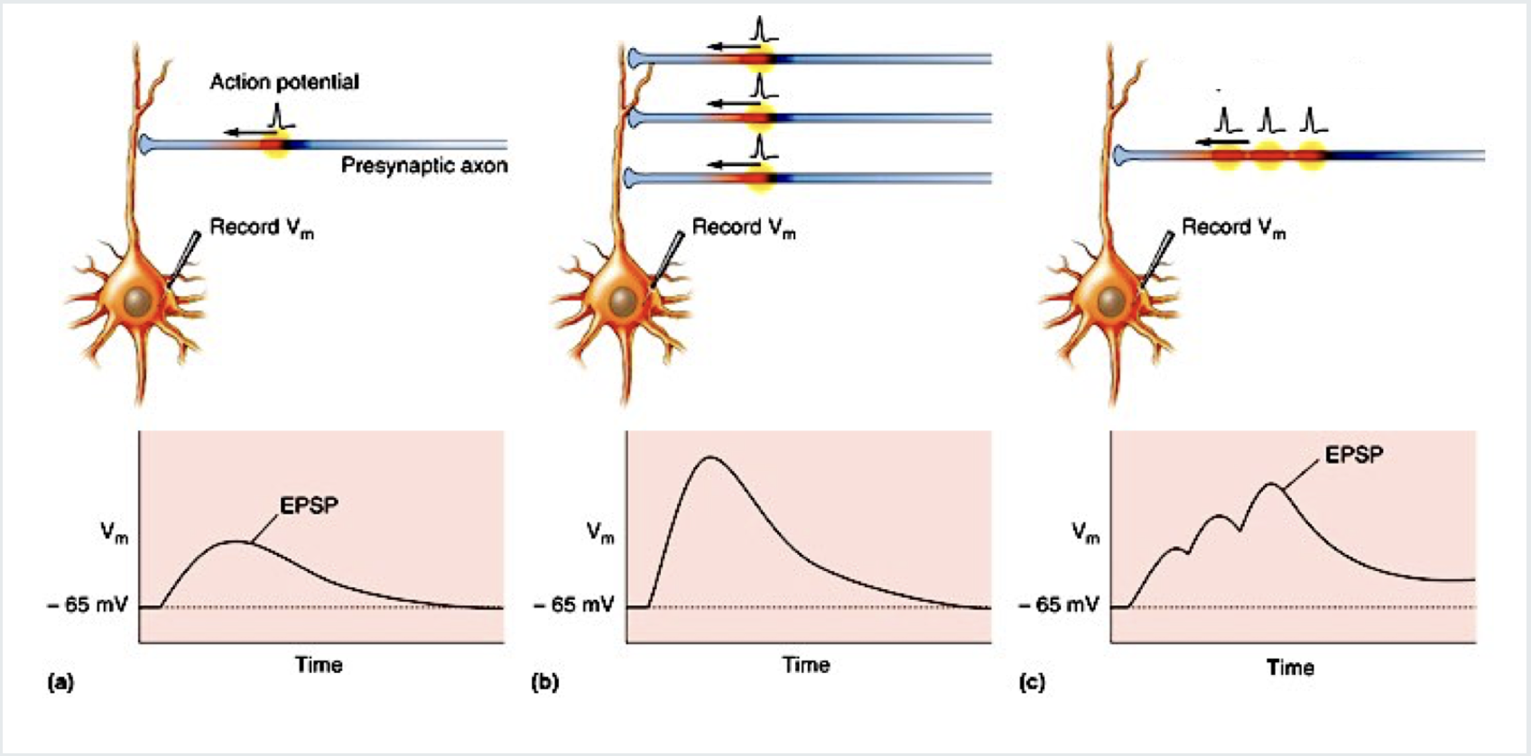
  • Spatial summation:

  • It occurs when action potentials on several presynaptic neurons arrive at the postsynaptic neuron simultaneously to result in a large compound EPSP, resulting in voltage that is enough to reach the threshold potential and trigger an action potential

  • Temporal summation:

  • It occurs when multiple action potentials travel down a presynaptic neuron one after another within a short time frame (it is time-dependent), causing them to summate/ add up at the axon hillock of the postsynaptic neuron.
<p><strong>EPSP summation:</strong></p><ul><li><p>The combined effect of various potentials</p><ul><li><p>The voltage changes from 2+ potentials and adds them</p></li></ul></li><li><p>Due to a single EPSP not being enough to reach the threshold potential and create an action potential</p><ul><li><p>Also, IPSPs reduced the likelihood of this happening.</p></li></ul></li><li><p>Summation occurs in the axon hillock</p></li><li><p>It decreases in voltage as it travels along the cell body to the axon hillock</p><ul><li><p>But, it must be enough where the membrane potential reaches the threshold so the action potential is generated.</p></li></ul></li></ul><p></p><p><strong>Types:</strong></p><ul><li><p><strong>Spatial summation:</strong></p></li><li><p>It occurs when action potentials on several presynaptic neurons arrive at the postsynaptic neuron simultaneously to result in a large compound EPSP, resulting in voltage that is enough to reach the threshold potential and trigger an action potential</p></li><li><p><strong>Temporal summation:</strong></p></li><li><p>It occurs when multiple action potentials travel down a presynaptic neuron one after another within a short time frame (it is time-dependent), causing them to summate/ add up at the axon hillock of the postsynaptic neuron.</p><p></p></li></ul><p></p>
34
New cards
<p>State the event (b) occurring in the image.</p>

State the event (b) occurring in the image.

Spatial summation of EPSPs:

  • Two or more presynaptic inputs are active at the same time., their individual EPSPs add together.
35
New cards
<p>State the event (c) occurring in the image.</p>

State the event (c) occurring in the image.

Temporal summation of EPSPs:

  • The same presynaptic fibre fires action potentials in quick succession.

36
New cards

Global brain circuits

Interconnected networks of neurons that process and transmit information within the brain.
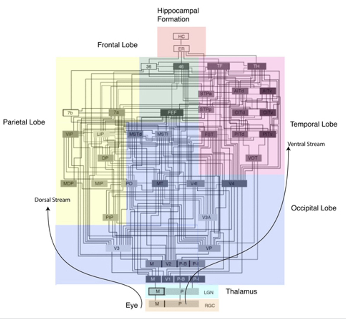
<p>Interconnected networks of neurons that process and transmit information within the brain.</p>
37
New cards

Association network

An area that integrates and interprets information from various sources to enable higher cognitive processes such as memory, learning, and decision-making.

38
New cards

Outline synaptic plasticity and how its examined.

  • Synaptic Plasticity:

    • Synaptic Conduction:

      • Can be strengthened or weakened based on past experiences.

    • Learning and Memory:

      • Synaptic plasticity represents forms of learning and memory.

    • Modifications:

      • Involves both presynaptic and postsynaptic modifications.

  • Mechanisms Examined:

    1. Changes in amount of neurotransmitter release.

    2. Biophysical changes to receptors and channels.

    3. Modulation by other transmitters, proteins, or channels.

    4. Morphological changes to the post-synapse (dendrites and spines).

    5. Synapse loss or sprouting.

    6. Changes in gene transcription.

39
New cards

State the various synaptic changes that occur during learning.

Synaptic changes:

  • Increased axonal transport

  • Increase in the number of synaptic vesicles

  • Change in size of synaptic cleft

  • Increase in terminal size or area

  • Increase in density of contact zones

  • Increase in spine size or area

  • Change in dendrite stem length and width

  • Increase in protein transport for spine construction

  • Changes in the number of spines and synapses
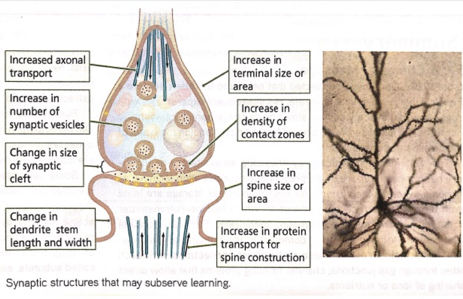
<p><span><strong>Synaptic changes:</strong></span></p><ul><li><p>Increased axonal transport</p></li><li><p class="p1">Increase in the number of synaptic vesicles</p></li><li><p class="p1">Change in size of synaptic cleft</p></li><li><p class="p1">Increase in terminal size or area</p></li><li><p class="p1">Increase in density of contact zones</p></li><li><p class="p1">Increase in spine size or area</p></li><li><p class="p1">Change in dendrite stem length and width</p></li><li><p class="p1">Increase in protein transport for spine construction</p></li><li><p class="p1">Changes in the number of spines and synapses </p></li></ul><p></p>
40
New cards

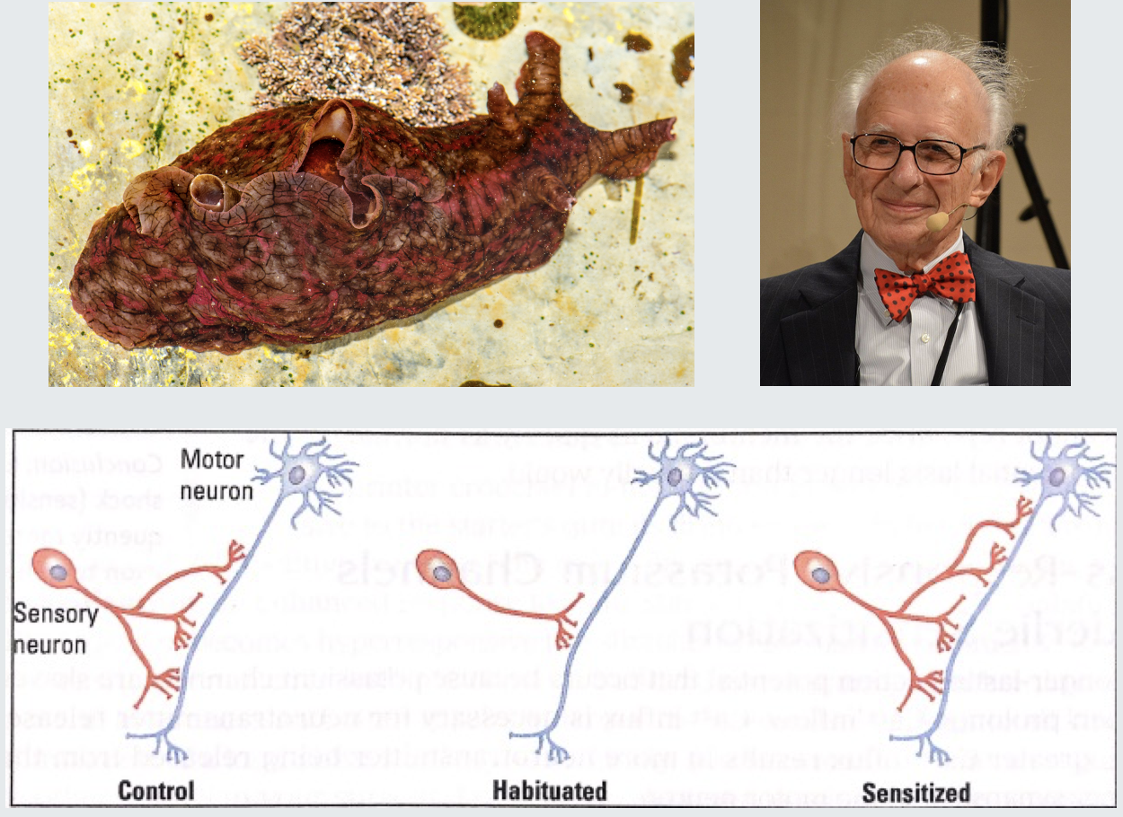
Outline the Eric Kandel’s discovery from Aplysia californica.

  • Discovery:

    • Researchers found that the number and size of sensory synapses change in well-trained, habituated, or sensitized Aplysia californica.

  • Synaptic Changes:

    • Habituated Animals:

      • Decrease in innate response to a repeated stimulus

      • Synapse number is decreased.

    • Sensitized Animals:

      • Increase in innate response to a repeated stimulus

      • Synapse number is increased.

<ul><li><p><strong>Discovery:</strong></p><ul><li><p>Researchers found that the number and size of sensory synapses change in well-trained, habituated, or sensitized Aplysia californica.</p></li></ul></li><li><p><strong>Synaptic Changes:</strong></p><ul><li><p><strong>Habituated Animals:</strong></p><ul><li><p>Decrease in innate response to a repeated stimulus</p></li><li><p>Synapse number is decreased.</p></li></ul></li><li><p><strong>Sensitized Animals:</strong></p><ul><li><p>Increase in innate response to a repeated stimulus</p></li><li><p>Synapse number is increased.</p></li></ul></li></ul></li></ul><p></p>
41
New cards

Outline the findings of the Kolb et al experiment on Drospohila.

Results:

  • They determined the cAMP was important for learning

  • Mutations that either increase or decrease cAMP levels reduce the learning capacity of drosophila.
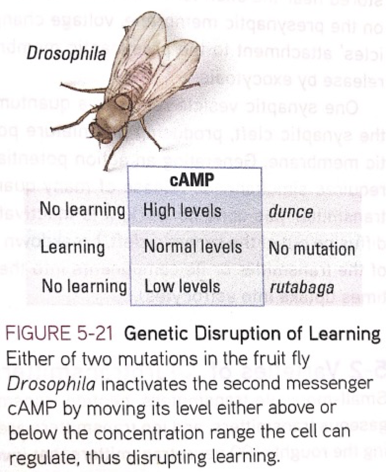
<p><strong>Results:</strong></p><ul><li><p>They determined the cAMP was important for learning</p></li><li><p>Mutations that either increase or decrease cAMP levels reduce the learning capacity of drosophila. </p></li></ul><p></p>
42
New cards

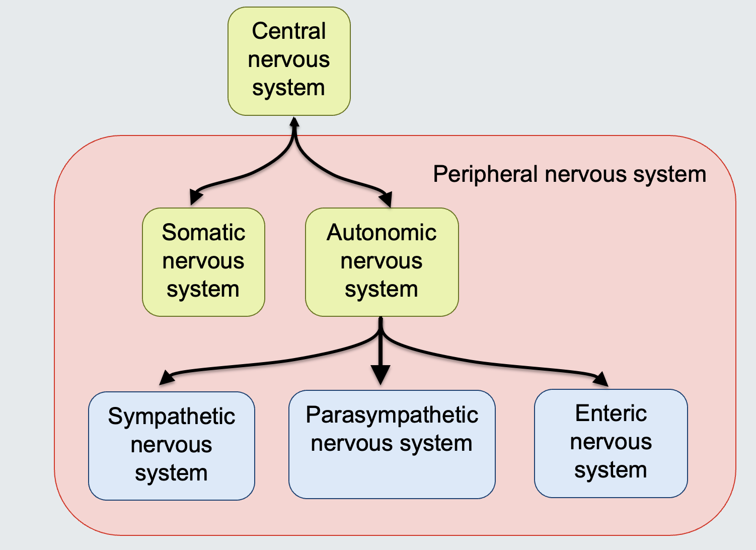
State the divisions of the nervous system.

Central Nervous System (CNS):

  • Subdivisions (Peripheral Nervous System):

    • Somatic Nervous System

    • Autonomic Nervous System

      • Sympathetic Nervous System

      • Parasympathetic Nervous System

      • Enteric Nervous System

<p>Central Nervous System (CNS):</p><ul><li><p>Subdivisions (Peripheral Nervous System):</p><ul><li><p>Somatic Nervous System</p></li><li><p>Autonomic Nervous System</p><ul><li><p>Sympathetic Nervous System</p></li><li><p>Parasympathetic Nervous System</p></li><li><p>Enteric Nervous System</p></li></ul></li></ul><p></p><p></p><p></p><p></p></li></ul><p></p>
43
New cards

Outline the structure and function of the nervous system divisions and draw their connections.

Central Nervous System (CNS):

  • Structure: Brain and spinal cord

  • Function: Integrative and control centers

Peripheral Nervous System (PNS):

  • Structure: Cranial nerves and spinal nerves

  • Function: Communication lines between the CNS and the rest of the body

Sensory (afferent) division:

  • Structure: Somatic and visceral sensory nerve fibers

  • Function: Conducts impulses from receptors to the CNS

Motor (efferent) division:

  • Structure: Motor nerve fibers

  • Function: Conducts impulses from the CNS to effectors (muscles and glands)

Autonomic nervous system (ANS):

  • Structure: Visceral motor (involuntary)

  • Function: Conducts impulses from the CNS to cardiac muscles, smooth muscles, and glands

Somatic nervous system:

  • Structure: Somatic motor (voluntary)

  • Function: Conducts impulses from the CNS to skeletal muscles

Sympathetic division:

  • Function: Mobilizes body systems during activity ("fight or flight")

Parasympathetic division:

  • Function: Conserves energy

  • Promotes "housekeeping" functions during rest
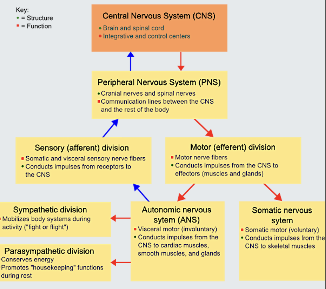
<p><strong>Central Nervous System (CNS):</strong></p><ul><li><p><strong>Structure: </strong>Brain and spinal cord</p></li><li><p><strong>Function:</strong> Integrative and control centers</p></li></ul><p></p><p><strong>Peripheral Nervous System (PNS):</strong></p><ul><li><p><strong>Structure: </strong>Cranial nerves and spinal nerves</p></li><li><p><strong>Function:</strong> Communication lines between the CNS and the rest of the body</p></li></ul><p></p><p><strong>Sensory (afferent) division:</strong></p><ul><li><p><strong>Structure: </strong>Somatic and visceral sensory nerve fibers</p></li><li><p><strong>Function:</strong> Conducts impulses from receptors to the CNS</p></li></ul><p class="p1"></p><p class="p1"><strong>Motor (efferent) division:</strong></p><ul><li><p><strong>Structure: </strong>Motor nerve fibers</p></li><li><p><strong>Function:</strong> Conducts impulses from the CNS to effectors (muscles and glands)</p></li></ul><p></p><p><strong>Autonomic nervous system (ANS):</strong></p><ul><li><p><strong>Structure: </strong>Visceral motor (involuntary)</p></li><li><p><strong>Function:</strong> Conducts impulses from the CNS to cardiac muscles, smooth muscles, and glands</p></li></ul><p class="p1"></p><p class="p1"><strong>Somatic nervous system:</strong></p><ul><li><p><strong>Structure: </strong>Somatic motor (voluntary)</p></li><li><p><strong>Function:</strong> Conducts impulses from the CNS to skeletal muscles</p></li></ul><p></p><p><strong>Sympathetic division:</strong></p><ul><li><p class="p1"><strong>Function:</strong> Mobilizes body systems during activity ("fight or flight")</p></li></ul><p></p><p><strong>Parasympathetic division:</strong></p><ul><li><p class="p1"><strong>Function:</strong> Conserves energy</p></li><li><p class="p1">Promotes "housekeeping" functions during rest</p></li></ul><p></p>
44
New cards

Outline the function and the vital role of the Autonomic Nervous System (ANS).

  • Function:

    • Regulates internal functions such as:

      • Keeping the heart beating

      • Releasing glucose from the liver

      • Adjusting pupils to light

  • Necessity:

    • Without the ANS, life would quickly cease.

    • It functions without conscious control, ensuring these processes continue during sleep.

  • Influence of Conscious States:

    • Sometimes, conscious states can affect the ANS.

    • For example, stress can lead to a raised heartbeat and other physiological changes.

45
New cards

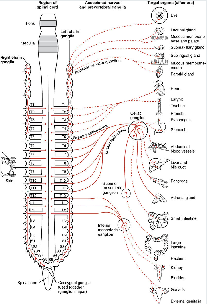
Outline the function of the sympathetic nervous system.

Function:

  • Fight or flight response

  • Constantly active at a basal level of homeostasis
<p><strong>Function:</strong></p><ul><li><p><span>Fight or flight response</span></p></li><li><p><span>Constantly active at a basal level of homeostasis</span></p></li></ul><p></p>
46
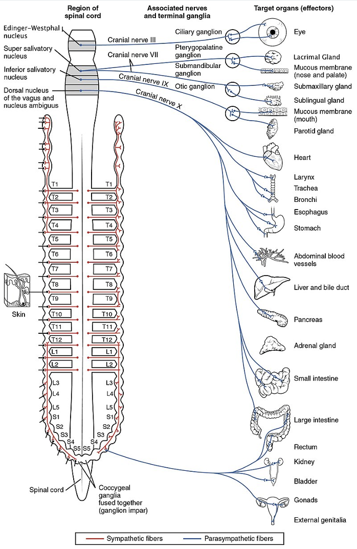
New cards

Outline the function of the parasympathetic nervous system.

Function:

  • Responsible for stimulation of "rest-and-digest" or "feed and breed“

  • Activities at rest, especially after eating, sexual arousal, salivation, lacrimation (tears), urination, digestion and defecation.

  • Involve the cranial nerves, the vagus nerve and pelvic splanchnic nerves.
<p><strong>Function:</strong></p><ul><li><p><span>Responsible for stimulation of "rest-and-digest" or "feed and breed“</span></p></li><li><p><span>Activities at rest, especially after eating, sexual arousal, salivation, lacrimation (tears), urination, digestion and defecation.</span></p></li><li><p><span>Involve the cranial nerves, the vagus nerve and pelvic splanchnic nerves.</span></p></li></ul><p></p>
47
New cards

Outline the organisation of neural outputs in the autonomic nervous system.

Somatic motor:

  • From CNS to skeletal muscle

Sympathetic (ANS):

  • From CNS, synapse in the autonomic (sympathetic) ganglion, to smooth muscle, cardiac muscle, gland cells

  • Contains pre and post-ganglionic fibres.
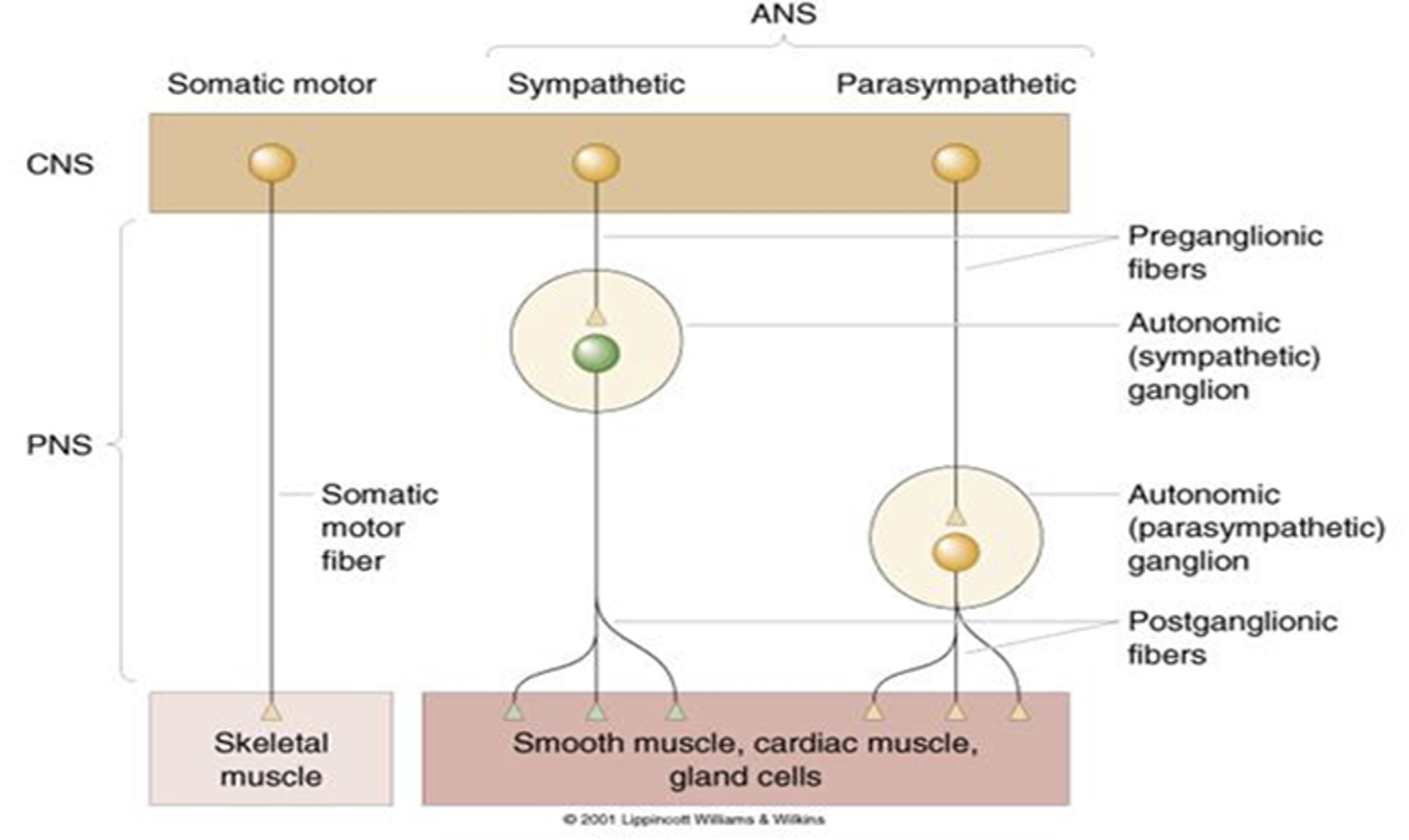
Parasympathetic (ANS):

  • From CNS, synapse in the autonomic (parasympathetic) ganglion, to smooth muscle, cardiac muscle, gland cells

  • Contains pre and post-ganglionic fibres.

<p><strong>Somatic motor:</strong></p><ul><li><p>From CNS to skeletal muscle</p></li></ul><p></p><p><strong>Sympathetic (ANS):</strong></p><ul><li><p>From CNS, synapse in the autonomic (sympathetic) ganglion, to smooth muscle, cardiac muscle, gland cells</p></li><li><p>Contains pre and post-ganglionic fibres. </p></li></ul><p></p><p><strong>Parasympathetic (ANS):</strong></p><ul><li><p>From CNS, synapse in the autonomic (parasympathetic) ganglion, to smooth muscle, cardiac muscle, gland cells</p></li><li><p>Contains pre and post-ganglionic fibres. </p></li></ul><p></p><p></p><p></p>
48
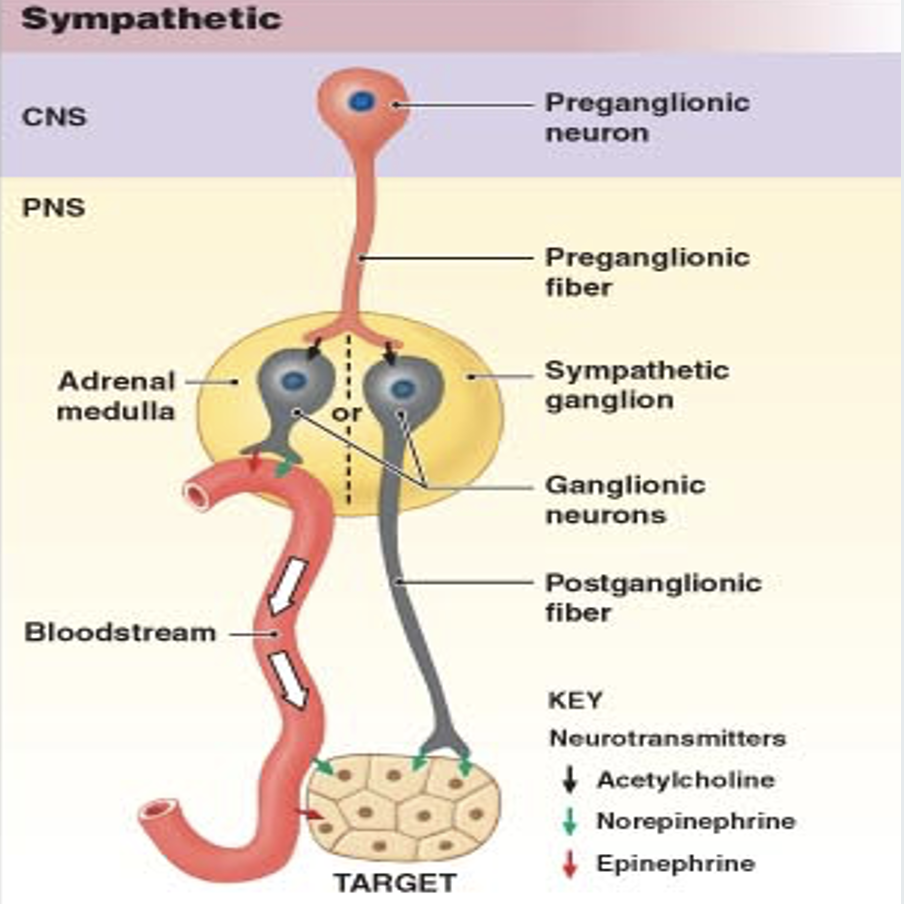
New cards

Describe neurotransmission in the sympathetic nervous systems.

First pathway for fight or flight response:

  1. Preganglionic Neurons:

    • Release acetylcholine (ACh).

  2. Postganglionic Neurons:

    • Express nicotinic ACh receptors.

    • Stimulation causes the release of norepinephrine.

  3. Norepinephrine Activation:

    • Activates adrenergic receptors on peripheral targets.

Second pathway for fight or flight response:

  1. Adrenal Medulla:

    • Stimulation releases norepinephrine and epinephrine into the bloodstream.
<p><strong>First pathway for fight or flight response:</strong></p><ol><li><p><strong>Preganglionic Neurons:</strong></p><ul><li><p>Release acetylcholine (ACh).</p></li></ul></li><li><p><strong>Postganglionic Neurons:</strong></p><ul><li><p>Express nicotinic ACh receptors.</p></li><li><p>Stimulation causes the release of norepinephrine.</p></li></ul></li><li><p><strong>Norepinephrine Activation:</strong></p><ul><li><p>Activates adrenergic receptors on peripheral targets.</p></li></ul></li></ol><p></p><p><strong>Second pathway for fight or flight response:</strong></p><ol><li><p><strong>Adrenal Medulla:</strong></p><ul><li><p>Stimulation releases norepinephrine and epinephrine into the bloodstream.</p></li></ul></li></ol><p></p>
49
New cards

Describe neurotransmission in the parasympathetic nervous system

  1. Preganglionic Neuron:

    • Releases acetylcholine (ACh) at the ganglion.

    • ACh acts on nicotinic receptors of postganglionic neurons.

  2. Postganglionic Neuron:

    • Releases ACh to stimulate the muscarinic receptors of the target organ.

  3. Muscarinic Receptors:

    • Different muscarinic receptors are expressed by different target organs.
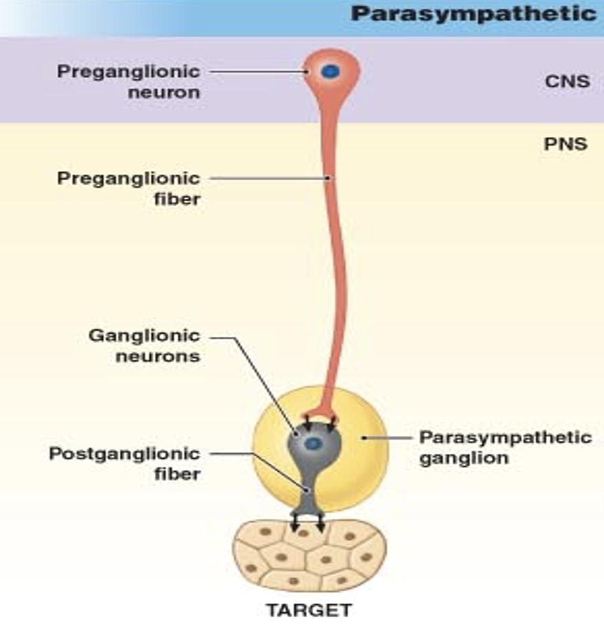
<ol><li><p><strong>Preganglionic Neuron:</strong></p><ul><li><p>Releases acetylcholine (ACh) at the ganglion.</p></li><li><p>ACh acts on nicotinic receptors of postganglionic neurons.</p></li></ul></li><li><p><strong>Postganglionic Neuron:</strong></p><ul><li><p>Releases ACh to stimulate the muscarinic receptors of the target organ.</p></li></ul></li><li><p><strong>Muscarinic Receptors:</strong></p><ul><li><p>Different muscarinic receptors are expressed by different target organs.</p></li></ul></li></ol><p></p>
50
New cards

Describe the role of summation, calcium and Hebb’s Postulate on synaptic plasticity.

  • Hebb’s Postulate:

    • "Cells that fire together, wire together."

  • Summation and Depolarisation:

    • Lots of summation leads to lots of depolarisation.

    • This activates NMDA receptors and allows calcium entry.

  • Calcium's Role:

    • Calcium triggers processes that strengthen synapses.

    • This makes it easier for these synapses to make the post-synaptic neuron fire an action potential in the future.

  • Experience and Synaptic Strength:

    • The strength of some synapses changes with experience.

  • Synaptic Weakening:

    • Connections between cells that don’t fire together become weaker.
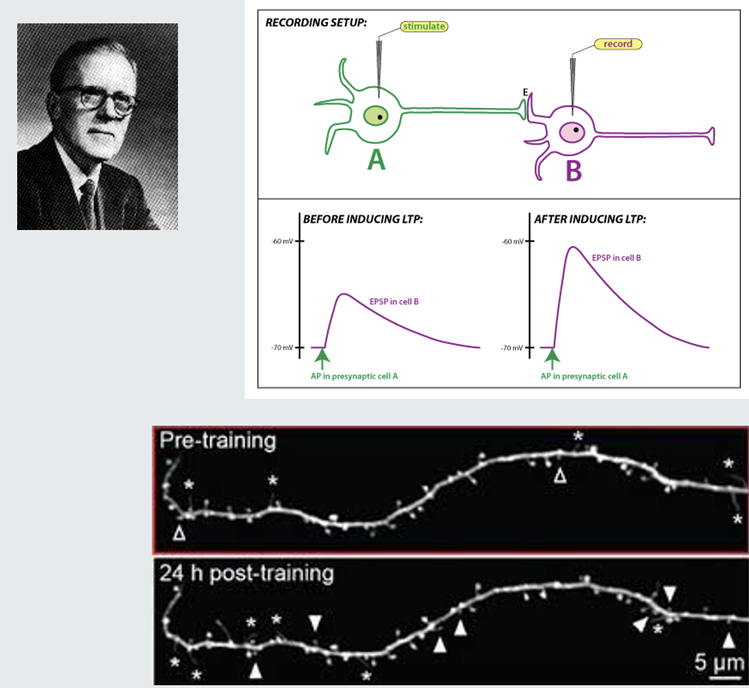
<ul><li><p><strong>Hebb’s Postulate:</strong></p><ul><li><p>"Cells that fire together, wire together."</p></li></ul></li><li><p><strong>Summation and Depolarisation:</strong></p><ul><li><p>Lots of summation leads to lots of depolarisation.</p></li><li><p>This activates NMDA receptors and allows calcium entry.</p></li></ul></li><li><p><strong>Calcium's Role:</strong></p><ul><li><p>Calcium triggers processes that strengthen synapses.</p></li><li><p>This makes it easier for these synapses to make the post-synaptic neuron fire an action potential in the future.</p></li></ul></li><li><p><strong>Experience and Synaptic Strength:</strong></p><ul><li><p>The strength of some synapses changes with experience.</p></li></ul></li><li><p><strong>Synaptic Weakening:</strong></p><ul><li><p>Connections between cells that don’t fire together become weaker.</p></li></ul></li></ul><p></p>